Browsed by
Category: Cancer

A Hidden Killer in Your Blood: The Truth About Leukemia

A Hidden Killer in Your Blood: The Truth About Leukemia

Leukemia is a type of blood cancer that affects the body’s white blood cells, weakening the immune system and making it harder to fight infections. Although it can sound frightening, early diagnosis and modern treatments have significantly improved survival rates. In this guide, you’ll learn everything about leukemia—its symptoms, causes, types, and the latest treatment options.


🧬 What Is Leukemia?

Leukemia develops in the bone marrow, where blood cells are produced. In this condition, the body creates abnormal white blood cells that don’t function properly. These unhealthy cells multiply quickly and crowd out healthy blood cells, leading to serious health issues.


⚠️ Common Symptoms of Leukemia

Leukemia symptoms can vary depending on the type and stage, but some early warning signs include:

  • Persistent fatigue and weakness
  • Frequent infections
  • Fever or chills
  • Easy bruising or bleeding
  • Unexplained weight loss
  • Swollen lymph nodes
  • Bone or joint pain

👉 These symptoms are often mild at first, so many people ignore them. Early detection is key to successful treatment.


🔍 Types of Leukemia

Leukemia is mainly classified into four major types:

1. Acute Lymphocytic Leukemia (ALL)

  • Common in children
  • Develops rapidly and requires immediate treatment

2. Acute Myeloid Leukemia (AML)

  • Affects both adults and children
  • Progresses quickly

3. Chronic Lymphocytic Leukemia (CLL)

  • Mostly seen in older adults
  • Develops slowly over time

4. Chronic Myeloid Leukemia (CML)

  • Associated with a genetic mutation
  • Progresses gradually but can become aggressive

🧪 Causes and Risk Factors

The exact cause of leukemia is not always known, but several risk factors may increase the chances:

  • Genetic mutations
  • Exposure to high levels of radiation
  • Smoking
  • Previous cancer treatments (chemotherapy or radiation)
  • Family history of blood cancers

👉 While these factors increase risk, leukemia can also occur in people with no known risk factors.


🩺 Diagnosis of Leukemia

Doctors use several tests to diagnose leukemia:

  • Blood tests to check abnormal cell counts
  • Bone marrow biopsy for confirmation
  • Imaging tests like CT scans
  • Genetic testing to identify specific mutations

Early diagnosis helps doctors choose the most effective treatment plan.


💊 Latest Treatment Options

Treatment depends on the type and severity of leukemia. Modern medicine offers several effective options:

1. Chemotherapy

The most common treatment that kills cancer cells or stops their growth.

2. Targeted Therapy

Drugs that specifically attack cancer cells without harming healthy cells.

3. Immunotherapy

Boosts the body’s immune system to fight leukemia.

4. Radiation Therapy

Uses high-energy rays to destroy cancer cells.

5. Stem Cell Transplant

Replaces damaged bone marrow with healthy cells.

👉 Advances in medical science have made these treatments more effective and less harmful than before.


🥗 Lifestyle and Supportive Care

While medical treatment is essential, lifestyle also plays an important role:

  • Eat a balanced, nutritious diet
  • Maintain hygiene to avoid infections
  • Stay physically active as per the doctor’s advice
  • Manage stress through meditation or relaxation techniques

Support from family and mental health care is equally important during recovery.


❤️ Can Leukemia Be Prevented?

There is no guaranteed way to prevent leukemia, but you can reduce risk by:

  • Avoiding smoking
  • Limiting exposure to harmful chemicals
  • Maintaining a healthy lifestyle

Regular health checkups can also help in early detection.


📝 Final Thoughts

Leukemia is a serious condition, but it is not unbeatable. With early diagnosis, advanced treatments, and proper care, many patients are living long and healthy lives. The key is awareness—recognizing symptoms early and seeking medical help without delay.

👉 If you notice any unusual symptoms, consult a healthcare professional immediately. Your health is your greatest asset—protect it.


References:

https://www.webmd.com/cancer/lymphoma/understanding-leukemia-basics
https://www.hopkinsmedicine.org/health/conditions-and-diseases/leukemia
https://my.clevelandclinic.org/health/diseases/4365-leukemia
https://www.medicinenet.com/leukemia/article.htm

Medications that have been suggested by doctors worldwide are available on the link below
https://mygenericpharmacy.com/category/disease/cancer

7 Brain Cancer Facts: Warning Signs & Proven Treatments

7 Brain Cancer Facts: Warning Signs & Proven Treatments

Brain Cancer Warning Signs You Should NEVER Ignore!

Brain cancer is a serious and life-threatening condition that affects thousands of people worldwide. Understanding the brain cancer causes and treatment options can help in early detection and better recovery. In this guide, we will explore key facts, warning signs, and proven treatments you should know.


What Is Brain Cancer?

Brain cancer occurs when abnormal cells grow uncontrollably in the brain. These tumors can be either benign (non-cancerous) or malignant (cancerous). Malignant tumors tend to grow quickly and can spread to other parts of the brain.

There are two main types:

  • Primary brain tumors (start in the brain)
  • Secondary brain tumors (spread from other organs)

7 Major Causes of Brain Cancer

Understanding the brain cancer causes and its treatment begins with identifying the risk factors. Here are seven key causes:

  1. Genetic mutations
  2. Family history of cancer
  3. Exposure to radiation
  4. Weak immune system
  5. Environmental toxins
  6. Age-related risks
  7. Previous cancer treatments

While these factors increase risk, not everyone exposed will develop brain cancer.


Early Warning Signs You Should Not Ignore

Recognizing symptoms early can save lives. Common warning signs include:

  • Persistent headaches
  • Frequent nausea or vomiting
  • Vision or speech problems
  • Seizures
  • Memory loss
  • Difficulty balancing

If these symptoms persist, it is important to consult a doctor immediately.


Types of Brain Tumors

There are several types of brain tumors, including:

  • Gliomas
  • Meningiomas
  • Pituitary tumors
  • Medulloblastomas

Each type varies in severity and requires a different approach to brain cancer causes and treatment.


Proven Treatment Options for Brain Cancer

Advancements in medicine have made treatment more effective. The most common options include:

Immunotherapy
It boosts the body’s immune system to fight cancer naturally.
Early diagnosis significantly improves treatment success rates.

Surgery
Doctors remove as much of the tumor as possible. This is often the first step.

Radiation Therapy
High-energy rays are used to kill cancer cells and shrink tumors.

Chemotherapy
Drugs are used to destroy cancer cells or stop their growth.

Targeted Therapy
This modern approach targets specific cancer cells without harming healthy tissue.


Prevention Tips & Risk Reduction

Although not all cases can be prevented, you can reduce risk by:

  • Avoiding unnecessary radiation exposure
  • Maintaining a healthy lifestyle
  • Eating a balanced diet
  • Exercising regularly
  • Avoiding toxic chemicals

These steps support overall brain health and reduce cancer risk.


When to See a Doctor

You should seek medical attention if symptoms like headaches, seizures, or vision problems persist. Early diagnosis plays a critical role in successful brain cancer causes and treatment outcomes.


Conclusion

Brain cancer is a complex condition, but awareness can make a big difference. By understanding the brain cancer causes and its treatment, recognizing early warning signs, and seeking timely care, you can improve survival chances and quality of life.

References:

https://my.clevelandclinic.org/health/diseases/6149-brain-cancer-brain-tumor
https://www.mayoclinic.org/diseases-conditions/brain-tumor/symptoms-causes/syc-20350084
https://www.cancer.org.au/types-of-cancer/brain-cancer

Medications that have been suggested by doctors worldwide are available on the link below
https://mygenericpharmacy.com/category/disease/cancer

The Truth About Malignant Tumors: Symptoms, Causes & Best Treatments

The Truth About Malignant Tumors: Symptoms, Causes & Best Treatments

Malignant Tumors: Symptoms, Causes & Treatments

Malignant tumors are a serious health concern that can affect any part of the body. These tumors are cancerous and can spread rapidly if not treated early. Therefore, understanding the symptoms, causes, and best treatments for malignant tumors is essential for early diagnosis and improved survival. In this guide, you will learn everything you need to know clearly and concisely.


What Are Malignant Tumors?

Malignant tumors are abnormal growths of cells that invade nearby tissues. Unlike benign tumors, they can spread to other parts of the body through the bloodstream or lymphatic system. In other words, malignant tumors are aggressive and require immediate medical attention. Early detection can significantly improve treatment outcomes.


Common Symptoms of Malignant Tumors

Recognizing symptoms early is very important. Although symptoms vary, some common warning signs include:

  • Unusual lumps or swelling
  • Persistent pain
  • Unexplained weight loss
  • Fatigue
  • Changes in skin or moles

Moreover, symptoms may worsen over time if ignored. Therefore, it is important to consult a doctor if you notice anything unusual.


Main Causes of Malignant Tumors

Understanding the causes helps in prevention. While the exact cause is not always known, several risk factors can increase the chances of developing malignant tumors.

Key Causes Include:

  • Genetic mutations
  • Family history of cancer
  • Exposure to radiation
  • Tobacco and alcohol use
  • Environmental toxins

Additionally, lifestyle choices play a major role. For instance, a poor diet and lack of exercise can increase cancer risk.


How Malignant Tumors Spread

Malignant tumors can spread through a process called metastasis. In this process, cancer cells travel to other organs and form new tumors. As a result, treatment becomes more complex. That is why early diagnosis is crucial for controlling the disease.


Best Treatment Options for Malignant Tumors

Fortunately, modern medicine offers several effective treatments. The choice depends on the type and stage of cancer.

Immunotherapy
Immunotherapy helps the immune system fight cancer naturally. It is a newer and promising option.

Surgery
Surgery is often the first step. Doctors remove the tumor and surrounding tissues.

Chemotherapy
Chemotherapy uses drugs to destroy cancer cells. It is commonly used when cancer has spread.

Radiation Therapy
Radiation therapy uses high-energy rays to kill cancer cells. It is often combined with other treatments.

Targeted Therapy
This treatment targets specific cancer cells without harming healthy ones. Therefore, it has fewer side effects.


Prevention Tips You Should Follow

Although not all tumors can be prevented, you can reduce your risk by following healthy habits.

  • Eat a balanced diet
  • Exercise regularly
  • Avoid tobacco and alcohol
  • Limit exposure to harmful chemicals
  • Get regular health checkups

In addition, staying informed can help you take early action.


When to See a Doctor

You should consult a doctor if symptoms persist for more than a few weeks. Early diagnosis increases the chances of successful treatment. Do not ignore warning signs, even if they seem minor.


Conclusion

Malignant tumors are dangerous but manageable if detected early. By understanding malignant tumor symptoms, causes, and best treatments, you can take control of your health. Ultimately, awareness, early diagnosis, and proper treatment can save lives.

References:

https://my.clevelandclinic.org/health/diseases/22319-malignantneoplasm
https://www.verywellhealth.com/what-does-malignant-and-benign-mean-514240
https://www.cancer.org/cancer/understanding-cancer/what-is-cancer/neoplasms-and-tumors.html
https://medical-dictionary.thefreedictionary.com/Malignant+tumors

Medications that have been suggested by doctors worldwide are available on the link below
https://mygenericpharmacy.com/category/disease/cancer

A New Era in Gastroenterology: AI Breakthrough for Early Cancer Detection

A New Era in Gastroenterology: AI Breakthrough for Early Cancer Detection

The field of gastroenterology is undergoing a powerful transformation, thanks to the rapid advancement of artificial intelligence (AI). One of the most exciting developments is the use of AI in early cancer detection—particularly for gastrointestinal cancers such as colorectal cancer, which remains a leading cause of cancer-related deaths worldwide. By enhancing diagnostic accuracy and enabling earlier intervention, AI is paving the way for a new era in patient care.


Why Early Cancer Detection Matters

Early detection is critical in improving survival rates for gastrointestinal cancers. When cancers are identified at an early stage, treatment is more effective, less invasive, and associated with better outcomes. However, traditional diagnostic methods such as endoscopy and colonoscopy, while effective, can sometimes miss subtle or early-stage abnormalities.


The Role of AI in Gastroenterology

AI is transforming how doctors detect and diagnose gastrointestinal diseases. Using advanced algorithms and machine learning, AI systems can analyze medical images, identify patterns, and detect abnormalities with high precision. These systems are particularly useful in procedures like colonoscopy, where detecting small or flat lesions can be challenging.


How AI Enhances Cancer Detection

1. Real-Time Image Analysis

AI tools can analyze endoscopic images in real time, flagging suspicious areas for further examination.

2. Improved Detection Rates

Studies show that AI-assisted procedures significantly increase the detection of polyps and early-stage cancers.

3. Consistent Accuracy

Unlike human interpretation, AI provides standardized analysis, reducing variability among clinicians.

4. Faster Decision-Making

AI helps clinicians make quicker and more informed decisions during procedures.


Applications in Gastrointestinal Diseases

AI is being used across a range of gastrointestinal conditions, including:

  • Colorectal cancer screening
  • Detection of polyps during colonoscopy
  • Monitoring patients with Ulcerative Colitis
  • Identifying early signs of esophageal and gastric cancers

Patients with Ulcerative colitis are at a higher risk of developing colorectal cancer, making AI-assisted screening especially valuable for this group.


Benefits of AI in Early Cancer Detection

1. Higher Accuracy

AI can detect abnormalities that may be missed by the human eye, improving diagnostic precision.

2. Early Intervention

Earlier detection allows for timely treatment, increasing survival rates.

3. Reduced Miss Rates

AI-assisted colonoscopy has been shown to reduce the number of missed lesions.

4. Better Patient Outcomes

Improved diagnosis leads to more effective treatment and improved quality of life.


What Does Research Say?

Research supported by organizations like the National Institutes of Health shows that AI can significantly improve the detection of colorectal polyps and early cancers.

Studies published in journals such as The Lancet highlight the growing role of AI in enhancing diagnostic performance in gastroenterology.

Additionally, the World Health Organization emphasizes that early detection is one of the most effective strategies for reducing cancer mortality worldwide.


Challenges and Limitations

Despite its promise, AI in healthcare faces several challenges:

  • High implementation costs
  • Need for large, high-quality datasets
  • Integration with existing healthcare systems
  • Ethical and data privacy concerns

Importantly, AI is designed to assist—not replace—healthcare professionals.


The Future of AI in Gastroenterology

The future of AI in gastroenterology is incredibly promising. Potential advancements include:

  • Personalized screening programs based on individual risk
  • Predictive analytics for disease progression
  • AI-driven treatment planning
  • Integration with wearable health technologies

As research continues, AI will likely become a standard part of gastrointestinal care.


Final Thoughts

The integration of AI into gastroenterology marks a significant milestone in modern medicine. By improving the early detection of cancers and enhancing diagnostic accuracy, AI is helping save lives and reshape the future of healthcare.

For high-risk patients, especially those with conditions like Ulcerative colitis, AI offers new hope for earlier diagnosis and better outcomes. As technology and medicine continue to evolve together, the promise of smarter, faster, and more effective healthcare is becoming a reality.


References:

  1. National Institutes of Health – AI in cancer detection research
  2. The Lancet – Studies on AI in gastroenterology
  3. World Health Organization – Cancer prevention and early detection
  4. American Gastroenterological Association – Screening and clinical guidelines
  5. Mayo Clinic – Gastrointestinal disease insights

Medications that have been suggested by doctors worldwide are available on the link below:
https://mygenericpharmacy.com/category/disease/cancer

Could It Be Bladder Cancer? Screening and Diagnosis You Need to Know

Could It Be Bladder Cancer? Screening and Diagnosis You Need to Know

Bladder Cancer Screening & Diagnosis: Early Detection GuideLearn about bladder cancer screening and diagnosis, key symptoms, tests, and why early detection is crucial for better treatment outcomes and survival.

Blood in your urine. A persistent urgency to go. A dull ache in your lower abdomen. These symptoms are easy to dismiss—perhaps a urinary tract infection, or simply “getting older.” But for thousands of men and women each year, these subtle signs mark the beginning of a bladder cancer diagnosis.

Bladder cancer is the 10th most common cancer worldwide, and it ranks even higher among men. The good news? When caught early, bladder cancer has a five-year survival rate of over 90%. The challenge lies in knowing when to seek screening and what to expect from the diagnostic process.

Who Is at Risk?

Understanding your risk factors is the first step toward early detection. While bladder cancer can affect anyone, certain populations face significantly higher odds:

  • Age: The risk increases dramatically after age 55. Most diagnoses occur in people aged 65 and older.
  • Gender: Men are 3 to 4 times more likely to develop bladder cancer than women.
  • Smoking: This is the single greatest risk factor. Smokers are 2 to 4 times more likely to develop bladder cancer than non-smokers. Approximately half of all bladder cancer cases are linked to smoking.
  • Chemical Exposures: Chronic exposure to industrial chemicals, arsenic in drinking water, and certain dyes or rubber products increases risk.
  • Chronic Bladder Inflammation: Recurrent infections or long-term catheter use can elevate risk for certain types of bladder cancer.

Recognizing the Warning Signs

Bladder cancer does not always announce itself loudly. The most common symptom—and often the first—is hematuria (blood in the urine).

Key Symptoms to Watch For:

  • Blood in the urine: This may appear bright red, rust-colored, or only detectable under a microscope. Importantly, bleeding is often painless and intermittent—it may come and go, leading many to assume the problem has resolved.
  • Changes in urination: Frequent urination, urgency, or a burning sensation that mimics a UTI.
  • Pelvic or back pain: Discomfort in the lower abdomen, pelvis, or flank area may indicate advanced disease.
  • Unexplained weight loss or fatigue: These systemic symptoms typically appear in later stages.

If you experience any of these symptoms, particularly blood in the urine, do not wait. Prompt evaluation is essential.

Screening: Is There a Standard Test?

Unlike breast, colon, or prostate cancer, there is no routine screening test for bladder cancer recommended for the general population. However, for high-risk individuals—such as long-term smokers or those with occupational exposures—doctors may recommend periodic urinalysis or other assessments.

If symptoms are present or suspicion is high, your healthcare provider will move to diagnostic testing.

The Diagnostic Pathway

Diagnosing bladder cancer involves a combination of tests. Understanding this process can help reduce anxiety and prepare you for what lies ahead.

1. Urinalysis and Urine Cytology

A simple urine test can detect the presence of blood and infection. Urine cytology goes a step further, examining urine under a microscope to look for cancer cells. While useful, cytology is not always definitive—some cancers do not shed detectable cells into the urine.

2. Cystoscopy: The Gold Standard

If bladder cancer is suspected, a cystoscopy is the definitive diagnostic procedure. A thin, flexible tube with a camera (cystoscope) is inserted through the urethra into the bladder. This allows the urologist to directly visualize the bladder lining and identify any abnormal growths, tumors, or suspicious areas.

Cystoscopy is typically performed in an office setting with local anesthesia. While it may cause some discomfort, it is the most reliable method for detecting bladder cancer.

3. Biopsy and Transurethral Resection of Bladder Tumor (TURBT)

If a suspicious area is found during cystoscopy, a biopsy is performed. In most cases, this involves a procedure called TURBT, where the urologist removes the abnormal tissue through the cystoscope. This serves two purposes: it provides tissue for pathological diagnosis, and it can be therapeutic if the tumor is completely removed.

The biopsied tissue is then examined by a pathologist to confirm the presence of cancer, determine the grade (how aggressive the cells appear), and assess the stage (whether it has invaded the muscle layer of the bladder wall).

4. Imaging Studies

If cancer is confirmed, imaging such as CT urography, MRI, or ultrasound may be used to evaluate the upper urinary tract (kidneys and ureters) and determine whether the cancer has spread beyond the bladder.

The Importance of Early Detection

Bladder cancer is broadly divided into two categories:

  • Non-muscle invasive bladder cancer (NMIBC): Confined to the inner lining of the bladder. This represents about 70-75% of cases at diagnosis. While it requires long-term surveillance and often recurring treatment, it is highly manageable.
  • Muscle-invasive bladder cancer (MIBC): Has penetrated the muscular wall of the bladder. This is more aggressive and requires more intensive treatment, including surgery, chemotherapy, or immunotherapy.

The distinction between these two stages hinges entirely on early diagnosis. Delaying evaluation allows tumors that might have been confined to the lining to invade deeper tissues.

What to Do Next

If you are experiencing any symptoms or fall into a high-risk category, do not wait for symptoms to worsen. Schedule an appointment with your primary care provider or a urologist. Be prepared to discuss:

  • Any visible or microscopic blood in your urine
  • Changes in urinary habits
  • Your smoking history
  • Occupational exposures to chemicals

Support Your Health Journey

At My Generic Pharmacy, we are committed to supporting patients through every stage of their health journey. While we do not provide diagnostic services, we offer a range of supportive products to help manage overall wellness before, during, and after treatment.

Reference:

https://www.cancer.org/cancer/types/bladder-cancer/detection-diagnosis-staging/how-diagnosed.html
https://www.hopkinsmedicine.org/health/conditions-and-diseases/bladder-cancer/bladder-cancer-screening-and-diagnosis
https://www.mayoclinic.org/diseases-conditions/bladder-cancer/symptoms-causes/syc-20356104
https://www.webmd.com/cancer/bladder-cancer/do-i-have-bladder-cancer
https://www.cancerresearchuk.org/about-cancer/bladder-cancer/getting-diagnosed/screening

Medications that have been suggested by doctors worldwide are available on the link below
https://mygenericpharmacy.com/category/disease/cancer


Disclaimer: This article is for educational purposes only and does not constitute medical advice. Bladder cancer screening, diagnosis, and treatment should be discussed with a qualified healthcare provider. If you are experiencing symptoms such as blood in your urine, seek medical evaluation promptly.

Colorectal Cancer: Warning Signs That Could Save Your Life

Colorectal Cancer: Warning Signs That Could Save Your Life

Colorectal cancer—cancer of the colon or rectum is the third most common cancer diagnosed in both men and women in the United States. It’s also one of the most preventable and treatable cancers when caught early. Yet despite advances in screening and treatment, many people delay seeing a doctor until symptoms become impossible to ignore.

The tragedy is that colorectal cancer often develops slowly, over several years, from precancerous polyps that can be removed before they become cancerous. Knowing the warning signs and acting on them promptly could literally save your life.

Why Early Detection Matters

Colorectal cancer has a 90% survival rate when caught early (localized stage). However, once it spreads to distant organs, the survival rate drops dramatically to around 14% . This stark difference underscores why recognizing warning signs and getting screened is so critical.

Key statistics:

  • About 1 in 24 people (4.2%) will be diagnosed with colorectal cancer in their lifetime
  • An estimated 153,000 new cases are diagnosed annually in the US
  • Screening could prevent 60% of colorectal cancer deaths

The Warning Signs You Should Never Ignore

Colorectal cancer often causes no symptoms in its earliest stages. That’s why screening is so important. But as the disease progresses, certain warning signs may appear.

1. Changes in Bowel Habits

This is one of the most common early warning signs. Pay attention to:

  • Diarrhea or constipation that lasts more than a few days
  • Narrow or ribbon-like stools (may indicate a tumor narrowing the colon)
  • Feeling that you haven’t completely emptied your bowel after a movement
  • Alternating constipation and diarrhea without a clear cause

2. Blood in Stool

Blood can appear in several ways:

  • Bright red blood on toilet paper or in the toilet bowl (may indicate bleeding near the rectum)
  • Dark red or maroon stools (suggest bleeding higher in the colon)
  • Black, tarry stools (indicate bleeding in the upper digestive tract)

Important: While blood in stool is often caused by hemorrhoids or anal fissures, never assume it’s harmless without medical evaluation.

3. Persistent Abdominal Discomfort

  • Cramping or pain in the lower abdomen
  • Gas pains or bloating that don’t resolve
  • Feeling full or bloated even after eating small amounts

4. Unexplained Weight Loss

Losing 10 pounds or more without trying can be a sign that cancer is affecting your body’s metabolism or causing inflammation.

5. Fatigue and Weakness

Persistent tiredness that doesn’t improve with rest may result from:

  • Internal bleeding causes anemia (low red blood cells)
  • The body’s energy is being diverted to fight cancer
  • Nutritional deficiencies from poor absorption

6. Iron Deficiency Anemia

Often discovered during routine blood tests, iron deficiency anemia without an obvious cause (like heavy periods) should always prompt investigation for colon cancer, especially in men and postmenopausal women.

When to See a Doctor

Don’t wait for multiple symptoms to appear. See a healthcare provider if:

  • YDo youhave any of these symptoms lasting more than a few days
  • You notice blood in your stool—even once
  • You have unexplained weight loss or fatigue
  • You’re due for screening (age 45 or older, or earlier with risk factors)
  • You have a family history of colorectal cancer or polyps

Who Is at Risk?

Non-Modifiable Risk Factors

  • Age: Risk increases significantly after 45 (new guidelines recommend starting screening at 45, down from 50)
  • Family history: Having a first-degree relative (parent, sibling, child) with colorectal cancer doubles your risk
  • Personal history: Previous colorectal polyps or cancer
  • Inflammatory bowel disease: Crohn’s disease or ulcerative colitis increases the risk
  • Genetic syndromes: Lynch syndrome, familial adenomatous polyposis (FAP)

Modifiable Risk Factors

  • A diet high in red and processed meats
  • Low fiber intake (fruits, vegetables, whole grains)
  • Sedentary lifestyle
  • Obesity (especially excess abdominal fat)
  • Smoking (increases risk by 20-30%)
  • Heavy alcohol use (more than 2 drinks daily for men, 1 for women)
  • Type 2 diabetes

Screening: Your Best Defense

Because colorectal cancer often develops without symptoms, screening is essential. Screening can find precancerous polyps that can be removed before they turn into cancer.

Screening Options

TestFrequencyWhat It Does
ColonoscopyEvery 10 yearsImaging of colon requires prep but no sedation
FIT (Fecal Immunochemical Test)YearlyDetects hidden blood in stool
Stool DNA test (Cologuard)Every 3 yearsDetects blood and DNA markers
CT colonography (virtual colonoscopy)Every 5 yearsImaging of the colon requires prep but no sedation

Gold standard: Colonoscopy remains the most comprehensive screening because it allows both detection and removal of polyps in one session.

Prevention Strategies

Diet and Lifestyle

  • Eat more fiber: Aim for 25-35 grams daily from fruits, vegetables, legumes, and whole grains.
  • Limit red meat: No more than 18ounces off cooked red meat weekly; avoid processed meats.
  • Exercise regularly: 150 minutes of moderate activity weekly reduces risk by 24%
  • Maintain a healthy weight: Obesity increases risk by 30-40%
  • Limit alcohol: If you drink, do so in moderation
  • Don’t smoke: Quitting reduces colorectal cancer risk

Medications

Some studies suggest aspirin and NSAIDs may reduce colorectal cancer risk, but the benefits must be weighed against bleeding risks. Discuss with your doctor.

What to Expect During Diagnosis

If screening or symptoms lead to suspicion of cancer, your doctor will typically:

  1. Perform a colonoscopy with biopsy of any suspicious areas
  2. Order imaging (CT scans) to see if cancer has spread
  3. Conduct blood tests, including CEA (carcinoembryonic antigen), a tumor marker

If cancer is confirmed, staging determines how far it has spread and guides treatment.

Treatment Overview

Treatment depends on cancer stage, location, and overall health:

Localized Cancer (Stage I-III)

  • Surgery: Removal of the tumor and nearby lymph nodes
  • Chemotherapy: Often, after surgery,y to kill remaining cells
  • Radiation: May be used for rectal cancer before or after surgery

Advanced Cancer (Stage IV)

  • Chemotherapy as primary treatment
  • Targeted therapy: Drugs targeting specific cancer mutations
  • Immunotherapy: For certain genetic subtypes
  • Surgery: May remove isolated metastases (liver, lung)

Quality of Life

  • Ostomy (colostomy): Some patients need temporary or permanent ostomy bags; nurses provide comprehensive training
  • Support services: Nutritionists, social workers, support groups

The Bottom Line

Colorectal cancer is highly preventable and treatable—but only if you pay attention to warning signs and get screened. If you’re 45 or older, talk to your doctor about screening. If you’re younger but have symptoms or risk factors, don’t wait.

Remember:

  1. Know the warning signs—changes in bowel habits, blood in stool, unexplained weightlosss
  2. Don’t ignore symptoms—early evaluation saves lives
  3. Get screened on schedule—screening prevents cancer
  4. Know your family history—and share it with your doctor
  5. Adopt healthy habits—diet, exercise, no smoking, limited alcohol

Colorectal cancer is one cancer where you have significant power—through prevention, early detection, and prompt treatment—to change the outcome.


References:

  1. American Cancer Society. Colorectal Cancer Facts & Figures.
  2. Centers for Disease Control and Prevention. Colorectal Cancer Statistics.
  3. Mayo Clinic. Colon cancer – Symptoms and causes.
  4. National Cancer Institute. Colorectal Cancer Prevention.
  5. American College of Gastroenterology. Colorectal Cancer Screening Guidelines.

Medications that have been suggested by doctors worldwide are available on the link below
https://mygenericpharmacy.com/category/disease/cancer


Disclaimer: This article is for educational purposes only. If you have symptoms or concerns about colorectal cancer, consult a healthcare provider immediately.

Deadly Brain Cancer: Understanding Glioblastoma and the Fight for Survival

Deadly Brain Cancer: Understanding Glioblastoma and the Fight for Survival

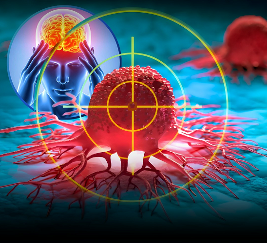
Glioblastoma: Symptoms, Risks & Treatment Guide

Learn about glioblastoma, an aggressive form of brain cancer. Discover its symptoms, causes, diagnosis, and treatment options to better understand this serious condition.

When we talk about deadly brain cancer, we’re primarily talking about glioblastoma multiforme (GBM) —the most aggressive and lethal form of brain cancer in adults. Accounting for approximately 45% of all primary malignant brain tumors, glioblastoma has a devastating prognosis that has remained stubbornly poor despite decades of research .

The statistics are sobering: the five-year survival rate for glioblastoma is only 8% , and median survival after diagnosis is just 14-18 months with standard treatment . In the United States alone, an estimated 24,820 new cases of brain and other nervous system tumors will be diagnosed in 2025, with approximately 18,330 deaths expected .

This article explores what makes glioblastoma so deadly, current treatment approaches, and promising research that offers hope for better outcomes.

What Makes Glioblastoma So Deadly?

Aggressive Growth Pattern

Glioblastoma is classified as a Grade IV tumor by the World Health Organization (WHO)—the highest grade possible. These tumors are characterized by:

  • Rapid proliferation: GBM cells divide quickly and uncontrollably
  • Diffuse infiltration: The tumor sends tentacle-like projections into surrounding brain tissue, making complete surgical removal nearly impossible
  • Extreme heterogeneity: Even within a single tumor, cancer cells vary dramatically, making them moving targets for treatment 
  • Angiogenesis: GBM stimulates growth of new blood vessels to fuel its expansion

Treatment-Resistant Nature

Several factors make GBM exceptionally difficult to treat:

The blood-brain barrier protects the brain from infection but also blocks many cancer drugs from reaching tumor cells . Even when drugs penetrate this barrier, GBM cells employ multiple resistance mechanisms to survive chemotherapy and radiation.

The tumor microenvironment is described as immunologically “cold” —it actively suppresses the immune system’s ability to recognize and attack cancer cells . This explains why immunotherapies that revolutionized other cancers have struggled against GBM.

Molecular Complexity

GBM harbors numerous genetic mutations that influence treatment response. The most clinically relevant include:

  • IDH1/IDH2 mutations: Present in some gliomas; patients with these mutations have better prognoses 
  • MGMT promoter methylation: Predicts better response to temozolomide chemotherapy
  • EGFRvIII mutation: Occurs in 25-30% of GBM patients and is associated with treatment resistance 
  • 1p/19q codeletion: Associated with better outcomes in certain glioma types 

Recognizing the Signs: Symptoms of Brain Cancer

Symptoms depend on the tumor’s size and location but commonly include :

  • Headaches (especially persistent or worse in the morning)
  • Seizures (occurring in about 20% of patients at presentation and up to 70% over the disease course)
  • Nausea and vomiting
  • Vision changes (blurred vision, double vision)
  • Weakness or numbness on one side of the body
  • Difficulty walking or balance problems
  • Speech and language difficulties
  • Memory loss and confusion
  • Personality changes

If you experience any of these symptoms persistently, consult a healthcare provider immediately.

Current Treatment Landscape

Standard of Care: The Stupp Protocol

For newly diagnosed GBM, the established treatment approach includes:

  1. Maximal safe surgical resection: Removing as much tumor as possible while preserving neurological function 
  2. Radiation therapy: Typically 60 Gy delivered over 6 weeks
  3. Temozolomide chemotherapy: The only FDA-approved drug for GBM, given concurrently with radiation and then as maintenance therapy 

Despite this aggressive approach, tumors nearly always recur.

Surgical Innovations

Awake brain surgery (brain mapping) allows neurosurgeons to remove tumors near critical brain regions controlling speech, language, and movement. Patients remain awake during part of the procedure, providing real-time feedback that helps preserve vital functions .

Supratotal resection —removing more tissue than just the visible tumor—has shown promising results in some studies .

Emerging Treatments

Tumor Treating Fields (TTFields): This innovative therapy uses low-intensity electric fields to disrupt cancer cell division. It’s approved for both newly diagnosed and recurrent GBM and has shown improved survival when combined with temozolomide .

Laser Interstitial Thermal Therapy (LITT): A minimally invasive technique that uses heat to destroy tumor cells, particularly useful for recurrent or hard-to-reach tumors .

Breakthrough Research: Hope on the Horizon

Immunotherapy Advances

A groundbreaking 2019 study (with long-term follow-up reported in 2025) showed that administering the immunotherapy drug pembrolizumab (Keytruda) before surgery significantly improved survival in recurrent GBM patients .

Key findings:

  • Patients receiving neoadjuvant pembrolizumab had median survival of 13.9 months versus 7.5 months for those receiving it after surgery
  • The pre-surgery approach allows T cells to recognize and attack a larger tumor burden, creating a broader and more durable immune response
  • Tumors from the neoadjuvant group showed increased T-cell infiltration and suppression of cancer proliferation pathways 

While experts caution that larger trials are needed before this becomes standard of care, the results represent one of the most promising advances in years.

Targeted Therapies for EGFRvIII-Positive GBM

For the 25-30% of GBM patients with EGFRvIII mutations, a 2025 network meta-analysis identified rindopepimut (a targeted vaccine) combined with bevacizumab as the most promising regimen for recurrent disease . This combination showed the best outcomes for overall survival, progression-free survival, and objective response rate, with the lowest incidence of severe side effects.

Personalized Medicine Through AI

Researchers at Georgetown Lombardi Comprehensive Cancer Center have developed scFOCAL —a computational framework that uses single-cell RNA sequencing to predict how individual tumors will respond to different treatments . This approach could eventually enable truly personalized therapy, identifying drug combinations tailored to each patient’s unique tumor cell populations.

Immune-Stimulating Wafers

Scientists at the University of Cincinnati are developing a delayed-release wafer containing interleukin-15 (IL-15) that would be placed in the surgical cavity after tumor removal. IL-15 activates immune cells to recognize and kill residual cancer cells . The team is testing this approach using “glioblastoma-on-a-chip” technology before moving to human trials.

Organ-on-a-Chip Technology

This innovative platform creates 3D models of human brain tumors that incorporate immune cells—something traditional cell cultures lack. It allows researchers to test therapies more accurately before reaching patients and could eventually predict individual patient responses to immunotherapy .

Risk Factors and Prevention

Unlike many cancers, there are no known lifestyle-related or environmental risk factors for primary brain tumors . This means there’s currently no proven way to prevent them.

Known Risk Factors

  • Radiation exposure: Therapeutic radiation to the head (rare)
  • Family history: Certain inherited syndromes increase risk:
    • Neurofibromatosis types 1 and 2
    • Li-Fraumeni syndrome
    • Tuberous sclerosis
    • Von Hippel-Lindau disease 

Factors with uncertain or unproven effects include cell phone use, exposure to vinyl chloride, and certain occupational chemicals .

Living with Glioblastoma: Support and Outlook

Prognostic Factors

Several factors influence individual prognosis :

  • Age: Younger patients generally fare better
  • Tumor genetics: IDH mutations and MGMT methylation predict better outcomes
  • Extent of resection: More complete removal improves survival
  • Performance status: Overall health and function at diagnosis

The Importance of Clinical Trials

Given the limited treatment options, all GBM patients should consider clinical trial participation at every stage of their disease. Trials are investigating:

  • Novel drug combinations
  • Immunotherapy approaches
  • Targeted therapies based on tumor genetics
  • Innovative delivery methods to bypass the blood-brain barrier

Support Resources

A glioblastoma diagnosis affects the entire family. Important resources include:

  • Neuro-oncology teams that include social workers and patient navigators
  • Support groups for patients and caregivers
  • Palliative care to manage symptoms and maintain quality of life
  • Rehabilitation services (physical, occupational, speech therapy)

Conclusion: Progress Amidst Challenges

Glioblastoma remains one of medicine’s most formidable challenges. Its aggressive nature, treatment resistance, and molecular complexity have thwarted countless therapeutic attempts. Yet the research landscape is shifting.

From immunotherapy timing that makes biological sense to personalized AI-driven treatment selection, from immune-stimulating wafers to targeted vaccines—genuine progress is being made. While we don’t yet have a cure, each study brings us closer to understanding how to outsmart this deadly cancer.

For patients and families facing this diagnosis, the message is one of cautious hope: research is accelerating, clinical trials are expanding, and the scientific community remains relentlessly committed to finding better treatments.


References:

  1. National Foundation for Cancer Research. Brain Cancer Statistics and Facts. 2024.
  2. Comparative efficacy and safety of therapeutic strategies for EGFRvIII positive recurrent glioblastoma. ScienceDirect. 2025.
  3. Georgetown University Medical Center. Method developed to identify best treatment combinations for glioblastoma. EurekAlert! 2026.
  4. National Cancer Institute. Central Nervous System Tumors Treatment (PDQ®). 2025.
  5. Neurology Today. Immunotherapy Administered Before Surgery for Recurrent Glioblastoma Leads to Better Survival Outcomes. 2025.
  6. Drug Target Review. Organ-on-a-chip tests immune wafer for glioblastoma. 2026.
  7. NUHS. Brain Cancer – Symptoms, Diagnosis, and Treatment. 2025.
  8. Editora da Universidade de Vassouras. Glioblastoma multiforme: What’s new in relation to established therapeutics? 2025.

Disclaimer: This article is for educational purposes only and does not constitute medical advice. If you or someone you know is experiencing symptoms of a brain tumor, consult a healthcare provider immediately. Treatment decisions should always be made in consultation with qualified medical professionals.

AI Breakthrough in Ulcerative Colitis Cancer Detection

AI Breakthrough in Ulcerative Colitis Cancer Detection

Artificial intelligence (AI) is rapidly transforming healthcare — and now it may help detect colorectal cancer risk earlier in patients with ulcerative colitis (UC). A new AI-powered tool shows promising potential in identifying subtle warning signs of cancer before they become life-threatening.

For people living with ulcerative colitis, this innovation could represent a major step forward in cancer prevention and early intervention.


Understanding the Link Between Ulcerative Colitis and Colorectal Cancer

Ulcerative colitis is a chronic inflammatory bowel disease (IBD) that causes long-term inflammation and ulcers in the colon. While many patients manage symptoms effectively with medication and lifestyle adjustments, prolonged inflammation increases the risk of developing colorectal cancer.

Research shows that the risk of colorectal cancer rises significantly in patients who:

  • Have had ulcerative colitis for more than 8–10 years
  • Experience severe or widespread colon inflammation
  • Have a family history of colorectal cancer
  • Have primary sclerosing cholangitis (PSC)

Because of this elevated risk, regular colonoscopic surveillance is recommended. However, detecting early cancerous changes can be challenging — even for experienced specialists.


The Challenge of Early Cancer Detection

Traditional colonoscopy relies heavily on visual inspection and tissue biopsies. While effective, this method has limitations:

  • Precancerous lesions may be subtle or flat
  • Dysplasia (abnormal cell changes) can be difficult to detect
  • Human error or variability in interpretation may occur

Early-stage cancer or precancerous changes may be microscopic and easily missed during routine screening. This is where artificial intelligence enters the picture.


How AI Is Changing Colorectal Cancer Screening

The newly developed AI tool uses advanced machine learning algorithms trained on thousands of colonoscopy images and pathology samples. By analyzing patterns that may not be visible to the human eye, the system can:

  • Identify abnormal tissue changes
  • Highlight suspicious areas in real-time during colonoscopy
  • Predict which patients are at higher risk of developing colorectal cancer

Some AI systems integrate deep learning technology to assess inflammation severity, mucosal irregularities, and subtle dysplastic changes.

In early trials, AI-assisted screening demonstrated improved accuracy in detecting precancerous lesions compared to standard methods alone.


Why This Matters for Ulcerative Colitis Patients

For individuals living with ulcerative colitis, early detection is critical. Colorectal cancer developing from chronic inflammation can progress silently. By the time symptoms appear, the disease may already be advanced.

AI-assisted tools could:

  • Improve early diagnosis
  • Reduce missed lesions
  • Enhance surveillance precision
  • Lower long-term cancer risk
  • Potentially decrease the need for aggressive surgery

This technology does not replace physicians — instead, it acts as a powerful support system that enhances decision-making and diagnostic confidence.


Clinical Trial Findings: Promising but Ongoing

Recent research suggests that AI integration during colonoscopy increases detection rates of dysplasia in patients with inflammatory bowel disease. Some early studies report higher sensitivity and improved consistency in identifying early cancer markers.

However, experts emphasize that larger, long-term clinical trials are still needed before widespread adoption.

The current evidence is promising, but AI tools must undergo rigorous validation to ensure safety, accuracy, and reliability across diverse populations.


Benefits of AI in Gastroenterology

Beyond ulcerative colitis, AI is reshaping multiple aspects of gastrointestinal care. Potential advantages include:

1. Real-Time Analysis

AI can analyze colonoscopy footage instantly, providing immediate feedback during procedures.

2. Reduced Human Error

Machine learning models can detect patterns consistently without fatigue or subjective bias.

3. Personalized Risk Prediction

AI may help stratify patients based on individual risk levels, enabling tailored surveillance plans.

4. Improved Efficiency

Automated image analysis may streamline workflow and improve healthcare resource utilization.


Limitations and Considerations

While exciting, AI-based detection tools are not without challenges:

  • Data privacy concerns
  • Need for standardized validation
  • Integration into existing healthcare systems
  • Cost and accessibility
  • Risk of over-reliance on technology

Healthcare professionals stress that AI should function as an adjunct tool — not a replacement for clinical expertise.


The Future of Cancer Prevention in Ulcerative Colitis

The integration of artificial intelligence into colonoscopy screening represents a major advancement in preventive medicine. As technology continues to evolve, AI could become a routine part of inflammatory bowel disease management.

Future developments may include:

  • AI-driven biopsy guidance
  • Predictive models for flare severity
  • Automated pathology interpretation
  • Personalized surveillance scheduling

If validated through large-scale trials, AI tools could significantly reduce colorectal cancer mortality in high-risk populations such as ulcerative colitis patients.


What Patients Should Do Now

If you have ulcerative colitis:

  • Follow recommended colonoscopy screening guidelines
  • Maintain regular follow-ups with your gastroenterologist
  • Manage inflammation through prescribed treatments
  • Discuss emerging screening technologies with your doctor

AI-assisted screening may soon enhance routine care, but traditional monitoring remains essential today.


Final Thoughts

Artificial intelligence is opening new possibilities in early cancer detection. For ulcerative colitis patients, this breakthrough could mean earlier warnings, better outcomes, and improved long-term survival.

While research continues, one thing is clear: the future of colorectal cancer prevention may be powered by both medical expertise and intelligent technology working together.

Early detection saves lives — and AI may soon make that detection even more precise.

Reference:

https://today.ucsd.edu/story/artificial-intelligence-predicts-colorectal-cancer-risk-in-ulcerative-colitis-patients
https://www.2minutemedicine.com/artificial-intelligence-predicts-colorectal-cancer-in-ulcerative-colitis-with-99-accuracy/
https://pmc.ncbi.nlm.nih.gov/articles/PMC12543294/
https://www.medicalnewstoday.com/articles/ai-tool-predicts-colorectal-cancer-risk-ulcerative-colitis-patients
https://www.sciencedirect.com/science/article/pii/S1542356525006160

Medications that have been suggested by doctors worldwide are available on the link below
https://mygenericpharmacy.com/category/disease/cancer

The Cancer Compass: Navigating ALL Types of Cancer with Knowledge and Hope

The Cancer Compass: Navigating ALL Types of Cancer with Knowledge and Hope

Welcome to Your Comprehensive Cancer Resource

Cancer is not one disease but hundreds—each with unique characteristics, treatments, and journeys. Whether you’re newly diagnosed, a long-term survivor, a caregiver, or proactively focused on prevention, this blog is your evidence-based guide through the complex world of oncology. Here, we translate science into understanding, fear into empowerment, and isolation into community.

Understanding Cancer: The Universal Threads

What Is Cancer, Really?

At its core, cancer is a disease of uncontrolled cell division. Normal cells follow a life cycle of growth, division, and death. Cancer cells ignore these rules due to genetic mutations, multiplying uncontrollably and potentially spreading (metastasizing).

The Hallmarks of Cancer (What Makes Cancer, Cancer):

  1. Sustaining proliferative signaling
  2. Evading growth suppressors
  3. Resisting cell death
  4. Enabling replicative immortality
  5. Inducing angiogenesis (creating a blood supply)
  6. Activating invasion and metastasis
  7. Deregulating cellular metabolism
  8. Avoiding immune destruction

Cancer Categories: Understanding the Landscape

Carcinomas (80-90% of cancers)

  • Originate in: Epithelial cells (skin, organ linings)
  • Examples: Breast, lung, prostate, colorectal, pancreatic, ovarian, bladder, kidney
  • Subtypes: Adenocarcinoma, squamous cell, basal cell

Sarcomas (<1% of adult cancers)

  • Originate in: Connective tissues (bone, muscle, fat, cartilage)
  • Examples: Osteosarcoma, liposarcoma, leiomyosarcoma, GIST
  • Characteristics: Often appear in younger patients

Leukemias (Blood/bone marrow cancers)

  • Originates in: Blood-forming tissues
  • Examples: ALL, AML, CLL, CML
  • Characteristics: “Liquid tumors,” often detected via blood tests

Lymphomas

  • Originates in: Lymphatic system
  • Examples: Hodgkin lymphoma, Non-Hodgkin lymphoma
  • Characteristics: Often present with swollen lymph nodes

Central Nervous System Cancers

  • Originates in the brain and the spinal cord
  • Examples: Glioblastoma, astrocytoma, meningioma
  • Characteristics: Rarely spreads outside the CNS but locally aggressive

Other Types:

  • Myeloma: Plasma cells in bone marrow
  • Melanoma: Pigment-producing cells (skin, eyes)
  • Germ cell tumors: Testicular, ovarian
  • Neuroendocrine tumors: Carcinoids, pancreatic NETs

Prevention & Risk Reduction: Your Power to Act

Lifestyle Factors Within Your Control

  • Tobacco: Causes ~30% of all cancer deaths (lung, bladder, pancreatic, etc.)
  • Diet & Weight: 18% of US cancers linked to excess weight, poor nutrition, inactivity
  • Alcohol: Increases risk of breast, colorectal, liver, and esophageal cancers
  • Sun Exposure: Primary cause of melanoma and non-melanoma skin cancers

Screening & Early Detection Saves Lives

Proven screening tools:

  • Breast: Mammography (starting 40-50, depending on risk)
  • Cervical: Pap smear/HPV testing
  • Colorectal: Colonoscopy (starting at 45), FIT tests
  • Lung: Low-dose CT scan for high-risk smokers/ex-smokers
  • Prostate: PSA discussion starting at 50-55 (earlier if high-risk)
  • Skin: Regular self-exams and dermatologist visits

Genetic Testing & Family History:

  • 5-10% of cancers are hereditary
  • Common syndromes: BRCA1/2 (breast/ovarian), Lynch (colorectal), FAP, Li-Fraumeni
  • When to consider testing: Multiple relatives withthe same cancer, early-onset, rare cancers

Diagnosis & Staging: The Roadmap for Treatment

The Diagnostic Journey

  1. Imaging: CT, MRI, PET, ultrasound, X-ray
  2. Biopsy: Needle, endoscopic, surgical
  3. Pathology: Microscopic examination, molecular testing
  4. Staging: Determines extent of disease (Stage 0-IV)

Modern Diagnostic Tools

  • Liquid biopsies: Detecting cancer DNA in blood
  • Genomic profiling: Identifying targetable mutations
  • Artificial intelligence: Improving imaging interpretation
  • Minimally invasive techniques: Reducing diagnostic risks

Treatment Modalities: The Evolving Arsenal

Surgery

  • Goal: Remove tumor with clear margins
  • Advances: Robotic-assisted, laparoscopic, organ-preserving techniques
  • Considerations: Often first-line for solid tumors

Radiation Therapy

  • Types: External beam, brachytherapy, proton therapy, SBRT
  • Goal: Damage cancer cell DNA while sparing healthy tissue
  • Advances: IMRT, image-guided, stereotactic radiosurgery

Systemic Therapies

Chemotherapy:

  • Traditional cytotoxic agents
  • Often used in combinations (regimens)
  • Can be neoadjuvant (before surgery), adjuvant (after), or palliative

Targeted Therapy:

  • Attacks specific molecular targets
  • Examples: TKIs for EGFR, ALK, BRAF mutations
  • Requires biomarker testing
  • Often has different side effect profiles than chemo

Immunotherapy:

  • Harnesses the immune system to fight cancer
  • Checkpoint inhibitors: PD-1/PD-L1, CTLA-4 inhibitors
  • CAR-T cell therapy: Genetically engineering patients’ T-cells
  • Cancer vaccines: Preventive (HPV) and therapeutic

Hormone Therapy:

  • Blocks hormones that fuel certain cancers
  • Examples: Breast (tamoxifen, aromatase inhibitors), Prostate (ADT)

Other Approaches:

  • Angiogenesis inhibitors: Cut off the tumor’s blood supply
  • PARP inhibitors: For cancers with DNA repair deficiencies (BRCA)
  • Antibody-drug conjugates: Targeted chemo delivery

Transplantation

  • Stem cell transplant: For leukemias, lymphomas, myelomas
  • Types: Autologous (own cells), allogeneic (donor cells)

Supportive & Palliative Care

  • Essential at ALL stages (not just end-of-life)
  • Manages symptoms, side effects, and quality of life
  • Integrative oncology: Evidence-based complementary approaches

Side Effect Management: Living Well During Treatment

Common Challenges & Solutions

  • Fatigue: Energy conservation, graded exercise, addressing anemia
  • Nausea: Multiple medication classes, dietary modifications, acupressure
  • Neuropathy: Medication adjustments, physical therapy, supplements (α-lipoic acid)
  • Skin reactions: Specialized skincare, radiation dermatitis protocols
  • Emotional distress: Counseling, support groups, mindfulness, medication when needed

Nutritional Support

  • During treatment: Maintain weight, manage symptoms, preserve muscle mass
  • Post-treatment: Cancer-preventive dietary patterns
  • Working with a registered dietitian specialized in oncology is invaluable

Survivorship: Life During and After Cancer

The “New Normal”

  • Physical changes: Scarring, lymphedema, early menopause, sexual health impacts
  • Emotional journey: Fear of recurrence, PTSD, gratitude, renewed perspective
  • Cognitive effects: “Chemo brain” – often improves but can persist
  • Financial toxicity: Treatment costs, lost income, insurance battles

Survivorship Care Plans Should Include:

  1. Treatment summary
  2. Surveillance schedule for recurrence
  3. Screening for secondary cancers
  4. Management of long-term/late effects
  5. Health promotion strategies
  6. Psychosocial resources

Pediatric vs. Adult Cancers: Critical Differences

Pediatric Cancers:

  • Often embryonal or sarcomas (vs. carcinomas in adults)
  • Typically, more treatment-responsive but with greater long-term toxicity concerns
  • Require specialized pediatric oncology centers
  • Most common: Leukemias, brain tumors, lymphomas, neuroblastoma

Special Considerations:

  • Fertility preservation: Discussion essential before treatment
  • Growth & development impacts: Lifelong monitoring needed
  • Psychosocial needs: Age-appropriate support is crucial

Global Perspectives & Disparities

Inequities in Cancer Care

  • Access: Screening, treatment, and palliative care availability
  • Outcomes: Higher mortality in marginalized communities
  • Research participation: Lack of diversity in clinical trials
  • Social determinants: Income, education, environment, discrimination

Addressing Disparities Through:

  • Community outreach and education
  • Patient navigation programs
  • Policy advocacy
  • Diverse research recruitment

Research Frontiers: Reasons for Hope

Precision Medicine Revolution

  • Comprehensive genomic profiling: Matching tumors to targeted therapies
  • Minimal residual disease detection: Predicting relapse earlier
  • Biomarker development: Predicting response to immunotherapy

Early Detection Innovations

  • Multi-cancer early detection tests (MCEDs): Blood tests detecting multiple cancers
  • Improved imaging: Higher resolution with lower radiation
  • AI-assisted screening: Improving accuracy and access

Treatment Advances

  • Bispecific antibodies: Engaging immune cells directly to cancer
  • Next-generation cellular therapies: Off-the-shelf CAR-T, CAR-NK cells
  • Viral therapies: Engineered viruses attacking cancer cells
  • Epigenetic therapies: Reprogramming gene expression

Supportive Care Research

  • Prehabilitation: Optimizing health before treatment
  • Digital health tools: Remote monitoring, symptom management apps
  • Novel antiemetics: Better nausea control
  • Cardio-oncology: Protecting heart health during/after treatment

Advocacy & Empowerment: Finding Your Voice

Being Your Own Best Advocate

  1. Get organized: Treatment binder, questions list, calendar
  2. Bring support: Second set of ears to appointments
  3. Ask about clinical trials at every decision point
  4. Know your insurance benefits and appeal rights
  5. Connect with advocacy organizations specific to your cancer type

Finding Community

  • Disease-specific organizations: American Cancer Society, Leukemia & Lymphoma Society, etc.
  • Online communities: Smart Patients, CancerCare, Inspire
  • Local support groups: Hospital-based or community centers
  • Peer mentoring programs: Connecting with someone who’s been there

Critical Disclaimer

This blog provides educational information about cancer in general, but cannot address individual circumstances. Always consult with your oncology team for personalized medical advice, diagnosis, and treatment decisions. Information about treatments is not an endorsement, and treatment options should be thoroughly discussed with your healthcare providers.

Bladder Cancer Screening and Diagnosis

Bladder Cancer Screening and Diagnosis

Several tests can determine the diagnosis of bladder cancer. A variety of tests may be used to make an exact diagnosis of bladder cancer.

Screening typically begins with pathology tests, where samples of fluid and tissue are examined by a pathologist in a laboratory. The most efficient, noninvasive, and inexpensive test is a urinalysis/cytology. Here, a sample of urine is taken from the patient and evaluated for cancer cells, red and white blood cells (which fight urinary tract infections), and microscopic hematuria or infection. Hematuria (blood in the urine) is also a sign of a possible urinary tract infection.

If abnormalities are found in the urine, a biopsy will be performed, in which a pathologist examines tissue for the presence of cancer cells. If the urine culture fails to show abnormalities, a biopsy or other tests still may be ordered — especially if there are symptoms of concern.

Imaging tests may be used to locate blockages and tumors, as well as to determine whether cancer has spread to other organs.
An intravenous pyelogram is an imaging test during which the patient is injected with dye, and the radiologist observes with an X-ray the movement of that dye through the urinary tract. This X-ray will look at the collecting system of the kidneys to determine the presence of any irregularities. This is good for seeing small cancer locations and the upper urinary tract, and especially for detailing the kidneys, ureters, and bladder.

CT or CAT scans are another form of X-ray, which creates a more detailed image of the body and organs. This is used to locate kidney or bladder blockages, and to determine staging, recommended therapy, and whether the bladder cancer has metastasized (spread to other parts of the body).

MRI is another imaging form that creates very high-quality and detailed images of bladder tumors in addition to adjacent organs, such as the chest, pelvis, and abdomen, to locate any metastasis.

Ultrasound imaging, without side effects or radiation, is noninvasive and looks primarily at the bladder and kidneys. It can locate small tract blocks and stones, and also measures the bladder wall thickness.

The gold standard for the evaluation of the lower urinary tract is a routine outpatient procedure called a cystoscopy. In the same way that a colonoscopy allows doctors to see inside the lower digestive tract, a cystoscopy provides a visual of the lower urinary tract and bladder lining.

During the procedure, a specialized instrument called a cystoscope is passed through the urethra into the bladder. Cystoscopes are either rigid or flexible. Fiber optics allows for images of the bladder lining to be created. If abnormalities, such as tumors, stones, or patches of abnormal-appearing tissue, are discovered during cystoscopy, a biopsy may be taken. The biopsy specimen will then be evaluated by a pathologist for the presence of cancerous cells.

Occasionally, the urologist will take a biopsy during a transurethral resection of bladder tumor procedure, which will be scheduled for a future appointment. This is a minimally invasive procedure that does not involve making an incision in the body. The entire removal of a bladder-confined tumor can be accomplished through an operative scope, which passes through the urethra into the bladder.

Reference:
https://www.hopkinsmedicine.org/health/conditions-and-diseases/bladder-cancer/bladder-cancer-screening-and-diagnosis

Medications that have been suggested by doctors worldwide are available on the link below
https://mygenericpharmacy.com/category/products/disease/cancer