Browsed by
Month: January 2023

Important parameters of Cervical cancer women need to know.

Important parameters of Cervical cancer women need to know.

What Is Cervical Cancer?

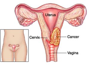
Women’s cervix, which connects the uterus and vagina, is where cervical cancer develops when cells in the cervix alter. The deeper tissues of their cervix may be affected by this cancer, and it has the potential to metastasis (spread to other parts of the body), most frequently the lungs, liver, bladder, vagina, and rectum.

Human papillomavirus (HPV) infection, which is avoidable with a vaccine, is the main cause of cervical cancer. Since cervical cancer develops slowly, it is typically detectable and treatable before it poses a major threat. Thanks to better screening through Pap tests, it claims fewer and fewer lives of women every year.

The majority of cases are women between the ages of 35 and 44. However, women over 65 make up more than 15% of new cases, particularly those who haven’t been undergoing routine exams.

Different Types of Cervical Cancer

Cervical cancer comes in several different forms.

  • Squamous cell carcinoma. This develops in your cervix’s lining. Up to 90% of cases have it.
  • Adenocarcinoma. This develops in the mucus-producing cells.
  • Mixed cancercarcinoma. This possesses traits from the other two categories.

Cervical cancer stages

Your doctor will determine the stage of your cancer after a diagnosis has been made. The stage reveals if and how far the cancer has spread if it has. Your doctor can identify the best course of treatment for you by staging your cancer.

There are four phases of cervical cancer:

  • Stage 1: A little cancer. There’s a chance the lymph nodes were affected. It hasn’t spread to other body areas.
  • Stage 2: The tumour has grown. It can have reached the lymph nodes or spread beyond the uterus and cervix. It hasn’t yet spread to other areas of your body.
  • Stage 3: The malignancy has gone to the pelvic or the lower vagina. The ureters, which are tubes that transfer urine from the kidneys to the bladder, may be blocked as a result. It hasn’t spread to other body areas.
  • Stage 4: The cancer may have spread to other organs, such as your lungs, bones, or liver, from the pelvis.

Signs and symptoms of cervical cancer

Early stages of cervical cancer are typically difficult to diagnose because they lack symptoms. It may take several years before cervical cancer symptoms appear. The greatest strategy to prevent cervical cancer is to find abnormal cells during testing for the disease.

Stage 1 cervical cancer symptoms and signs might include:

  • Vaginal discharge that is either bloody or watery, may be heavy, and may smell bad.
  • Vaginal bleeding following sex, in between cycles, or following menopause.
  • Periods of menstruation could be heavier and longer than usual.

Symptoms of cancer that has spread to adjacent tissues or organs include:

  • urination that is painful or difficult, occasionally with blood in the pee.
  • diarrhoea, abdominal pain, or bleeding when you poop.
  • fatigue, weight loss, and appetite loss
  • a state of general disease
  • a dull backache or leg swelling.
  • abdominal and pelvic pain

You should have a comprehensive gynaecological exam, which includes a Pap test, if you suffer abnormal bleeding, vaginal discharge, or any other unexplained symptoms.

Cervical cancer causes

The sexually transmitted human papillomavirus is the primary factor in most occurrences of cervical cancer (HPV). Genital warts are brought on by the same virus.

There are over 100 distinct HPV strains. Cervical cancer is only caused by specific types. HPV-16 and HPV-18 are the two strains that cause cancer the most frequently.

Cervical cancer is not a guarantee even if you have an HPV cancer-causing strain. Most HPV infections are cleared up by your immune system, frequently within two years.

In both men and women, HPV can lead to other malignancies. These consist of:

  • vulvar cancer
  • vaginal cancer
  • penile cancer
  • anal cancer
  • rectal cancer
  • throat cancer

Cervical cancer risk factors

The greatest risk factor for cervical cancer is HPV. Additional elements that may raise your risk include:

  • HIV
  • chlamydia
  • smoking
  • obesity
  • a history of cervical cancer in the family
  • consuming little fruit and veg
  • using contraceptive tablets
  • being pregnant three times at term
  • being under the age of 17 when you first became pregnant

You are not destined to develop cervical cancer even if you have one or more of these risk factors.

How is cervical cancer treated?

One member of the team treating cervical cancer is a gynecologic oncologist (a doctor who specialises in cancers of female reproductive organs). The stage of the disease, your age and general health, and whether or not you intend to have children in the future all play a role in the recommended course of therapy for cervical cancer.

Radiation, chemotherapy, surgery, targeted therapy, and immunotherapy are all options for treating cervical cancer.

Radiation Therapy

Your cervix’s cancerous cells are destroyed by energy beams used in radiation therapy. Radiation therapy is available in two different forms:

  • External beam radiation therapy (EBRT) uses a machine outside the body to direct powerful radiation towards tumours.
  • Radiation is applied directly to or near a malignancy during brachytherapy.

Chemotherapy

Chemotherapy (chemo) kills cancer cells by administering medications by injection into your veins or oral ingestion. It enters your circulation and kills cells effectively throughout your body. Chemotherapy uses a variety of medications, some of which can be combined. Cycles of chemotherapy are frequently administered.

Surgery

Cervical cancer is treated with a variety of surgical procedures. The most typical procedures used to treat cervical cancer include:

  • Laser procedure
  • conical biopsy
  • an easy hysterectomy
  • Trachelectomy
  • Pelvic enlargement
  • Targeted treatment

Specific cancer cells are eliminated by targeted medication therapy without harming healthy cells. It functions by focusing on proteins that regulate how cancer cells proliferate and spread.

Immunotherapy

In immunotherapy, drugs are used to activate your immune system’s capacity to detect and eliminate cancer cells. Cancer cells can also signal to avoid being attacked by your immune system. Targeting these signals with immunotherapy makes it so cancer cells can’t deceive your body into believing they are healthy cells.

Clinical trials are yet another form of treatment. Some people supplement their cancer therapy with complementary therapies like nutrition, herbs, acupuncture, and other practises. Speak with your healthcare practitioner about alternative practises that promise to lessen the symptoms of cancer. Some may be beneficial, while others may be dangerous.

Cervical cancer prevention

Screening with a Pap smear or a hrHPV test on a regular basis is one of the simplest strategies to avoid cervical cancer. Precancerous cells are detected during screening so they can be treated before they progress to malignancy.

Most occurrences of cervical cancer are caused by HPV infection. With the help of the vaccines Gardasil and Cervarix, the illness can be avoided. The best time for vaccination is before a person starts acting sexually. Boys and girls can both receive the HPV vaccine.

You can lessen your risk of HPV and cervical cancer by doing the following additional things:

  • Do not have too many sexual partners.
  • When engaging in vaginal, oral, or anal intercourse, you should always use a condom or another barrier device.

You may have precancerous cells in your cervix if your Pap smear results are abnormal.

REFERENCES:

For more details, kindly visit below.

Expert note on causes of teeth sensitivity & its treatment.

Expert note on causes of teeth sensitivity & its treatment.

When you have sensitive teeth, it can occasionally be uncomfortable to eat and drink. However, certain natural therapies might help lessen tooth sensitivity.

The condition of sensitive teeth is frequent and curable. One in eight patients who went to dental offices had sensitive teeth, according to a 2013 research.

What is Teeth Sensitivity?

Dentin hypersensitivity, also known as tooth sensitivity, is what it means like. The teeth that become painful or uncomfortable in reaction to particular stimuli, such as hot or cold temperatures, causes sensitivity.

It can affect one tooth, many teeth, or all of the teeth in one person, and it can be a short-term or long-term issue. While sensitive teeth might have a variety of causes, the majority of them can be readily remedied by altering your oral hygiene routine.

Symptoms of sensitive teeth

People who have sensitive teeth may feel pain or discomfort when certain triggers are present. This discomfort may be felt at the tooth roots of the troubled teeth. Among the most typical triggers are:

  • piping hot food and drink
  • chilly foods and drinks
  • chilly air
  • sweet dishes and drinks
  • sour foods and drinks
  • especially while performing routine dental cleanings, cold water
  • flossing or brushing your teeth
  • Alcoholic mouthwashes

Over time, your symptoms could fluctuate without any apparent cause. They could be light or strong.

What causes tooth sensitivity?

The occurrence of sensitive teeth can be caused by a variety of circumstances, such as:

Brushing too strongly:

Over time, vigorous brushing or the use of a toothbrush with a firm bristle can wear down the enamel and expose the dentin. It might also result in gum recession (when your gum tissue pulls away from your teeth).

Gum recession:

Some individuals have thin gum tissue as a genetic predisposition. Periodontal disease also causes gum recession in other people. Gum recession occurs when the gum line recedes from the teeth, exposing the roots.

Gum disease:

Inflamed and painful gum tissue may result in sensitivity because the ligaments that hold the gums in place have been removed, exposing the root surface that directly connects to the tooth’s nerve.

Cracked teeth:

Broken or chipped teeth may allow plaque-containing germs to enter the pulp and cause irritation.

Grinding or clenching:

Doing this to your teeth can wear away the enamel and reveal the dentin beneath.

Products for teeth whitening:

These products are a main cause of tooth discomfort. Speak to a dentist about teeth whitening for sensitive teeth if you wish to improve your smile.

Age:

Between the ages of 25 and 30, teeth sensitivity is at its peak.

Plaque accumulation:

Root surface sensitivity may be brought on by the presence of plaque.

Use of mouthwash:

If you have exposed dentin, several over-the-counter mouthwashes include acids that might exacerbate tooth sensitivity (the middle layer of the tooth). The dentin layer of the tooth is further harmed by the acids. Consult your dentist about using a neutral fluoride solution if you experience dentin sensitivity.

Acidic foods:

Erosive enamel can result from routinely consuming foods with a high acid content, such as citrus fruits, tomatoes, pickles, and tea.

Recent dental procedures:

Following fillings, teeth cleanings, and dental restoration placement, patients may experience sensitive teeth. Dental procedure-related sensitivity is transient and often goes away in four to six weeks.

Sensitive teeth diagnosis

Make an appointment with your dentist if this is the first time you’ve noticed tooth sensitivity. You may make a dental appointment with a professional in your neighbourhood. They can examine the condition of your teeth and search for any issues that might be causing the discomfort, such as cavities, loose fillings, or receded gums.

During your regular teeth cleaning, your dentist can perform this. They’ll perform a visual inspection and clean your teeth. They might use dental tools to gently touch your teeth to check for sensitivity, and they might also request an X-ray to rule out other possibilities like cavities.

Treatment of teeth sensitivity

Your dentist can employ a variety of techniques to reduce your discomfort once the issue has been identified, including:

  • toothpaste for teeth that are sensitive
  • The fluoride gel
  • coverings for exposed roots
  • Sealants
  • You can purchase desensitising pastes from your dentist (not to be used with a toothbrush).
  • If you grind your teeth, wear a mouthguard.
  • A root canal may be recommended by your dentist if your condition is serious.

Additionally, it’s critical to avoid avoiding dental care due to tooth pain. Ignoring your teeth can exacerbate the situation. Twice-daily brushing and flossing will maintain your smile healthy and pain-free. Additionally, go to the dentist twice a year for a checkup.

Prevention tips for sensitive teeth

People can lessen tooth sensitivity and safeguard their tooth enamel by:

  • using a fluoride toothpaste to wash your teeth twice a day
  • once daily flossing
  • taking pauses from tooth whitening
  • minimising intake of foods that are high in sugar, carbohydrates, and acids
  • reducing alcohol consumption
  • using a mouthguard at night to avoid clenching and grinding
  • regular dental visits
  • giving up smoking

REFERENCES:

For more details, kindly visit below.

Quick peek of study in the Deep Vein Thrombosis (DVT).

Quick peek of study in the Deep Vein Thrombosis (DVT).

Blood clots commonly form in a deep vein in the leg, which is where deep vein thrombosis occurs. The disorder can also affect pelvic veins in addition to leg veins. The condition known as venous thromboembolism includes the conditions of deep vein thrombosis (DVT) and pulmonary embolism (PE).

A medical emergency is DVT. According to the Centers for Disease Control and Prevention (CDC), 10–30% of persons with DVT in the leg die within a month of their diagnosis due to severe consequences.

What is Deep Vein Thrombosis (DVT)?

Blood clotting known as DVT occurs in deep veins, frequently in the pelvis or leg. Doctors refer to a thrombus or blood clot breaking off as an embolus. A PE can develop if emboli reach the lung.

As in the case of those who have Paget-Schotter illness, clots can also form in the arm veins. DVT is the most frequent reason for maternal death in the industrialised world, according to a 2017 analysis.

DVT in children is incredibly uncommon. The most recent statistics, according to a 2016 study, indicate that 0.30 of every 100,000 children under the age of nine and 0.64 of every 100,000 children between the ages of 10 and 19 get DVT.

DVT symptoms

Only about half of DVT patients, according to the Centers for Disease Control and Prevention (CDC), experience symptoms. Typical DVT signs include:

  • swelling on one side of your leg, ankle, or foot
  • cramping pain, which typically starts in the calf, in the affected leg.
  • acute, irrational foot and ankle pain
  • a patch of skin that feels warmer than the rest of the body
  • depending on skin tone, the skin over the affected area becomes pale, reddish, or bluish in hue.

People who have an arm blood clot or an upper extremity DVT may not exhibit any symptoms. If they do, typical signs include:

  • a stiff neck
  • shoulders hurt
  • the hand or arm swelling
  • deeper or bluer tinge to the skin
  • from the arm to the forearm moving discomfort
  • fragility in the hand

It’s possible for someone to discover they have DVT only after receiving emergency care for a pulmonary embolism (blood clot in the lung).

A DVT clot that has entered the lung from the arm or leg may cause a pulmonary embolism. It is life threatening and necessitates immediate medical attention when an artery in the lung becomes blocked.

Causes and risk factors

A blood clot is the root cause of DVT. The clot prevents blood from flowing through a vein and into your body normally. Various things can cause a DVT or raise the chance of getting one.

They consist of:

  • Injury: Blood flow can be restricted or blocked when a blood vessel’s wall is damaged. As a result, a blood clot may develop.
  • Surgery: Blood vessels may be harmed during surgery, which may cause a blood clot to form. Following surgery, bed rest with minimal to no activity may potentially raise your chance of developing a blood clot.
  • Reduced mobility or inactivity: Blood can accumulate in your legs, especially the lower ones, when you sit regularly, such on a lengthy flight. The blood circulation in your legs may dwindle if you are immobile for a lengthy amount of time. This may lead to the formation of a clot.
  • A few drugs: Some medications make it more likely for your blood to clot. These include antidepressants, glucocorticoids, hormone treatment medications, and birth control pills.
  • Age: DVT can occur at any age, however it is more common as people get older. DVT only occurs in 1 in 10,000 people under the age of 20, whereas it occurs in 1 in 100 people beyond the age of 80.
  • Trauma: A blood clot may form as a result of a wound that weakens your veins, such as a bone fracture.
  • Obesity. Being overweight might increase strain on the veins in your pelvis and legs.
  • Pregnancy: DVT risk is higher during pregnancy. Actually, compared to non-pregnant people, pregnant people have a 5–10 times higher risk of developing DVT.
  • Family history: If you have DVT, you may be more prone to get it.
  • Catheter: Inserting a catheter into a vein can make it more likely for a blood clot to form.
  • Smoking: This is linked to an increased risk of DVT.

Additionally, a number of other medical disorders can raise the risk of DVT.

These consist of

Complications of DVT

Pulmonary embolism is one of the main side effects of DVT. If a blood clot travels to your lungs and clogs a blood vessel, you could have a pulmonary embolism.

Your lungs and other organs could suffer severe harm as a result. Immediately seek medical attention if you experience any pulmonary embolism symptoms. These indicators include:

  • dizziness
  • sweating
  • coughing or taking big breaths causes chest pain that intensifies
  • quickly breathing
  • spitting blood
  • quick heartbeat

Treatments of DVT

Some DVT sufferers may require inpatient treatment. Others might be eligible for outpatient care.

Compression stockings, elevating the affected leg(s) during the day, and taking anticoagulant drugs (blood thinners) are all forms of treatment. In rare instances, intrusive therapies (catheter-based procedures) may be necessary when the DVT is severe.

The main objectives of treatment include:

  • Ensure that the clot doesn’t spread or include other veins.
  • Avoid having a venous clot fragment escape and travel to your lungs.
  • reduce the possibility of a new blood clot.
  • Prevent long-term complications from the blood clot (like chronic venous insufficiency).

Prevention of DVT

Many required modifications are included into a healthy lifestyle to stop blood clots from forming. This entails increasing physical activity, giving up smoking, and maintaining a healthy weight. Your risk of developing DVT can also be decreased by:

  • control of blood pressure
  • quitting smoking
  • keeping a healthy weight

In order to reduce your risk of getting clots following surgery, take whatever blood thinners your doctor recommends. When you sit for longer than four hours, your chance of getting DVT increases.

When you’ve been sitting for a while, moving your legs around will also keep your blood circulating. After being confined to bed, getting up and moving around can stop clots from developing. During long travels, get out of the vehicle and take regular breaks to stretch.

REFERENCES:

For more details, kindly visit below.

Get liberty from painful and frustating frozen shoulder.

Get liberty from painful and frustating frozen shoulder.

The shoulder stiffens and loses mobility as a result of the common condition known as frozen shoulder. Additionally called sticky capsulitis.

Although these two illnesses are unrelated, the term “frozen shoulder” is sometimes used improperly to refer to arthritis. Arthritis can apply to one or more joints, whereas frozen shoulder particularly refers to the shoulder joint.

It often affects adults between the ages of 40 and 60, and women are more prone to develop it than males. About 3% of persons are thought to be affected. One or both shoulders may be impacted.

Three bones that make up your shoulder connect together in a ball and socket fashion. They are your collarbone, shoulder blade, and upper arm (humerus) (clavicle). Additionally, the tissue that surrounds your shoulder joint holds everything in place. The shoulder capsule is what we refer to as.

When a shoulder is frozen, the capsule grows to be extremely thick and rigid. Bands of scar tissue develop, and the synovial fluid needed to keep the joint lubricated is reduced. These things further restrict motion.

Symptoms of frozen shoulder

The major signs of a frozen shoulder are pain and stiffness, which make moving it challenging or impossible.

You’ll probably have a dull or aching pain in one shoulder if you have frozen shoulder. The shoulder muscles that surround the top of your arm may also be painful. Your upper arm can have the similar sensation. It might be difficult to fall asleep at night if your pain gets severe.

Typically, a frozen shoulder will go through three stages. Each has its own own timing and set of symptoms.

Freezing phase:

  • Every time you move your shoulder, a pain (sometimes quite intense) develops there.
  • Over time, it gradually grows worse and could hurt more at night.
  • This may last for six to nine months.
  • Your shoulder’s range of motion is constrained.

Frozen stage:

  • Although your pain may lessen, your stiffness will only get worse.
  • It gets increasingly challenging to move your shoulder and more challenging to carry out normal tasks.
  • 4 to 12 months are possible during this stage.

Thawing phase:

  • You begin to regain your normal range of motion.
  • It could take anywhere from six months to two years to complete.

Causes of Frozen shoulders

Three bones make up the shoulder: the humerus, which is the upper arm bone, the collarbone, and the shoulder blade. A ball and socket joint is present in the shoulder. The upper arm bone’s rounded head slides into this socket.

The shoulder capsule is a band of connective tissue that encircles the joint. The joint can move freely thanks to synovial fluid.

The formation of scar tissue in the shoulder is hypothesised to cause frozen shoulder. As a result, there is less movement possible due to the capsule of the shoulder joint becoming thicker and more rigid. It could become unpleasant and stiff to move.

The actual cause is not always known and cannot always be determined. But the majority of those who have frozen shoulder have been immobile recently due to an accident or fracture. Patients with diabetes frequently experience the condition.

Who is at risk?

  • Adults, typically between the ages of 40 and Adults, typically between the ages of 40 and 60.
  • Gender: More prevalent in women than in men.
  • Recent shoulder injury: Any operation or damage to the shoulder that necessitates immobilisation (by using a shoulder brace, sling, shoulder wrap, etc.). A rotator cuff tear and fractures of the shoulder blade, collarbone, or upper arm are two examples.
  • Diabetes: Frozen shoulder affects between 10 and 20 percent of those with diabetes mellitus.
  • Stroke, hypothyroidism (an underactive thyroid gland), hyperthyroidism (an overactive thyroid gland), Parkinson’s disease, and heart disease are among the other illnesses and ailments. Because a stroke may impede arm and shoulder movement, it is a risk factor for frozen shoulder. The risk of having a frozen shoulder is higher in certain illnesses and circumstances is not clear.

How is frozen shoulder diagnosed?

In order to identify frozen shoulder, your doctor will:

  • Review your medical history and talk about your symptoms.
  • Examine your shoulders and arms physically:
    • Your shoulder will be moved in all directions by the doctor to determine its range of motion and whether it hurts when you move it. Finding your “passive range of motion” involves an examination in which your doctor moves your arm rather than you.
    • In order to determine your “active range of motion,” the doctor will also watch you move your shoulder. The two motions are contrasted. The range of motion for those who have frozen shoulders is constrained, both actively and passively.
  • In order to confirm that another shoulder issue, like arthritis, is not the source of the symptoms, normal shoulder X-rays are also taken. In most cases, frozen shoulder can be diagnosed without the use of sophisticated imaging techniques like ultrasonography and magnetic resonance imaging (MRI). They might be examined to rule out other issues, like a torn rotator cuff.

Treatments for frozen shoulder

Until the early phase has passed, treatment typically consists of pain management techniques. If the issue continues, rehabilitation and surgery can be required to restore motion if it doesn’t happen naturally.

Several straightforward remedies are:

  • Compresses, both hot and cold. These aid in reducing swelling and pain.
  • Medications that lessen swelling and pain. These include nonsteroidal anti-inflammatory medicines (NSAIDs), which include acetaminophen (Tylenol®) and ibuprofen (Advil®, Motrin®). Your doctor might also recommend more painkillers and anti-inflammatory medications. Injections of steroids may be used to treat more severe pain and edoema. Direct injection of a corticosteroid, such as cortisone, into the shoulder joint.
  • Physical treatment. Exercises for stretching and range of motion given by a physical therapist.
  • Exercise regime at home. Maintain your home workout routine.
  • Transcutaneous electrical nerve stimulation (TENS). Using a tiny battery-powered device to block nerve impulses and so lessen pain.

After roughly a year of testing, if these straightforward treatments haven’t reduced discomfort and shoulder stiffness, alternative techniques may be explored. These comprise:

  • Anesthesia-induced manipulation: During this procedure, your doctor will make your shoulder move by forcibly sedated you. As a result, the joint capsule will either stretch or rip, releasing the tension. The range of motion will consequently grow.
  • During a shoulder arthroscopy, your doctor will make incisions through your joint capsule’s tight spots (capsular release). Your shoulder is sliced in small places, and tiny pencil-sized instruments are implanted.

For better outcomes, these two techniques are frequently combined.

Can frozen shoulder be prevented?

Physical therapy should be started as soon as possible following any shoulder injury that causes painful or problematic shoulder movement in order to prevent or at least reduce the likelihood of developing a frozen shoulder. An exercise regimen can be created by your physical therapist or orthopaedic physician to suit your individual requirements.

REFERENCES:

For more details, kindly visit below.

Important note on gum diseases and its treatment.

Important note on gum diseases and its treatment.

A non-destructive form of periodontal disease is gingivitis. This condition is sometimes referred to as gum disease. Early gum disease known as gingivitis often has minimal symptoms.

The main forms of gingivitis are two. Dental plaque buildup irritates the gums, causing swelling, discolouration, and pain. This condition is known as plaque-induced gingivitis.

In contrast, bacterial, viral, or fungal infections can cause non-plaque-induced gingival lesions. This kind of gingivitis can also be brought on by infections, allergies, and reactions to foreign objects like dentures.

If either kind of gingivitis is not appropriately treated, it might develop into periodontitis. A more serious condition, periodontitis, can cause additional problems, like tooth loss.

Types of gum infections

In most cases, tooth plaque buildup is what causes gingivitis. Inadequate dental hygiene is only one of several potential contributing variables, which also include:

  • a few drugs, including oral or injectable birth control, cyclosporine, calcium channel blockers, and phenytoin (these medications can cause gingivitis or make it worse because they can lead to an overgrowth of gum tissue and make plaque hard to remove)
  • severe vitamin C deficiency (this is rare in the United States)
  • hormonal changes, such as those that occur during pregnancy and menopause
  • leukaemia
  • nickel, a hefty metal that is used in various jewellery
  • exposure to bismuth, a substance present in several cosmetics

Some gum infections that result in gingivitis are unrelated to plaque accumulation. These consist of:

  • some viral or fungal illnesses, like thrush
  • a tooth that is impacted, or one that doesn’t fully emerge (if this happens, the flap of gum over the tooth can trap debris and cause gingivitis)

What causes gingivitis?

In contrast to what we see, your gums actually join your teeth at a place below the gum line. This creates a little void known as a sulcus. An accumulation of food and plaque in this area might result in gingivitis or a gum infection.

Bacterial thin film is known as plaque. It continuously develops on your teeth’s surface. Tartar develops as plaque hardens over time. Plaque that penetrates below the gum line might cause gum infection.

Gingivitis can cause the gums to split from the teeth if it is not treated. This may lead to:

  • harm to the teeth’s supporting bone and soft tissue
  • the tooth to become fragile and loose
  • the tooth’s eventual loss if the illness spreads

Risk factors for gingivitis

Risk factors for gingivitis and periodontitis include the following:

  • Using tobacco to smoke or chew
  • diabetes
  • several drugs, including:
    • oral conceiving pills
    • steroids
    • anticonvulsants
    • blockers of calcium channels
    • chemotherapy
  • Uneven teeth
  • an inadequate fit for dental appliances
  • faulty fillings
  • pregnancy
  • genetic influences
  • weakened immunity, such as that caused by HIV

Symptoms of gingivitis and periodontitis

The majority of people are unaware they have gum disease. Gingivitis can exist without showing any symptoms. However, the following signs of gum disease can occur:

  • Symptoms of red, painful, or swollen gums
  • bleeding gums during tooth brushing or flossing
  • gums that are no longer attached to your teeth
  • tooth decay
  • a modification in the way your teeth bite together (malocclusion)
  • pus between the gums and teeth
  • difficulty chewing
  • vulnerable teeth
  • no longer fitting partial dentures
  • bad breath that persists even after brushing your teeth

How is Gum disease diagnosed?

Your gums will be prodded with a small ruler during a dental examination.

  • By probing, you can examine the area for inflammation.
  • Any pockets around your teeth are measured. 1 to 3 millimetres is a normal depth.
  • X-rays may also be requested by your dentist to check for bone loss.

Discuss your symptoms and the causes of your gum disease with your dentist. This could be used to identify gingivitis. If gingivitis is present, a periodontist may be consulted. A dentist who focuses on the treatment of gum disorders is known as a periodontist.

How is gingivitis treated?

To treat gingivitis, you must maintain good oral hygiene. Additionally, if you smoke, you should reduce your intake, and if you have diabetes, you should take care of it. Even though giving up smoking can be challenging, a doctor can assist you in creating a strategy that will work for you.

Other therapies comprise:

  • dental hygiene routine
  • antibacterial drugs
  • surgery
  • brushing teeth

Numerous methods can be utilised to thoroughly clean your teeth without surgery. To avoid causing gum inflammation, they all eliminate plaque and tartar:

  • Scaling. Tartar from both above and below the gum line is removed by teeth scaling.
  • Root thinning. By doing so, the root surface’s rough patches are smoothed down and plaque and tartar are removed.
  • Lasers. In comparison to scaling and root planing, this procedure may eliminate tartar with less discomfort and blood.

Medications

Gum disease can be treated with a variety of drugs:

  • Chlorhexidine-containing antiseptic mouthwash can be used to clean your mouth.
  • After root planning, pockets can be filled with timed-release antiseptic antiseptic chips containing chlorhexidine.
  • After scaling and planing, antibiotic microspheres produced with minocycline can be placed into pockets.
  • Inflammation of the gums that persists can be treated with oral antibiotics.
  • An antibiotic called doxycycline helps prevent enzymes from harming teeth.

Surgery

You may require surgery if your gingivitis is severe, especially if it has led to any bone or gum tissue loss. A periodontist can perform several different kinds of gum surgery, including:

  • flap surgery During a flap surgery, the gums are pulled back and tartar and plaque are removed from deeper places. The gums are then positioned around the tooth and secured with sutures.
  • tissue and bone transplants. When your teeth and jaw are too damaged to repair, grafting may be necessary.
    • To conceal the exposed tooth root, gum graft surgery takes tissue from your mouth’s roof. This lessens further bone and gum deterioration.
    • Your doctor will begin a bone graft operation similarly to a flap procedure, but they will insert a bone graft to help your body replace any missing jaw bone.
  • lengthening of dental crowns. There may be more gum tissue in some gingivitis sufferers. A periodontist can remodel your gum and bone tissue in this situation so that more of your teeth are visible. Additionally, it could be required prior to some dental restoration or cosmetic operations.

REFERENCES:

For more details, kindly visit below.

Issues in an individual’s life dealing with thalassemia

Issues in an individual’s life dealing with thalassemia

What is thalassemia?

A blood illness called thalassemia causes the body to produce an abnormal kind of haemoglobin. The protein component in red blood cells that carries oxygen is called haemoglobin.

Anemia is brought on by the disorder’s excessive red blood cell oxidation. Your body doesn’t produce enough normal, healthy red blood cells, which is a condition known as anaemia.

Since thalassemia is hereditary, at least one of your parents must have the condition. It results from either a genetic mutation or the loss of specific important gene segments.

A less serious variation of the condition is called thalassemia minor. There are primarily two more severe kinds of thalassemia. At least one of the alpha globin genes has a mutation or other aberration in alpha thalassemia. The beta globin genes are impacted by beta thalassemia.

Different subtypes of these thalassemias exist. The severity of your symptoms and your prognosis will depend on the specific kind you have.

Types of thalassemia

Both the severity of the ailment and the portion of the haemoglobin damaged can be used to classify thalassemia. An individual’s particular kind of thalassemia is typically caused by genetic factors.

Alpha and Beta

Hemoglobin is made up of two beta-globin and four alpha-globin protein chains. Alpha and beta thalassemia are the two primary kinds.

Both parts of the haemoglobin can be impacted by thalassemia, which is sometimes referred to as that part. The term “alpha thalassemia” designates an abnormality of the hemoglobin’s alpha globin chain.

Severity

The degree of the condition is denoted by the phrases “trait,” “minor,” “intermedia,” and “major.” For instance, whereas serious thalassemia may result in severe symptoms necessitating substantial blood transfusions, having the underlying genetic feature for the condition may not cause any symptoms.

Defined terms for identification

The haemoglobin does not create enough alpha protein in people with alpha thalassemia.

Four genes, two on each of the 16 chromosomes, are required for the production of alpha-globin protein chains. Each father gives two to each child. Alpha thalassemia will develop from the absence of one or more of these genes.

The number of mutated genes determines how severe thalassemia is.

  • The individual with one mutant gene shows no symptoms. A carrier is a healthy person who has a child exhibiting thalassemia signs. The name for this variety is alpha thalassemia minimum.
  • Two genes were altered: He or she has a slight anaemia. It is referred to as minor alpha thalassemia.
  • Three genes were altered: Hemoglobin H illness, a form of chronic anaemia, affects the person. Throughout their lifecycle, they will require frequent blood transfusions.
  • Four genes were changed: The most severe type of alpha thalassemia is called major. It is understood to induce hydrops fetalis, a dangerous disorder in which fluid builds up in various regions of a fetus’s body.

Four defective genes make it impossible for a foetus to create healthy haemoglobin, making survival even with blood transfusions difficult.

In southern China, Southeast Asia, India, the Middle East, and Africa, alpha thalassemia is a more prevalent form of thalassemia.

A beta thalassemia

For a person to produce beta-globin chains, two globin genes are required, one from each parent. Beta thalassemia will develop if one or both genes are mutated.

How many genes are altered determines the severity.

  • An altered gene: It’s known as beta thalassemia mild.
  • Two genes were altered: Moderate or severe symptoms are both possible. This is referred to as major thalassemia. It was formerly known as Colley’s anaemia.

People with a Mediterranean background have a higher prevalence of beta thalassemia. In West Asia and North Africa, the prevalence is higher.

Symptoms of thalassemia

Depending on the type of thalassemia, there are different thalassemia symptoms. Symptoms typically start to show in newborns with beta thalassemia and some forms of alpha thalassemia about 6 months of age. This is due to foetal haemoglobin, a unique kind of haemoglobin seen in newborns.

After six months, “normal” haemoglobin begins to replace foetal haemoglobin, and symptoms may start to show. These consist of:

  • pale skin and jaundice
  • sluggishness and exhaustion
  • a chest ache
  • breathing difficulty
  • quick heartbeat
  • sluggish growth
  • nausea and lightheadedness
  • higher propensity to contract infection

As the body strives to manufacture more bone marrow, skeletal deformations may occur. The iron from blood transfusions may build up. The spleen, heart, and liver can all suffer from too much iron.

Gallstones and an enlarged spleen are more common in people with haemoglobin H, an alpha thalassemia-related blood type. Thalassemia problems that are left untreated have the potential to cause organ failure.

Causes of thalassemia

When one of the genes responsible for producing haemoglobin is faulty or mutated, thalassemia develops. Your parents pass on this genetic aberration to you.

You could develop a condition known as thalassemia minor if only one of your parents is a thalassemia carrier. If it does, you’ll most likely not show any symptoms, but you’ll still be a carrier. Mild symptoms might sometimes appear in patients with thalassemia minor.

You have a higher probability of acquiring a more severe form of the disease if both of your parents are thalassemia carriers.

People from Asia, the Middle East, Africa, and Mediterranean nations like Greece and Turkey are those who have thalassemia the most frequently.

Diagnosis of thalassemia

Your doctor will probably request a sample of your blood to diagnose thalassemia. This sample will be sent to a lab for testing on anaemia and abnormal haemoglobin. Additionally, a lab technician will examine the blood under a microscope to check for abnormally shaped red blood cells.

Thalassemia is indicated by red blood cells with abnormal shapes. The haemoglobin electrophoresis test may also be carried out by the lab technician. By separating the various components in the red blood cells, this test can determine which kind is abnormal.

A physical examination might also assist your doctor in making a diagnosis of thalassemia, depending on the kind and severity of the condition. For instance, your doctor would suspect haemoglobin H illness if you have a substantially enlarged spleen.

Treatment options for thalassemia

The kind and severity of the underlying disease will determine the course of treatment for thalassemia. The optimal course of treatment for your unique situation will be recommended to you by your doctor.

Several of the therapies consist of:

Your doctor might advise against taking iron-containing vitamins or supplements. This is particularly true if you require blood transfusions because those who do so build excess iron that the body finds difficult to eliminate. The accumulation of iron in tissues has the potential to be deadly.

Chelation therapy might also be required if you’re undergoing blood transfusions. Typically, a chemical that binds with iron and other heavy metals is injected into the body in order to do this. This aids in getting rid of superfluous iron from your body.

REFERENCES:

For more details, kindly visit below.

Innovative ways to treat and prevent the Bad Breath.

Innovative ways to treat and prevent the Bad Breath.

According to estimates, 25% of people have bad breath. Halitosis may be brought on by a variety of factors, but dental hygiene accounts for the great majority of them.

Halitosis and fetor oris are other names for it. Although halitosis can be a substantial source of worry, humiliation, and anxiety, it is rather simple to treat.

What is halitosis (Bad breath)?

A common issue that can lead to serious psychological anguish is bad breath. There are numerous probable reasons and available therapies. Bad breath can affect anyone. According to estimates, 1 in 4 persons regularly have terrible breath.

After gum disease and tooth decay, halitosis is the third most prevalent reason people seek dental care. Often, the problem can be resolved with a few straightforward home remedies and lifestyle modifications like better tooth care and giving up smoking. To check for underlying causes, however, it is advised to see a doctor if bad breath persists.

Causes of bad breath

Among the potential reasons for foul breath are:

Tobacco:

Tobacco products have unique mouth odours of their own. They also raise the risk of gum disease, which can lead to poor breath.

Food:

Odors may be caused by the breakdown of food particles lodged in the teeth. Some foods, like garlic and onions, can also contribute to bad breath. Once they have been digested, the blood carries the breakdown products to the lungs where they can impact breathing.

Dry mouth:

The mouth is naturally cleaned by saliva. Odors can accumulate if the mouth is normally dry or dry as a result of a specific illness, like xerostomia.

Dental hygiene:

Brushing and flossing ensure that minute food particles that can accumulate and slowly decompose, causing odour, are removed. If you don’t brush regularly, plaque—a coating of bacteria—builds up. Periodontitis, an inflammation between the teeth and gums, can be brought on by this plaque irritating the gums. Additionally, dentures that are not properly or routinely cleaned may contain halitosis-causing germs.

Crash diets:

Halitosis can be brought on by fasting and low-carbohydrate diets. This is a result of the breakdown of fats, which releases molecules known as ketones. The fragrance of these ketones is potent.

Drugs:

Some medicines can decrease saliva production, which elevates smells. As other medications break down and release substances into the breath, they can emit scents. Nitrates used to treat angina, some chemotherapeutic compounds, and some tranquillizers like phenothiazines are a few examples. Large vitamin supplement users may also be more susceptible to developing foul breath.

Mouth, nose, and throat ailments:

On occasion, tiny stones covered in bacteria can develop on the tonsils at the back of the throat and emit a foul odour. Halitosis can also result from nasal, throat, or sinus infections or inflammation.

Foreign body:

In children especially, having a foreign body stuck in the nasal cavity might result in bad breath.

Illnesses:

Because of the unique chemical combinations that some malignancies, liver failure, and other metabolic diseases produce, halitosis can be brought on by these conditions. Due to the continuous reflux of stomach acid, gastroesophageal reflux disease (GERD) can result in poor breath.

Symptoms of bad breath

Depending on the underlying reason of the issue, the particular smell of breath can change. As it might be challenging to judge your mouth odour, it is preferable to ask a close friend or relative to do so.

If no one is around, you can check the smell by licking your wrist, letting it dry, and then sniffing it. You may have halitosis if you have an unpleasant odour on this part of your wrist.

Even though they may not have much or any mouth odour, some people worry about their breath. Halitophobia is a syndrome that can make cleaning your mouth an obsession.

How is breath odor diagnosed?

Your dentist will check your breath and inquire about the issue. They could advise you to make an appointment early in the day, before you brush your teeth.

You can anticipate being asked about your eating habits, how frequently you brush and floss, and any allergies or illnesses you may have. Inform your doctor about your drug intake, how frequently you snore, and when the problem first occurred.

Your doctor will smell your mouth, nose, and tongue to try to identify the source of the odour in order to diagnose what is causing your bad breath. In order to rule out an underlying ailment, your dentist will advise that you visit your family doctor if the odour doesn’t appear to be coming from your mouth or teeth.

Treatment options for breath odor

A dental cleaning may be the solution if plaque accumulation is the cause of bad breath. If you have periodontal disease, you could need a thorough dental cleaning.

Breath odour can also be improved by treating underlying medical conditions such a sinus infection or renal illness. If dry mouth is the root of your odour issue, your dentist may advise using an artificial saliva product and drinking plenty of water.

How can I prevent breath odor?

Twice daily tooth brushing is recommended (while taking care not to overbrush). Every day, floss, making careful to reach all of your teeth. Use antibacterial mouthwash every day to eliminate bacteria. Additionally, using a tongue scraper or toothbrush to brush your tongue can assist get rid of microorganisms.

Breath odour can frequently be reduced or avoided by staying hydrated. To flush away food residue and maintain moisture in your mouth, drink water. Quitting smoking can also keep your tongue fresh and odor-free if you currently smoke.

There are a number of practises that can help prevent bad breath:

  • Clean your retainers, mouth guards, and dentures every day.
  • Every three months, switch out your old toothbrush for a new one.
  • Every six months, make an appointment for a dental cleaning and checkup.

REFERENCES:

For more details, kindly visit below.

Lets explore the different stages of burn and treatment.

Lets explore the different stages of burn and treatment.

What are burns?

One of the most frequent injuries in the home is burns, especially for kids. The word “burn” refers to more than just the searing sensation this injury causes. Burns are defined by severe skin injury that results in the death of the impacted skin cells.

Depending on the source and severity of the damage, the majority of people can recover from burns without experiencing any severe health effects. To avoid complications and death, more severe burns require prompt emergency medical attention.

Stages of Burn

There are three layers to the skin, each of which serves as a barrier to bacteria and viruses entering the body. Which are:

  • the epidermis
  • the dermis
  • hypodermis or subcutaneous tissue

The epidermis

This is the skin’s apparent outer layer, which protects the body and aids in temperature regulation. There are no blood vessels in it.

Burns that are superficial or of the first degree simply damage the skin; it is unharmed. A first-degree burn is the least serious and is frequently cured at home.

Dermal layer

The dermis is the skin’s deeper layer. It is known as the papillary region and is made up of elastic fibres, nerve endings, collagen, and sweat glands. The skin’s deepest layer, it offers flexibility and strength.

A burn of the second degree involves the dermis. The severity of this exceeds that of a first-degree burn.

Hypodermis or subcutaneous layer

This is made up of adipose tissue, which stores energy as fat. Additionally, the body’s insulating and cushioning connective tissue.

A third-degree burn is one that penetrates the hypodermis and damages every layer of the skin. Burns of the third degree are serious and need to be treated right away.

Types of burns and their signs

All burns have the potential to hurt and show physical signs. When deciding how to proceed with medical care, it is crucial to comprehend the type and degree of the burn. Three tiers exist:

  • first-degree
  • second-degree
  • third-degree

First-degree burn

The most frequent kind of burn is one of the first degrees. These signs include:

  • minor edoema and dry skin
  • variations in skin tone
  • pain
  • itchiness
  • The capacity for touch

Blisters and peeling skin can sometimes happen. Skin could turn white when touched (lighten in color). Usually, the epidermis is unaltered.

Even though first-degree burns can hurt, they seldom cause long-term harm. Common causes of “superficial burns,” as they are also termed, include

  • slight sunburn
  • overturned hot liquids
  • a hot bath
  • cooking utensils
  • heated devices like a stovetop or iron
  • Skin-to-hard-surface friction can occur on carpets, floors, highways, sports fields, or other similar terrain.

Burns of the first degree frequently recover on their own after a week. In the event that the burn covers a sizable portion of skin, medical attention might be necessary. Ask a healthcare professional for guidance.

Second-degree burn

Second-degree burns can cause severe pain and harm deeper skin layers than first-degree burns.

Both the epidermis and dermis are affected, and the burn site frequently exhibits swelling and blistering. Additionally, the region may appear damp, and if the blisters rupture, a scab-like tissue may form. They are also known as partial-thickness burns by doctors.

Depending on where it is and how deep it is, a second-degree burn is more likely to require medical attention. Burns in the second degree can be caused by:

  • steaming water
  • fire’s blazing flames
  • warm stoves
  • lighting a candle
  • from an iron, steam
  • warm iron
  • serious incidences of sunburn across a vast area
  • toxic burns

Even though scar tissue might form, many second-degree burns recover in a few of weeks.

Third degree burn

The most serious burn type necessitates medical attention. The burn site frequently appears pale or burned due to nerve and blood vessel damage.

Due to damage to the nerve endings, third-degree burns are frequently painless despite their severity. They may be known as full-thickness burns by doctors.

Third-degree burns can be caused by:

  • boiling liquid
  • flames
  • an electric power supply
  • contact for a long time with a hot object
  • a substance source

The epidermis and skin follicles are destroyed with third-degree burns, thus new skin cannot regrow. Third-degree burn victims require emergency medical care.

Treatment

The degree, size, and location of a burn determine how it should be treated. Some burns can be treated at home, but more serious burns require emergency medical attention.

Treating first-degree burns

These are usually not serious, and the majority go away very soon. First-degree burns can be uncomfortable though. A video from the American Academy of Dermatology (AAD) offers instructions on how to handle first-degree burns.

Here’s a little explanation:

  • When the discomfort goes away, hold under cold water or apply a cool compress for 5–10 minutes.
  • Apply a sterile, non-stick bandage to the burn.
  • Gently wash the wound with lukewarm water.
  • everyday use of petroleum jelly
  • Ibuprofen is an over-the-counter (OTC) drug that can aid with pain and inflammation.

If the burn is on the face or body, providing a cool compress. Gently cleaning and bathing the burn – always wash your hands first. Wrapping loosely with a bandage if clothing or dirt is likely to irritate the burn.

Treatment for second-degree burns

The location and size of these burns will determine how they are treated. Second-degree burns can be brought on by hot water and objects, radiation, friction, electricity, or chemicals.

The skin may blanch when pushed, blister, and swell as symptoms. Within a few days, these burns go away.Home remedies consist of:

  • To relieve discomfort, run cool water over the burn; do not use ice because it could injure the surrounding tissue.
  • Taking off any jewellery, rings, or outfits that may become too constrictive around the swelling
  • If the burn is on the face or body, using a cool compress
  • Gently cleansing and cleaning the burn is always a good idea. If clothing or dirt is likely to irritate it, wrap it loosely with a bandage.
  • Applying lotion can be helpful, but make sure you follow all guidelines.
  • applying an ointment with an over-the-counter antibiotic
  • talking ibuprofen or acetaminophen as painkillers

Deeper partial-thickness burns can be brought on by hot oil, grease, or microwaved liquids. Since symptoms may not appear for several days, keeping an eye on the incision is essential to avoiding infection.

People who have second-degree burns that are more serious should seek medical attention. A course of antibiotics or an ointment may be recommended. In severe cases, a person can need a skin graft.

Treatment of third-degree burns

The most serious burns always require medical attention. A third-degree burn frequently damages the nerve endings, therefore the victim may not experience any pain while touching the region. The skin may become waxy and pallid, or raised, leathery, and dark brown.

Warmth and stillness should be provided for anyone who has third-degree burns. Possible complications include:

Skin grafts and intravenous antibiotics may be necessary for severe burns that cover a significant portion of the body. The length of recovery depends on the burn’s location.

Preventing all degrees of burns

The best method to treat burns is to avoid getting them in the first place. The majority of burns occur at home, despite the fact that some jobs put you at higher risk for them. Young children and infants are most susceptible to burns. Among the preventive steps you can take at home are:

  • Keep kids away from the kitchen while you are cooking.
  • Turn pot handles in the direction of the stove’s back.
  • Add a fire extinguisher to the kitchen or close by.
  • Every month, test the smoke detectors.
  • Every ten years, smoke detectors should be replaced.
  • Keep the temperature of the water heater below 120 degrees Fahrenheit.
  • Before using, check the temperature of the bath.
  • Lock up your lighters and matches.
  • Install covers for the outlets.
  • Check for exposed wires in electrical cords and throw them away.
  • When using chemicals, keep them out of the hands’ reach and wear gloves.
  • Always use sunscreen, and stay out of direct sunshine.
  • Make sure that all smoking materials are totally stubbed out.
  • Dryer lint traps should be cleaned frequently.

It’s also crucial to establish a fire escape plan and to rehearse it once a month with your family. Make sure to crawl under smoke if there is a fire. As a result, there will be less chance that you’ll pass out and end up in a burning building.

REFERENCES:

For more details, kindly visit below.

Quick survey on Paralysis and its different types.

Quick survey on Paralysis and its different types.

What is Paralysis?

Loss of muscle function in the body is referred to as paralysis. Sometimes paralysis is transitory, and occasionally it is permanent. While paralysis can affect any portion of the body, the majority of cases are seen in the limbs. Paralysis, both partial and total, can happen at any time. There is no immediate pain experienced by a patient who is paralysed.

The treatment plan is designed to either cure or treat the condition, depending on the nature of the underlying cause, in order to prevent the patient’s everyday lifestyle from being significantly impacted.

The most frequent underlying cause of partial or total paralysis in a patient is a stroke. Incomplete paralysis means the patient has no control over the afflicted muscle tissue while partial paralysis means the patient still has some control over the affected muscle.

Types and causes of Paralysis

The paralysis types that are best known to the public are:

  • Monoplegia
  • Hemiplegia
  • Paraplegia
  • Quadriplegia
  • lock-in Syndrome

Monoplegia:

Monoplegia is a form of paralysis in which a person is unable to control one limb. The paralysis frequently affects just one arm, or occasionally only particular arm muscles. A common side effect of cerebral palsy is monoplegia. The brain’s capacity to control some body muscles is lost in cerebral palsy. The majority of cerebral palsy patients are young children and teenagers.

Since the patient can still do his everyday activities with only one limb paralysed, monoplegia is regarded as a positive indicator in the prognosis of this illness.

Hemiplegia:

Hemiplegia is a form of paralysis in which one side of the body loses control. This usually only has an impact on one arm or limb, though it might occasionally also have an impact on the torso. Hemiparesis is a condition in which a person’s functional use of their limbs is greatly diminished in terms of strength and endurance, but they retain some degree of functional use. In a few situations, hemiparesis progresses to hemiplegia.

When the corpus callosum between the left and right sides of the brain is damaged, hemiplegia can result. It can also result from spinal cord injury. A stroke, which impairs one side of the brain’s functionality, can also result in hemiplegia.

Left and right hemiplegia are additional divisions of hemiplegia. The diagnosis of right or left hemiplegia depends on where the injury is located.

Paraplegia:

In the case of paraplegia, the patient is unable to control their muscles below the waist. Each person experiences paraplegia differently. The fundamental cause of paraplegia typically arises in the brain or spinal cord, and people who experience it have completely normal legs. Sometimes a lower torso limb only has partial paraplegia, while other times it occurs completely. Regular physical therapy and medication are frequently responsible for partial paraplegia.

Paraplegia frequently results from a patient suffering from a brain, spinal cord, or both types of injuries. The impulses that the brain sends to the lower body are not returned to the brain through the spinal cord in paraplegic patients. Patients who suffer from this absence of brain communication not only lose their ability to move, but also their ability to feel.

A patient with partial or incomplete paraplegia may still be able to use one leg, whereas a patient with complete paraplegia loses sensation and use of both legs.

Quadriplegia

All four limbs of the body are paralysed in a quadriplegia. In this scenario, the patient’s hands and legs become impaled as a result of the brain’s signals to the areas below the neck not being returned. Sending and receiving signals from the brain is the responsibility of the spinal cord. This stops working in quadriplegia, resulting in the condition.

Quadriplegia may begin as a result of spinal cord or brain injuries. The likelihood of recovery from this condition increases as the wounds to the afflicted area heal or as the brain inflammation subsides. It is impossible to provide an accurate forecast, though.

In addition to exhaustion, sudden spasms, lack of sensation below the neck, trouble passing urine from the body, respiratory distress, bedsores, and depression, patients with quadriplegia also experience fatigue.

Locked-in Syndrome:

In a condition known as “Locked-in Syndrome,” the patient is unable to control any area of their body with their under-eye muscles. Locked-in syndrome is primarily a consequence of a serious brain damage, a stroke, or brain cancer. A person suffering from locked-in syndrome won’t be able to move any of his body’s limbs or his lips, jaw, or up and down or side to side movements in the neck. However, those who experience locked-in syndrome are still able to blink and move their eyes up and down.

Doctors rely on the moment of the eyes to make a precise diagnosis because locked-in syndrome frequently mimics the symptoms of a coma.

To ensure that the patient gets all the nutrients needed, adequate nutrition is essential. However, the patient must be fed through a stomach tube, which must either be inserted directly into the stomach through an incision made in the small intestines or through the nose.

Patients who spend a lot of time in bed can develop pressure sores, blood clots, damaged muscles, and damaged nerves. By moving the patient, rubbing the muscles, rotating the joints along their axes, and using physiotherapy, care should be given to prevent pressure sores.

Complications of paralysis

Other physiological processes including respiration and heart rate might be impacted by paralysis. Other body systems in the affected area may also be affected by the illness. Depending on the kind of paralysis you have, you could be vulnerable to:

  • breathing issues, coughing, and pneumonia risk.
  • Deep vein thrombosis (DVT) and clots in the blood.
  • issues with speech or swallowing (dysphagia).
  • both anxiety and depression.
  • Sexual difficulties and erectile dysfunction.
  • Excessively high blood pressure (autonomic dysreflexia) or low blood pressure (orthostatic hypotension) and heart problems.
  • bowel incontinence and urinary incontinence.
  • Sepsis and pressure wounds (bedsores).

How is paralysis diagnosed?

Paralysis is frequently simple to diagnose, particularly when your lack of muscle function is visible. Your doctor may employ X-rays, CT scans, MRI scans, or other imaging techniques to examine inside body parts where paralysis is more challenging to detect.

If you suffer a spinal cord injury, your doctor might perform a myelogram to determine how you are doing. In this surgery, a particular dye will be injected into the spinal cord’s nerves. This will make it easier for them to see your nerves on X-rays. In addition, they might conduct an electromyography. They will utilise sensors to assess the electrical activity of your muscles during this operation.

How is paralysis treated?

The underlying cause of the paralysis as well as the symptoms present will determine a therapy strategy. For illustration, a physician would advise:

  • operation or potential amputation
  • physical exercise
  • Workplace therapy
  • wheelchairs, braces, portable scooters, and other mobility aids
  • If you have spastic paralysis, you may take drugs like Botox or muscle relaxants.

Paralysis is frequently incurable. However, a medical team might suggest a range of medications, equipment, and tactics to assist manage symptoms.

REFERENCES:

For more details, kindly visit below.

Know the mainstream causes & symptoms of colour blindness.

Know the mainstream causes & symptoms of colour blindness.

What is color blindness?

When issues with the color-sensing pigments in the eye result in difficulty or a lack of capacity to discern colours, colour blindness is the result.

Most colorblind people are unable to tell the difference between red and green. Although this type of colour blindness is less prevalent, it might be difficult to distinguish between yellows and blues. Instead of the reds, greens, and teals that others see in colour charts, most colour blind people see the following hues:

  • yellow
  • grey
  • beige
  • blue

Mild to severe cases of the illness are possible. Achromatopsia, or total colorblindness, causes a person to only be able to see in grey or black and white. This syndrome is extremely uncommon, though.

Although there is a continuum (spectrum) of colours that we can all perceive, which ones we do so depends on how effectively our photoreceptors work. Your eyes have cells called photoreceptors that react to particular light wavelengths. Each person perceives colour slightly differently, and certain age-related eye diseases like cataracts may cause our perception of colour to vary over time.

Why do we see different colors?

Distinct light wavelengths are what we actually perceive as different colours when we view them. Your eyes have photoreceptors, which are cells that interpret light entering the eye to aid in colour perception. Rods can distinguish between light and darkness. When the lighting is bright enough, cone cells may distinguish colours. A different colour of light in the visible spectrum is associated with each wavelength. The longest wavelengths are red, the middle ones are green, and the shortest ones are blue.

How common is color blindness?

In men, colour blindness is more prevalent. Men are more likely to inherit colour blindness, while women are more likely to possess the faulty chromosome that causes it.

Approximately 8% of white males and 0.5 % of all girls are born with colour vision deficiencies, according to the American Optometric Association.

According to a 2014 study on colour blindness in Southern California preschoolers, non-Hispanic white children are more likely to have the condition than Black children, who are less likely to have it. In the entire world, 1 in 30,000 persons have achromatopsia. Up to 10% of them are completely colour blind.

Types of color blindness

Different colour vision issues are brought on by various types of colour blindness.

Colour blindness to red and green

Red and green can be difficult to distinguish from one another due to the most prevalent type of colour blindness.

Red-green colour blindness comes in 4 different forms:

  • Deuteranomaly. The most typical kind of red-green colour blindness is this one. It intensifies the red in green. This kind is modest and typically doesn’t interfere with daily activities.
  • Protanomaly. Red appears less brilliant and more greenish as a result. This kind is modest and typically doesn’t interfere with daily activities.
  • Deuteranopia and Protanopia. They both render it impossible for you to distinguish between red and green at all.

Blue-yellow color blindness

It is challenging to distinguish between blue and green as well as between yellow and red when one has this less prevalent type of colour blindness.

Blue-yellow colour blindness comes in two varieties:

  • It is challenging to distinguish between yellow and red and blue and green due to tritanomaly.
  • You can’t distinguish between blue and green, purple and red, or yellow and pink if you have tritanopia. Colors also appear less vivid as a result.

Blindness to all colours

You cannot see any colours if you are completely colour blind. This is exceedingly rare and is also known as monochromacy. You might also have problems seeing clearly and be more sensitive to light, depending on the type.

What causes color blindness?

The retina, a light-sensitive layer of tissue in the back of the eye, is made up of cone-like nerve cells that help you see colours.

Different wavelengths of light are absorbed by three different types of cones, and each kind responds to either red, green, or blue light. To help the brain distinguish between hues, the cones transmit information.

Color perception will be compromised if one or more of these cones in your retina are damaged or absent.

Heredity

Most cases of colour vision impairment are inherited. Usually, a woman passes it on to her son. Blindness or other vision loss is not brought on by inherited colour blindness.

Diseases

A illness or injury to your retina can also cause colour blindness.

Glaucoma is characterised by an excessively high intraocular pressure, which is the pressure inside the eye. The optic nerve, which transmits messages from the eye to the brain so you can see, is damaged by the pressure. Your capacity to discriminate between hues could therefore deteriorate.

Since the late 19th century, it has been known that persons with glaucoma cannot discriminate between blue and yellow, according to the journal Investigative Ophthalmology & Visual Science.

The cones are found in the retina, which is harmed by macular degeneration and diabetic retinopathy. It may lead to colour blindness. It can sometimes result in blindness. The lens of your eye gradually transforms from translucent to opaque if you have a cataract. This could cause a fading of your colour vision.

Various other conditions that might impair vision include:

Medications

Changes in colour vision may result from taking certain drugs. These include thioridazine and chlorpromazine, two antipsychotic drugs.

Ethambutol (Myambutol), an antibiotic used to treat tuberculosis, can affect the optic nerve and make it difficult to perceive particular colours.

Other elements

Other causes may also contribute to colour blindness. Age is one of the factors. Age-related progressive vision loss and colour deficiencies are possible. Additionally, the loss of colour vision has been connected to harmful compounds like styrene, which can be found in some plastics.

Symptoms of color blindness

A change in your eyesight is the sign of colour blindness that occurs the most frequently. For instance, it could be challenging to tell a traffic light’s red from green. Colors can appear less vivid than previously. A color’s various hues could all appear to be the same.

Color blindness frequently becomes obvious in young toddlers as they begin to learn their colours. Some people’s issue is not noticed since they have developed an association between certain hues and particular objects.

For instance, they refer to the colour they perceive as green since they are aware that grass is green. A person might not be aware that they aren’t seeing specific colours if their symptoms are quite minor.

If you think you or your kid may be colorblind, you should talk to your doctor. They can confirm the diagnosis and rule out any other, more severe health problems.

Who’s at risk for color blindness?

The majority of colorblind people have the condition from birth. They have inherited it from their relatives. However, certain drugs, ocular diseases, and even injuries can cause colour blindness.

You could be more susceptible to colour blindness if you:

  • are men.
  • being white.
  • possess relatives that are also colorblind.
  • Take vision-altering drugs.
  • have eye conditions such cataracts, glaucoma, or age-related macular degeneration.
  • Experiencing diabetes, multiple sclerosis, or Alzheimer’s disease (MS).

How is color blindness diagnosed?

Color perception is individual. It is impossible to tell if you perceive reds, greens, and other hues in a similar manner to those who have excellent vision. Nevertheless, during a routine eye checkup, your eye doctor can check for the problem.

Pseudoisochromatic plates, a unique type of picture, will be used in testing. These pictures are composed of coloured dots with embedded numbers or symbols. These numerals and symbols can only be seen by those with normal vision.

You might not see the number or might see a different number if you have colorblindness. As a large portion of early childhood instructional materials focus on the identification of colours, it is crucial that kids are evaluated before they begin school.

REFERENCES:

For more details, kindly visit below.