Browsed by
Category: Skin health

The Truth About Itchy Skin: Causes, Conditions, and Relief Tips

The Truth About Itchy Skin: Causes, Conditions, and Relief Tips

Itchy skin, also known medically as pruritus, is a common condition that can range from mild irritation to severe discomfort. While occasional itching is normal, persistent or intense itching may signal an underlying skin issue or health condition. Understanding the causes, identifying symptoms, and knowing how to manage itchy skin can help you find relief and protect your overall health.


Pruritus refers to the sensation that makes you want to scratch. It can occur with or without visible changes to the skin.

Itching may affect a small area or the entire body and can be temporary or chronic.


1. Skin Conditions

Many skin disorders can lead to itching, including:

These conditions often cause redness, dryness, and rashes.


2. Dry Skin

Dry skin is one of the most common causes of itching, especially in colder weather or with excessive washing.


3. Allergic Reactions

Exposure to allergens such as pollen, certain foods, or chemicals can trigger itching and rashes.


4. Infections

Fungal, bacterial, or viral infections may also cause itching. For example:


5. Internal Health Conditions

Sometimes, itching may be linked to underlying health issues such as:

  • Liver disease
  • Kidney problems
  • Thyroid disorders
  • Diabetes

In these cases, itching may occur without a visible rash.


Itchy skin may present with:

  • Redness or rash
  • Dry, cracked skin
  • Bumps or blisters
  • Flaky or scaly patches

If itching is severe or persistent, it may interfere with sleep and daily activities.


1. Moisturize Regularly

Use fragrance-free moisturizers to keep the skin hydrated and reduce dryness.

2. Avoid Irritants

Stay away from harsh soaps, detergents, and chemicals that can worsen itching.

3. Take Cool Showers

Hot water can strip natural oils from the skin, increasing dryness and irritation.

4. Use Medications (When Needed)

Doctors may recommend:

These treatments help reduce inflammation and allergic reactions.


5. Wear Soft, Breathable Fabrics

Cotton clothing can help prevent irritation and allow the skin to breathe.


Seek medical attention if you experience:

  • Severe or persistent itching
  • No improvement with home remedies
  • Signs of infection (swelling, pus, fever)
  • Itching without a visible cause

Early diagnosis can help prevent complications.


To reduce the risk of itching:

  • Keep your skin moisturized
  • Stay hydrated
  • Maintain good hygiene
  • Avoid known allergens
  • Use gentle skincare products

Simple daily habits can go a long way in protecting your skin.


Itchy skin may seem like a minor issue, but it can sometimes indicate underlying conditions such as Eczema or Psoriasis. By understanding the causes and taking the right steps for prevention and treatment, you can manage symptoms effectively and maintain healthy skin. If itching persists or worsens, don’t ignore it—consult a healthcare professional for proper diagnosis and care.


  1. American Academy of Dermatology – Itchy skin causes and treatment
  2. Mayo Clinic – Pruritus symptoms and care
  3. National Institutes of Health – Skin health and itching research
  4. World Health Organization – Skin conditions and public health
  5. Cleveland Clinic – Diagnosis and management of itchy skin

Healthy, Radiant Skin: The Ultimate Guide to Skincare That Works

Healthy, Radiant Skin: The Ultimate Guide to Skincare That Works

Introduction: Why Skin Care Matters

Your skin is your body’s largest organ—a living, breathing barrier that protects you from environmental damage, regulates temperature, and reflects your overall health. Yet despite its importance, skin care is often misunderstood, oversimplified, or buried under marketing hype. The truth is, effective skin care doesn’t require a 12-step routine or expensive products. It requires understanding your skin’s needs and consistency in meeting them.

Know Your Skin Type

Before choosing products, identify your skin type—this determines what your skin needs :

Skin TypeCharacteristicsWhat It Needs
NormalBalanced, not too oily or dryMaintenance, protection
OilyShiny, enlarged pores, prone to acneOil control, lightweight hydration
DryFlaky, tight, rough textureRich moisturizers, gentle cleansing
CombinationOily in T-zone (forehead, nose, chin), dry elsewhereBalanced products, targeted care
SensitiveEasily irritated, red, itchyFragrance-free, soothing ingredients

The Essential Three-Step Routine

Dermatologists agree that most people need only three basic steps for healthy skin :

1. Cleanse (Morning and Evening)

Cleansing removes dirt, oil, makeup, and pollutants. Choose a gentle, pH-balanced cleanser that doesn’t strip your skin’s natural moisture barrier. Avoid harsh soaps that leave skin feeling tight or squeaky.

2. Moisturize (Morning and Evening)

Moisturizers hydrate and seal in moisture. Even oily skin needs hydration—look for oil-free, non-comedogenic (won’t clog pores) formulas. Dry skin benefits from richer creams with ingredients like ceramides and hyaluronic acid.

3. Protect (Morning Only)

Sunscreen is non-negotiable. UV radiation causes premature aging, dark spots, and skin cancer. Use broad-spectrum SPF 30+ daily, even when cloudy or indoors. Reapply every 2 hours when outdoors.

The Role of Active Ingredients

Once basics are covered, targeted ingredients address specific concerns :

For Anti-Aging

  • Retinoids (retinol, tretinoin): Boost collagen, speed cell turnover
  • Vitamin C: Antioxidant, brightens, protects from environmental damage
  • Peptides: Support collagen production
  • Niacinamide (vitamin B3): Improves elasticity, evens tone

For Acne-Prone Skin

  • Salicylic acid (BHA): Exfoliates inside pores
  • Benzoyl peroxide: Kills acne-causing bacteria
  • Adapalene: Prescription-strength retinoid for acne

For Hyperpigmentation

  • Vitamin C: Fades dark spots
  • Kojic acid, azelaic acid, tranexamic acid: Brightening agents
  • Hydroquinone: Prescription lightener (short-term use only)

For Dry or Sensitive Skin

  • Ceramides: Restore skin barrier
  • Hyaluronic acid: Attracts moisture
  • Centella asiatica (cica), oatmeal: Soothe irritation

Beyond Products: Lifestyle Factors

Hydration

Drink adequate water—dehydrated skin looks dull and feels tight. Aim for 6-8 glasses daily, more if active or in dry climates.

Nutrition

What you eat shows on your skin:

  • Antioxidant-rich foods: Berries, leafy greens, and nuts protect from damage
  • Healthy fats: Omega-3s (salmon, walnuts, flaxseed) support the skin barrier
  • Limit sugar and processed foods: Promote inflammation and breakouts

Sleep

During sleep, skin repairs damage and regenerates cells. Chronic sleep deprivation increases cortisol, which breaks down collagen and triggers breakouts.

Stress Management

Stress triggers inflammation and can worsen acne, eczema, and psoriasis. Incorporate stress-reducing activities: exercise, meditation, adequate rest.

Avoid Smoking and Limit Alcohol

Smoking accelerates aging by damaging collagen and constricting blood vessels. Alcohol dehydrates and dilates pores.

When to See a Dermatologist

Consult a dermatologist if you experience:

  • Persistent acne not responding to over-the-counter treatments
  • Skin growths that change, bleed, or grow rapidly
  • Severe eczema, psoriasis, or rosacea
  • Unexplained rashes or skin changes
  • Hair loss or nail problems

Building Your Routine: Start Simple

Morning:

  1. Gentle cleanser (or water rinse)
  2. Vitamin C serum (optional)
  3. Moisturizer
  4. Sunscreen

Evening:

  1. Cleanser (double cleanse if wearing makeup)
  2. Treatment (retinoid, acne treatment, or brightening serum)
  3. Moisturizer (richer at night)

Introduce new products one at a time and patch test first. Give products 4-6 weeks to show results.

Conclusion: Consistency Over Perfection

Healthy skin isn’t about achieving perfection—it’s about consistent care that respects your skin’s unique needs. A simple routine performed daily beats an elaborate one performed sporadically. Protect, hydrate, and nourish your skin, and it will serve you well for life.


References:
https://www.berlindermatology.com/blog/the-ultimate-guide-to-skincare-achieving-healthy-radiant-skin
https://www.mayoclinic.org/healthy-lifestyle/adult-health/in-depth/skin-care/art-20048237
https://www.aad.org/public/everyday-care/skin-care-secrets/routine/healthier-looking-skin
https://www.webmd.com/beauty/skin-care-basics

Medications that have been suggested by doctors worldwide are available on the link below
https://mygenericpharmacy.com/category/products/skin-care


Disclaimer: This article is for educational purposes. Consult a dermatologist for personalized skin care advice.

Don’t Ignore the Itch: How to Identify and Cure Scabies Effectively

Don’t Ignore the Itch: How to Identify and Cure Scabies Effectively

Introduction: The Uninvited Guest

Scabies is a highly contagious skin condition caused by the microscopic mite Sarcoptes scabiei var. hominis. Affecting over 200 million people worldwide at any given time, scabies has been dubbed the “seven-year itch” not because it lasts that long untreated, but because of the relentless, persistent itching it causes [1]. Despite its prevalence, scabies remains misunderstood, stigmatized, and often misdiagnosed. The good news? It’s completely curable with proper treatment.

The Mite: Know Your Enemy

The scabies mite is an arachnid, related to spiders and ticks. The female mite burrows into the outer layer of skin (stratum corneum), laying 2-3 eggs daily as she tunnels at an impressive rate of 0.5-5mm per day [2]. She lives for about 4-6 weeks, after which she dies at the end of her burrow.

The intense itching associated with scabies isn’t caused by the mite itself but by an allergic reaction to the mite’s saliva, eggs, and feces (scybala). This delayed-type hypersensitivity explains why first-time infestations may not itch for 2-6 weeks, while reinfestations trigger symptoms within 1-4 days [3].

Transmission: How Scabies Spreads

Scabies spreads through prolonged skin-to-skin contact. Brief handshakes or hugs rarely transmit the mite, but the following situations pose high risk:

  • Sexual contact: A common mode of transmission in adults
  • Household contacts: Living in close quarters
  • Institutional settings: Nursing homes, prisons, dormitories
  • Sharing bedding, clothing, or towels: Mites can survive off the host for 24-36 hours at room temperature [4]

Clinical Presentation: What to Look For

Classic Scabies

Distribution: Scabies favors specific body sites:

  • Web spaces between fingers (most common)
  • Flexor surfaces of wrists
  • Elbows and armpits
  • Waistline and beltline
  • Buttocks and genitalia (in men)
  • Nipples and areolae (in women)
  • Soles of feet (in infants)

Lesions:

  • Burrows: Thin, wavy, grayish-white lines (2-15mm) – pathognomonic but often excoriated away
  • Papules and vesicles: Red, raised bumps
  • Excoriations: From scratching
  • Secondary infection: Impetigo from bacterial superinfection

Symptoms:

  • Intense itching, worse at night (hallmark feature)
  • Family members or close contacts with similar symptoms

Crusted (Norwegian) Scabies

A severe form occurring in immunocompromised, elderly, or neurologically impaired individuals. Patients harbor thousands to millions of mites (vs. 10-15 in classic scabies) with thick, crusted lesions containing abundant mites. These patients are highly contagious [5].

Diagnosis: Confirming the Infestation

Diagnosis is primarily clinical, based on history and examination. Definitive diagnosis requires:

  • Microscopic examination: Mineral oil scraping of burrows reveals mites, eggs, or feces
  • Dermoscopy: Burrows appear asa “jet-with-contrail” pattern

Treatment: Eradicating the Mite

First-Line Therapies

Permethrin 5% Cream (Elimite):

  • Application: Apply to the entire body from the neck down (including under nails, between fingers/toes, genitals). Pay special attention to web spaces, wrists, elbows, axillae, and buttocks.
  • Duration: Leave on for 8-14 hours (overnight), then wash off
  • Repeat: Second application one week later
  • Efficacy: 90% cure rate with two applications [6]

Oral Ivermectin (Stromectol):

  • Dosing: 200 mcg/kg orally, repeated in 7-14 days
  • Indications: Alternative for patients who cannot tolerate topical therapy, institutional outbreaks, or crusted scabies
  • Note: Not FDA-approved for scabies but widely used off-label

Special Considerations

Crusted Scabies:
Requires combination therapy: topical permethrin + oral ivermectin (multiple doses over weeks), often with keratolytic agents to remove crusts [7].

Pregnancy and Lactation:
Permethrin is pregnancy category B and considered safe. Ivermectin is avoided in pregnancy.

Infants and Children:
Permethrin is safe. Treat the entire body, including scalp, face, and ears (common sites in infants).

Environmental Decontamination

To prevent reinfestation:

  1. Wash all bedding, clothing, and towels used in the past 3 days in hot water (≥60°C) and dry on high heat
  2. Items that cannot be washed should be sealed in plastic bags for 72-96 hours (mites die without a human host)
  3. Vacuum carpets and furniture – discard the vacuum bag immediately
  4. Treat all close contacts simultaneously, even if asymptomatic, to prevent ping-pong transmission

Managing the Itch

Antihistamines (cetirizine, diphenhydramine), calamine lotion, and topical corticosteroids can relieve itching. Important: Itching may persist for 2-4 weeks after successful treatment due to ongoing allergic reaction to dead mite debris.

When Treatment Fails

Treatment failure occurs in 5-10% of cases due to:

  • Incorrect application
  • Missed areas
  • Failure to treat contacts
  • Reinfestation from the environment
  • Permethrin resistance (rare)

A second course or switching to oral ivermectin is recommended for persistent cases.

Complications

  • Secondary bacterial infection: Impetigo, cellulitis, abscesses
  • Post-streptococcal glomerulonephritis: In developing countries
  • Eczema and lichenification: From chronic scratching

Prevention: Breaking the Cycle

  • Avoid skin-to-skin contact with infested individuals
  • Practice good hand hygiene
  • Avoid sharing bedding, clothing, or towels
  • In institutional settings, prompt diagnosis and mass treatment of exposed individuals

Conclusion: Itch No More

Scabies is an ancient affliction that remains remarkably common, but modern treatments make it readily curable. The keys to success are:

  1. Correct diagnosis with a high index of suspicion
  2. Meticulous application of scabicides
  3. Treating all close contacts simultaneously
  4. Environmental cleaning to prevent reinfestation
  5. Patience with post-treatment itching

With these steps, the “seven-year itch” can be eliminated in a matter of weeks.


References:
https://www.medicalnewstoday.com/articles/crusted-scabies
https://go.drugbank.com/articles/A2984
https://emedicine.medscape.com/article/1109204-overview
https://www.truemeds.in/diseases/skin/scabies-258
https://www.emedicinehealth.com/scabies/article_em.htm

Medications that have been suggested by doctors worldwide are available on the link below
https://mygenericpharmacy.com/category/products/skin-care/scabicide


Disclaimer: This article provides educational information about scabies. If you suspect scabies, consult a healthcare provider for proper diagnosis and treatment.

Food and Eczema Flares in Children

Food and Eczema Flares in Children

Food and Eczema Flares in Children: What Parents Need to Know

Eczema, also known as atopic dermatitis, is a common skin condition in children that causes dry, itchy, and inflamed skin. While genetics and environmental factors are major causes, certain foods can trigger or worsen eczema flares in some children.

If your child experiences frequent flare-ups, understanding the relationship between food and eczema can help you manage symptoms more effectively.


What Is Eczema in Children?

Eczema is a chronic inflammatory skin condition that often appears in infancy or early childhood. Symptoms may include:

  • Persistent itching
  • Red or inflamed skin
  • Dry, rough, or scaly patches
  • Oozing or crusting in severe cases

You can learn more about skin-related conditions in our detailed guide on common skin disorders and treatments.


How Food Can Trigger Eczema Flares

Not every child with eczema reacts to food, but in some cases, food allergies or sensitivities can cause immune reactions that lead to skin inflammation and itching.

Food-related eczema flares may occur:

  • Within minutes to hours after eating
  • As delayed reactions, appearing the next day

Common Food Triggers for Eczema in Children

1. Dairy Products:

Milk, cheese, and other dairy products can trigger eczema in children sensitive to cow’s milk protein.

2. Eggs:

Egg allergies are common in young children and may worsen eczema symptoms.

3. Nuts:

Peanuts and tree nuts are known allergens that can trigger eczema flares and allergic reactions.

4. Wheat and Gluten:

Some children experience flare-ups after consuming foods containing wheat.

5. Soy Products:

Soy milk, soy formula, and processed soy foods may trigger symptoms in sensitive children.

Related reading: Understanding Food Allergies in Children


Signs That Food May Be Triggering Eczema

Parents should look out for the following signs:

  • Eczema flare-ups soon after meals
  • Increased itching, especially at night
  • Digestive issues such as vomiting or diarrhea
  • Hives or swelling along with skin symptoms

How Food Triggers Are Diagnosed

Doctors may recommend:

  • Keeping a food and symptom diary
  • Elimination diets under medical supervision
  • Allergy testing (skin prick or blood tests)

Never remove major food groups without consulting a pediatrician or allergist. For treatment options, explore our page on allergy medications and management.


Diet Tips to Manage Eczema in Children

  • Introduce new foods one at a time
  • Focus on fresh, anti-inflammatory foods
  • Avoid highly processed foods
  • Ensure adequate hydration
  • Maintain balanced nutrition

Foods That May Help Reduce Eczema Symptoms

Some foods support skin health and may reduce inflammation:

  • Omega-3-rich foods (fatty fish)
  • Probiotics (if tolerated)
  • Leafy green vegetables
  • Fruits rich in antioxidants

When to See a Doctor

Consult a healthcare professional if:

  • Eczema is severe or persistent
  • Skin shows signs of infection
  • Diet changes affect growth
  • Symptoms worsen despite treatment

Food can play a role in triggering eczema flares in some children, but triggers vary from child to child. Identifying problem foods, following a balanced diet, and seeking medical guidance can significantly improve eczema management.

With proper care and awareness, children with eczema can enjoy a healthy and comfortable life.

A large study links vitamin D to the severity of psoriasis.

A large study links vitamin D to the severity of psoriasis.

An inflammatory skin condition called psoriasis is characterized by elevated, irritated, scaly areas of skin that can also be unpleasant and itchy.

From person to person, psoriasis severity varies widely. According to recent studies, having more severe psoriasis may be linked to having low vitamin D levels.

In the US, psoriasis is a disorder that affects more than 7.5 million people. Low vitamin D levels may be linked to more severe psoriasis, according to recent research from the Warren Alpert Medical School of Brown University.

Scientists believe that psoriasis is an autoimmune illness, which means that it results from the immune system mistakenly attacking your body instead of protecting it. The specific etiology of psoriasis is still unknown. In psoriasis, this immunological activity speeds up the production of new skin cells, which leads to the development of thick, scaly patches on the skin’s surface.

Psoriasis symptoms can range from minor to severe. The National Psoriasis Foundation reports:

  • Less than 3% of the body is affected with moderate psoriasis.
  • 3–10% of the body is affected by mild psoriasis.
  • More than 10% of the body is affected by severe psoriasis.

The connection between psoriasis and vitamin D

Experts enquired as to the biological relationship between vitamin D and psoriasis from Eunyoung Cho, ScD, research team head and associate professor of dermatology and epidemiology at Brown University.

Your skin’s keratinocytes, which are cells, have vitamin D receptors. Currently, topical vitamin D analogs are used to treat psoriasis because they bind to vitamin D receptors on keratinocytes and stop their proliferation. These analogs replicate the effects of vitamin D. Dr. Eunyoung Cho explained that this multiplication causes the thick plaques that are typical of psoriasis.

Italian, Brazilian, and Nepalese researchers found that psoriasis patients have significantly lower serum levels of vitamin D, and that these levels are correlated with the severity of the condition.

Dr. Cho and her associates wanted to determine whether this association would hold true in a sizable, nationally representative US population because the majority of earlier investigations have been carried out outside of the US.

Vitamin D deficiency associated with more severe psoriasis

Data from the National Health and Nutrition Examination Survey (NHANES) were utilised by Dr. Cho’s team to determine the number of psoriasis cases between 2003 and 2006 and between 2011 and 2014. Out of the 40,401 people that were evaluated, they discovered 491 cases, including 162 from 2003 to 2006 and 329 from 2011 to 2014.

The amount of vitamin D in the blood, the body surface area affected by psoriasis (a measurement of the severity of psoriasis on the body), and other details including age, gender, race, body mass index, and smoking habits were also recorded.

The researchers employed a mathematical technique known as “multivariate linear regression” to evaluate the connection between low vitamin D levels and the severity of psoriasis.

They discovered that the severity of psoriasis increased as blood levels of vitamin D declined. The mean serum vitamin D levels of those with the least amount of psoriasis-affected body surface area were highest (67 nmol/L), whereas those with the most amount of psoriasis-affected body surface area had the lowest levels (56 nmol/L).

When they separated the population into groups based on the body surface area affected by psoriasis and examined the proportion of individuals with vitamin D deficiency in each group, the researchers observed a similar trend. Vitamin D deficiency affected 39% of the group with the most severe psoriasis compared to 25% of the group with the least severe psoriasis.

The new study adds to our understanding of psoriasis.

Lim was reported in a press release as saying, “Only one prior study, published in 2013, used NHANES data to analyse the relationship between vitamin D and psoriasis.” Our results are more current and statistically significant than those obtained from previously accessible data because we were able to include more recent data, which more than tripled the number of psoriasis cases analysed.

The University of California, San Francisco’s Dr. Tina Bhutani, an associate professor of dermatology, co-director of the Psoriasis and Skin Treatment Centre, and head of the dermatology clinical research unit, noted that these findings are not new because “similar associations have been reported in the past.”

Nevertheless, “the advantage of NHANES is that it is likely to be more representative of the US population vs. other prior studies,” Dr. Bhutani noted.

The University of Pennsylvania Perelman School of Medicine’s James J. Leyden Professor of Dermatology and Epidemiology, Dr. Joel M. Gelfand, stated that the study “shows a modest association between vitamin D levels and psoriasis severity” but cautioned that it cannot be used to establish a causal relationship.

According to this study, “We cannot say whether slightly lower vitamin D levels cause more severe psoriasis or whether slightly higher vitamin D levels cause less severe psoriasis,” stated Dr. Gelfand.

What does this signify for those who have psoriasis?

Dr. Cho stated that even though “topical vitamin D analogs are already used to treat psoriasis, further research, such as large randomized clinical trials of oral vitamin D supplementation, is warranted before any firm medical recommendations are made on the use of oral vitamin D supplementation among psoriasis patients.”

Despite this, Dr. Cho advised that persons with psoriasis and vitamin D insufficiency “discuss this with their clinicians and treat the deficiency.”

Despite the correlation between vitamin D levels and the severity of psoriasis revealed by these data, Dr. Bhutani concurred that “we do not have enough information here to recommend the use of vitamin D supplementation in our psoriasis patients.”

Dr. Gelfand further stated that monitoring or augmenting vitamin D levels in psoriasis patients to treat or prevent psoriatic illness is not currently supported by sufficient levels or quality of data.

Drs. Bhutani and Gelfand both emphasised in their remarks that there have been conflicting outcomes from earlier research testing vitamin D supplementation for psoriasis.

According to Dr. Gelfand, a clinical trial that was conducted in 2022 “showed some evidence that vitamin D supplementation may marginally prevent the development of autoimmune diseases, with some evidence, though not statistically significant, that this includes prevention of psoriasis.”

However, a clinical trial that was released in 2023 revealed that vitamin D supplementation had no impact on the severity of psoriasis.

A cautionary tale is the experience of vitamin D and prevention of cancer and cardiovascular disease – after many years of intense investigation, large RCTs involving >25,000 patients showed no benefit of Vitamin D supplementation for preventing these major health outcomes,” noted Dr. Gelfand.

REFERENCES:

For Skin disease medications that have been suggested by doctors worldwide are available here https://mygenericpharmacy.com/index.php?therapy=27

How non-alcoholic wine is a Magnificent anti-aging tool?

How non-alcoholic wine is a Magnificent anti-aging tool?

Red wine has been used for its therapeutic properties for ages. Red, white, or rosé wines made from muscadine grapes are known to contain a significant amount of an antioxidant known as polyphenols.

A tiny amount of de-alcoholized wine prepared from muscadine grapes per day, according to University of Florida researchers, can help rejuvenate ageing skin.

Researchers have been debating the potential health advantages of wine, particularly red wine, for a long time. Since wine has been used medicinally for so long, some people think it may have been the earliest known “medicine.”

According to earlier research, drinking red wine may help to prevent heart disease, chronic inflammation, and cognitive deterioration. Additionally, additional studies indicate that consuming red wine may lengthen life and boost the number of beneficial bacteria in the gut microbiome.

Recent research from the University of Florida demonstrated how dealcoholized wine derived from muscadine grapes can help to improve ageing skin at NUTRITION 2023, the American Society for Nutrition’s premier annual meeting.

Skin that is more elastic and loses less water

Dr. Lindsey Christman, graduate research assistant in the University of Florida’s Department of Food Science and Human Nutrition, and her group gathered 17 women between the ages of 40 and 67 for this study. They were given the option of drinking either a wine that had been decaffeinated or a placebo that had no polyphenols.

Over the course of six weeks, study participants drank around two glasses of the given drink each day. After a three-week hiatus, individuals resumed drinking the beverage they had been abstaining from throughout the first six weeks of the study.

Each participant’s skin conditions and indicators of oxidative stress and inflammation were assessed at the start of the trial and at the conclusion of each six-week period.

Analysis revealed that participants’ skin elasticity had been greatly enhanced by consuming the de-alcoholized muscadine wine.

Dr. Christman, a co-author of this study, stated, “We were hoping that it would improve elasticity.” In dealcoholized muscadine wine, polyphenols such ellagic acid, anthocyanins, quercetin, and myricetin may lessen UVB-induced protease activation. These proteases are in charge of the elasticity loss and sagging that come along with ageing.”

Additionally, the wine was linked to a reduction in water loss from the skin’s surface, suggesting that the skin’s protective barrier was more effective.

The amount of participants’ skin wrinkles did not significantly alter throughout the trial, according to the researchers.

Furthermore, there was no discernible change in these variables between the dealcoholized wine and the placebo drink, despite the fact that there were some improvements in skin smoothness and reduced indications of inflammation and oxidative stress compared to baseline.

What makes muscadine grapes unique?

The grape species known as the muscadine grape (Muscadinia rotundifolia) is indigenous to the Southeast of the United States. They grow well in warm, humid areas, unlike other grape varietals.

Typically, these grapes are a deep purple or black colour. Red, rosé, or white wines can be made from the juice.

Polyphenols, a type of antioxidant generally found in plants, are known to be present in significant amounts in muscadine grapes.

Comparing the muscadine grape to other red wine types, researchers discovered that it has a distinctive polyphenolic profile. As a result, the biological activity may differ from that of other red wines, according to Dr. Christman.

Pre-clinical research employing cells from triple-negative breast cancer and prostate cancer has already investigated the impact of muscadine grapes on specific cancers.

Additionally, dealcoholized muscadine wine may be able to lessen the symptoms of inflammatory bowel disease, according to a mouse study that was published in June 2021.

Why is grape juice preferred over decaffeinated wine?

Wouldn’t muscadine grapes provide the same advantages given that they can also be used to manufacture alcoholic wine and grape juice? Not always, according to Dr. Christman.

These findings cannot be applied to wine that contains alcohol since alcohol introduces a new variable and could change the findings, she said. The procedure of decoholization may also have changed the wine’s overall chemical composition. Because of this, the findings also cannot be applied to juice.

However, Dr. Christman noted that the research “does suggest that muscadine wine polyphenols have the potential to improve skin conditions, so there may be a chance of the same results.”

However, a future study would need to be done with these products in order to confirm,” she continued.

An excellent source of antioxidants is muscadine grapes.

Dr. Alexis Livingston Young, a dermatologist of the Hackensack University Medical Centre, was also consulted by experts regarding this study.

The findings of the study, according to Dr. Young, were not unexpected given what we already know about the health advantages of consuming muscadine grapes.

She explained, “Muscadine wine is a good source of resveratrol, which is a potent antioxidant.”

Muscadine grapes have some of the greatest antioxidant levels of any fruit, and they contain more of this chemical than other varieties of grape. Antioxidants are known to help the body produce fewer free radicals,” she continued.

Free radicals are associated with several chronic conditions, including diabetes, heart disease, and aging-related cell and tissue damage. Therefore, the research demonstrated that the antioxidants in these grapes may definitely encourage improved skin and prevent the development of wrinkles,” according to Dr. Alexis Livingston Young.

Dr. Young stressed again how beneficial it would be to conduct more research on this issue.

Since this study was somewhat small, I would like to see additional research conducted with a larger sample size over a longer time frame. But I do believe that this is a fantastic place to start,” she added.

Ways to delay skin ageing

Your body’s largest organ is actually the skin that covers it.

A person’s skin has three layers:

  • The top layer is called the epidermis.
  • The middle layer, or dermis, is where the body’s blood vessels, nerves, and other crucial components are located.
  • The innermost skin layer, the hypodermis, includes fat cells.

The epidermal layer thins with age, making the skin appear more translucent. In parts of the epidermis that may have been harmed by excessive sun exposure earlier in life, dark age spots may start to appear.

Additionally, the collagen-containing connective tissue that holds the various skin layers together starts to deteriorate. The skin may start to sag and wrinkle as a result of this.

There are steps people may take to assist slow down the skin ageing process, even though it is impossible to stop the ageing process. The American Academy of Dermatology Association offers the following advice:

  • Put on sunscreen
  • daily use of a face moisturiser
  • Skip the tanning bed.
  • employ calming skin care products
  • examine retinol cream
  • maintain a healthy lifestyle.

REFERENCES:

For Alzheimer’s disease medications that have been suggested by doctors worldwide are available here https://mygenericpharmacy.com/index.php?cPath=77_328

Melanoma: Black man at 26% higher risk to die.

Melanoma: Black man at 26% higher risk to die.

Significant melanoma discrepancies between racial and ethnic groupings have been discovered by researchers.

Using the National Cancer Database, researchers discovered that Black males had the lowest survival rates for melanoma diagnoses and a 26% higher mortality risk than white men.

Put on sun-protective clothing, use sunscreen, and examine your skin once a month to protect yourself from melanoma.

While there are many studies on both male and female melanoma instances, there is little information on how race affects this skin disease, particularly in men.

A group of experts looked over the National Cancer Database to find out more. They looked at male non-Hispanic white, non-Hispanic black, non-Hispanic Asian, non-Hispanic American Indian/Alaska Native instances of primary cutaneous invasive melanoma.

Their data showed melanoma incidence differences between racial and ethnic groupings.

The trunk was the most typical site for melanoma in both white people and American Indian/Alaskan Native people. Men of color Black, Asian, and Hispanic had their lower extremities found to have melanoma, though.

The majority of stage 3 or stage 4 melanomas (48.6%) were seen in Black people. White guys (75.1%) and Black males (51.7%) had the highest 5-year overall melanoma survival rates.

According to research, black people with melanoma had a 26% higher mortality rate than white people with the same diagnosis.

Dr. Bianka Bubic, study author and a dermatology research fellow at The Ohio State University Wexner Medical Centre, said, “We hope that this study lays the foundation for future research to explore the reasons for why there are different presentations and survival among men of diverse racial groups in melanoma.”

Survival rates for melanoma vary by race

Researchers are currently looking into why Black people have a higher chance of developing severe melanoma. Pigmented lesions that may have variations in size, form, symmetry, or pattern can be early indicators of melanoma.

A board-certified dermatologist at Psoriasis Telehealth in Palo Alto, California, Dr. Faranak Kamangar, told that early detection of skin abnormalities in the Black community may be more challenging, thus postponing diagnosis.

She pointed out that the results emphasise the value of early cancer screening in many racial and ethnic groups.

Dr. Kamanger pointed out that socioeconomic issues such a lack of cheap insurance and medical treatment may disproportionately affect the severity of melanoma in Black communities, which could result in a diagnosis at a late stage.

The main tendency, that Black men are diagnosed with melanoma at later stages, making it less likely to be treated and probably leading to greater rates of morbidity and mortality for this population, has been known to us for some time. The research also confirms previously reported findings that Black men are more likely to develop acral lentiginous melanoma, a subtype of melanoma that is typically detected at a later stage and may occur in difficult-to-examine body regions. Bob Marley is a well-known illustration. He unfortunately had a late diagnosis of melanoma and passed away from it,” according to board-certified dermatologist Dr. Faranak Kamangar.

Acral lentiginous melanoma is the most prevalent melanoma subtype in Black people, but it is also more challenging to identify and diagnose early.

Dr. Wael Harb, a haematologist and medical oncologist at MemorialCare Cancer Institute at Orange Coast Medical Centre in Fountain Valley, California, said that acral lentiginous melanoma “typically appears on less noticeable or examined areas like the palms, soles, or under the nails.”

Are there genetic factors that influence melanoma risk?

Racial and ethnic inequalities in melanoma risk may also be influenced by genetics.

According to Dr. Kamanger, “Acral lentiginous melanoma has higher rates in this population due to genetic predispositions and, in general, is diagnosed at a later stage.”

We have now discovered genes that predispose to acral lentiginous melanoma, and this is the key factor contributing to greater risk among some groups. Diagnosis may be delayed if the nails and bottom of the feet are involved. Except for the amelanotic subtype, melanoma is often pigmented and brown in colour, according to Dr. Kamanger.

Dr. Harb emphasised that acral lentiginous melanoma frequently manifests in locations that are not as exposed to the sun. This may explain why certain body parts, such as the palms, soles, and areas under the nails, are particularly vulnerable.

Dr. Harb noted that “this type of melanoma frequently develops in areas with less melanin, which provides natural protection against UV damage.”

Dr. Harb contrasted this with the development of superficial spreading melanoma, which frequently appears as a new or changing mole or discoloured area on sun-exposed skin.

The different ways that melanoma manifests in Black and White people emphasizes the significance of thorough skin inspections that include all body parts, not just those that are regularly exposed to the sun.

Research on the prevalence of melanoma in various racial groups is still lacking.

The majority of research papers conducted so far focus on white people’s melanoma cases. Dr. Kamanger noted that as a result, the conclusions that may be drawn are limited by the tiny sample size of Black men.

The primary flaw with this study is that Black men make up less than 0.5% of the population. To obtain useful sub-data, this is a very small quantity, as Dr. Kamanger pointed out.

The study has some limitations, even if it offers insightful information. It does not take into consideration disease-specific survival, which limits our capacity to distinguish between melanoma mortality and death from other causes,” according to Dr. Harb.

Additionally, certain data were missing, which may have impacted the precision and thoroughness of the findings.

Additionally, compared to white people, there were significantly fewer instances of melanoma among ethnic minority groups. Dr. Harb continued that this can result in bias because the sample might not accurately reflect the entire population.

Taking steps to prevent melanoma

The first step in preventing skin cancer is to shield yourself from the sun. There is no safe level of ultraviolet light exposure, according to Dr. Kamanger, who described ultraviolet light as a real carcinogen.

“UPF clothing, SPF 30 and above sun protection, and seeking shade should be practised.”

Every part of your body, including your feet and nails, should be examined once a month, according to Dr. Kamanger.

When in doubt, schedule a yearly skin cancer test with a board-certified dermatologist, said Dr. Kamanger.

According to Dr. Bubic, “any lesions that may be changing, increasing in size, bleeding, or not healing appropriately should be evaluated.”

REFERENCES:

For Alzheimer’s disease medications that have been suggested by doctors worldwide are available here https://mygenericpharmacy.com/index.php?therapy=10

Improve melanoma treatment with fecal transplant?

Improve melanoma treatment with fecal transplant?

Using immune checkpoint inhibitors like pembrolizumab or nivolumab in conjunction with fecal transplants demonstrated the procedure’s safety in patients with advanced melanoma, according to a phase 1 clinical trial.

65% of the trial participants experienced a favorable response to immunotherapy. Following the fecal transplant, positive responders’ gut microbiomes revealed a rise in helpful bacteria and a decrease in dangerous bacteria.

Larger phase 2 trials will be carried out, and the use of faecal transplants in difficult-to-treat malignancies like pancreatic cancer will be investigated.

Numerous cancer patients have recently benefited from a type of treatment called immunotherapy, which uses the immune systems of the patients to identify and eliminate cancer cells.

Some immunotherapy medications, such as pembrolizumab (Keytruda) and nivolumab (Opdivo), function by preventing the mechanism by which cancer cells can conceal themselves from the immune system.

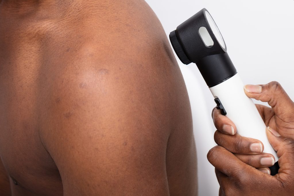
These immune checkpoint inhibitors, also known as anti-programmed death (PD-1) medications, are successful in treating roughly 50% of patients with melanoma, a kind of skin cancer.

Recently, researchers investigated whether patients with metastatic melanoma might respond better if immunotherapy and fecal microbiota transplants were combined.

This combination was not only risk-free but most patients responded well to the therapy, with some obtaining complete remission.

Phase 1 of the trial

Faecal transplants were coupled with the licenced medications pembrolizumab or nivolumab, which are already the standard of care for advanced melanoma, in the phase 1 MIMic trial.

The objective of the clinical experiment was to determine whether it is secure to combine these two medications in melanoma patients. As a supplementary goal, the impact of faecal transplants on the immune system and gut flora was evaluated.

Following a technique that was approved by Health Canada, healthy donors were carefully chosen. Then, capsules were created using the faeces of healthy donors.

Twenty metastatic melanoma patients were enrolled in the trial from Lawson Health Research Institute, the Jewish General Hospital (JGH), and the Centre Hospitalier de l’Université de Montréal (CRCHUM).

Each research subject was given capsules containing 80–100 mg of a fecal transplant from a single healthy volunteer donor. At least a week before receiving treatment with approved immunotherapy medications (either pembrolizumab or nivolumab), the fecal transplants were administered orally as capsules.

Is fecal transplantation plus immunotherapy safe?

The faecal transplantation operation was successfully completed by each of the 20 patients.

No major side effects were noticed prior to beginning immunotherapy, and no infections were spread through faecal transplantation. However, eight patients (40%) did have mild to moderate side effects from faecal transplantation, including diarrhoea, flatulence, and abdominal discomfort.

17 patients (85%) of the group encountered adverse immune-related events, the majority of which (70%) happened within the first three months of immunotherapy. Of these, five study participants (25%) experienced significant immune-related adverse effects, including nephritis (n = 1), arthritis (n = 2), exhaustion (n = 1), pneumonitis (n = 1), and arthritis (n = 2). These side effects forced the study participants to stop receiving their medication.

The researchers found no previously unreported adverse reactions to immunotherapy or faecal transplantation.

Did combined therapy lead to better results?

Four of the 20 participants in the trial (20%) experienced complete remission, making up 65% (13 out of 20) of the patients who responded favorably to the therapy.

All patients had strains of the donor’s bacteria in their gut microbiomes, according to analysis; however, this resemblance only got stronger over time in those patients who had a good response to the therapy. After receiving faecal transplants, respondents had higher levels of helpful bacteria and lower levels of dangerous bacteria.

The good impact of healthy donor faeces in boosting the efficiency of immunotherapy was further demonstrated in studies on mice by the researchers.

Fecal microbial transplantation: what is it?

Fecal transplantation, also known as fecal microbial transplantation (FMT), is a medical treatment in which the recipient’s intestines are filled with a healthy person’s donated poo (or feces).

In order to address medical disorders linked to abnormalities in gut bacteria, this method involves introducing healthy bacteria into the recipient’s intestines.

The effective treatment for recurrent Clostridium difficile infections is fecal transplantation. Fecal transplants are frequently administered via colonoscopy, however they can also be given as pills.

Gut and immune system interaction

So why do immune checkpoint inhibitors not work for everyone?

Recent research reveals that the bacteria in the gut may have an impact on how well the medications work. Immune checkpoint inhibitor-responsive individuals have a distinctive and healthy gut microbiome, often known as a “group of microorganisms in their gut.”

One of the study’s authors, Saman Maleki, Ph.D., assistant professor of oncology, pathology and laboratory medicine, and medical biophysics at Western University, as well as a researcher at the London Regional Cancer Programme and Lawson Health Research Institute, reasoned that altering a person’s gut microbiome to make it more diverse and healthy may enhance their response to immune checkpoint inhibitors.

Faecal microbial transplantation is one technique to modify the gut microbiota.

Will fecal transplants be used in the management of melanoma?

The principal study investigator, Dr. John Lenehan, a medical oncologist at the London Regional Cancer Programme, an associate scientist at the Lawson Health Research Institute, and an associate professor of family medicine and oncology at Western University, stated that the most significant finding in the study was that “none of the patients were harmed by the experimental treatment.”

Faecal transplants had been demonstrated to be beneficial by observational and pre-clinical studies, but “what happens in mice does not always translate to patients,” he noted. In fact, according to Dr. Lenehan, “more recent studies using similar therapies have shown harm, with patients having a worse response.”

He clarified that faecal transplantation was carried out differently in these other investigations than it was in the MIMic experiment.

“There are several factors, including bowel preparation, the number of FMTs required, the amount of stool required, and the identity of the donors. We had no idea if our approach would be secure or efficient. Thankfully, it appears that it was both! “, he exclaimed.

The director of the Supportive Oncology Research Group at the University of Adelaide and a research fellow at the Hospital Research Foundation Group, Hannah Wardill, Ph.D., who was not involved in this study, thinks this combination therapy strategy has the potential to be a successful treatment.

FMT is a reasonably accessible intervention, and this study shows it is safe and likely effective at improving immunotherapy response,” she added.

The combination of faecal transplants and immunotherapy results in an improved response rate in patients who would otherwise be unresponsive to immunotherapy, which indicates that “more people will benefit from immunotherapy,” according to Dr. Wardill.

REFERENCES:

For Cancer disease medications that have been suggested by doctors worldwide are available here https://mygenericpharmacy.com/index.php?therapy=10

How bacteria can occupy the skin and elevate eczema growth?

How bacteria can occupy the skin and elevate eczema growth?

Researchers looked into how bacteria might impact the histology of eczema in a recent study. They claimed that S. aureus bacteria change in eczema patches, speeding up their spread.

Eczema, the most prevalent type of atopic dermatitis, is an itchy, dry skin condition that is not communicable. In the US, 30% of the population is affected by the illness.

Although there is no known cure for eczema, there are drugs available to treat its symptoms. These include topical emollients, topical immunosuppressants, and topical corticosteroids.

Eczema is believed to result from a combination of hereditary and environmental factors. A flare-up of eczema may occur when the immune system is triggered by irritants found in soaps and surface cleansers, for instance.

Variants in the gene that makes the protein filaggrin may cause lower production in eczema patients. Filaggrin is crucial for boosting skin elasticity.

People who have eczema may have breaks in their skin, which bacteria can enter and develop in. The immune system may try to stop this colonization by escalating the inflammation, which aggravates the itching and further damages the skin.

The creation of new medications to treat eczema may be aided by knowing more about how germs grow into eczema sufferers’ skin and how it causes inflammation.

Recent studies looked into how Staphylococcus aureus adjusts to the skin of eczema sufferers. They claimed that the bacteria undergo alterations that cause them to lose their cellular capsule, allowing them to grow more quickly on the skin.

Dr. Alain Michon, the medical director of Project Skin MD Ottawa in Canada and a non-participant in the study, was consulted by specialists over the results.

What kind of bacteria is S. aureus?

According to earlier studies, S. aureus can frequently be found on the skin of eczema sufferers. Their eczema tends to be more severe the more bacteria they have.

By secreting toxins and drawing in immune cells, S. aureus is hypothesised to contribute to the pathophysiology of eczema and worsen the condition of the skin barrier.

S. Aureus is present in the nasal passages of up to 30% of persons. While the majority of infections are not serious, they can result in pneumonia, bone and joint infections, and serious bloodstream infections.

Information from the study on bacteria and eczema

The 23 children in Mexico between the ages of 5 and 15 who had moderate to severe eczema were the subjects of this longitudinal study by the researchers.

Standard medical care, such as topical steroids, emollient moisturisers, and bleach baths, were given to all of the subjects.

The children’s skin microorganisms were sampled by the researchers once per month for three months, and then again at nine months. Samples were collected from common eczema-affected areas such as the inside of the elbows and the backs of the knees. Additionally, they collected samples from the noses and forearms, which are often unaffected by the bacterium.

After that, the scientists cultivated S. aureus cells from every location, producing nearly 1,500 different colonies. This allowed them to more closely track the evolution of the certain cells.

At the end of the trial, they discovered that the majority of participants had only one lineage of S. aureus, indicating that new strains did not develop over time from the environment or other participants. However, they observed that throughout the trial, each lineage underwent significant mutation.

A gene called caps, which codes for an enzyme required for synthesizing polysaccharide a capsule-like shell that protects S. aureus from immune cells suffered several changes that lowered or abolished function, the researchers found in particular.

In a third of the subjects, the researchers discovered that capD mutations completely dominated the S. aureus microbiome population over the course of the study.

The researchers initially identified four distinct capD mutations in one youngster. By the time the trial was through, one of the variations had taken over and had expanded throughout the entire microbiome.

Increased eczema immunodetection

Dr. J. Wes Ulm of the National Institutes of Health, who was not involved in the study, was interviewed by Medical News Today about how mutations that make S. aureus more detectable by the immune system increase the spread of the bacteria and eczema on the skin.

Ulm remarked that from some angles, S. aureus becoming more readily identifiable by the immune system could appear to be a drawback. But he went on to say that if capD expression is lost or reduced, the bacteria may be better able to grow and spread since the energy that would have been used to create a useful capsule can now be used to fuel development.

Additionally, the absence of a capsule would make it simpler for the bacterium to adhere to the skin’s surface, improving its ability to spread throughout the skin.

Its lack of capD makes it easier for the immune system to detect and target the capD-deficient strain when it becomes more prevalent on the [skin’s] microbiome, Ulm said. Consequently, “and this, in turn, can enhance the immune response and magnify the inflammatory reaction giving rise to the characteristic rash and symptoms of eczema.”

Problems with the eczema research

The study’s tiny sample size, according to Michon, limits how broadly these results may be applied to other populations.

The results, he continued, might have been impacted by the fact that certain individuals’ microbiomes may have changed among those who took antibiotics both before and during the trial.

Other restrictions were also mentioned by Cameron K. Rokhsar, FAAD FAACS, a dermatologist and fellowship-trained cosmetic and laser surgeon in Manhattan and Long Island, New York, who was not associated with the study.

The drawback of these discoveries, according to Rokhsar, is that bacterial overgrowth only accounts for a portion of the overall puzzle. “The malfunctioning barrier specific to these people is the real problem with atopic dermatitis. Antibiotics are given to patients to reduce atopic dermatitis flare-ups, but they do not treat eczema.

REFERENCES:

For Skin disease medications that have been suggested by doctors worldwide are available here https://mygenericpharmacy.com/index.php?therapy=27

Can we manage chronic inflammation with psoriasis?

Can we manage chronic inflammation with psoriasis?

Psoriasis is regarded by medical professionals as an immune-mediated inflammatory illness even though the actual origin is uncertain. This indicates that the underlying cause of the disease is inflammation.

Psoriasis affects up to 3% of people in the US. It can affect other bodily components, such as the joints and eyes, and manifest signs on the skin, such as elevated plaques and discoloration.

According to experts, inflammation may be the common culprit affecting these various locations.

Psoriasis: What is it?

Skin inflammation is brought on by the autoimmune disease psoriasis. Psoriasis symptoms include thick patches of scale-covered, discolored skin. Plaques are the name for these scaly, thick patches.

As a chronic skin disorder with no known cure, psoriasis can flare up at any time.

Psoriasis comes in a variety of forms, including:

Plaque psoriasis: The most prevalent form of psoriasis is plaque psoriasis. Plaque psoriasis affects between 80% and 90% of those with psoriasis.

  • Inverse psoriasis: This kind develops in the creases of your skin. It results in tiny, scale-free plaques.
  • Guttate psoriasis: A streptococcal infection-related sore throat may be followed by the development of guttate psoriasis. It frequently affects children and young adults and appears as tiny, red, drop-shaped scaly patches.
  • Pustular psoriasis: This form of the condition features tiny, pus-filled lumps on top of plaques.
  • Erythrodermic psoriasis: This form of psoriasis is severe and affects a significant portion (greater than 90%) of your skin. Skin shedding and extensive skin discolouration are the results.
  • Sebopsoriasis: This kind often manifests as lumps and plaques with a greasy, yellow scale on your face and scalp. This is a hybrid of seborrheic dermatitis and psoriasis.
  • Psoriasis of the nails: Psoriasis of the nails can change your fingernails and toenails as well as the skin of your hands and feet.

What results in psoriasis inflammation?

Immune system malfunction in psoriasis patients leads to an accumulation of inflammatory cells in the dermis, the middle layer of skin. Additionally, the disease accelerates the proliferation of skin cells in the epidermis, the top layer of the skin.

Skin cells typically develop and slough off over the course of a month. In those with psoriasis, this process accelerates to only a few days. Skin cells accumulate on the skin’s surface instead of being shed, causing painful symptoms such elevated plaques, scales, edoema, and redness or discolouration.

Even though psoriasis is a skin disorder, the inflammation it causes affects the entire body. It can raise the risk of cancer, inflammatory bowel disease, psoriatic arthritis, heart disease, and others.

Is inflammation curable in any way?

Although immune system dysregulation is the cause of the inflammation in psoriasis, research indicates that patients can lessen this inflammation by making dietary and lifestyle adjustments. This may aid in symptom reduction and quality-of-life enhancement.

Many psoriasis sufferers can sustain remission—a prolonged period without having psoriasis symptoms—using these techniques.

In addition, certain psoriasis treatments work by lowering inflammation. Topical corticosteroids, biologics for injection, and oral drugs are some of these.

Psoriasis affects people differently. Some patients will need longer-term care than others.

Managing inflammation

Although there is presently no cure for psoriasis, the following behaviors may lessen inflammation caused by psoriasis and raise a person’s chances of going into remission.

Consuming a wholesome diet

Diet and systemic inflammation are closely related. According to studies, some inflammatory food habits might worsen psoriasis symptoms and increase the likelihood of developing the condition.

Everybody’s definition of a healthy diet is unique. However, the actions listed below could assist someone in establishing one:

Avoiding pro-inflammatory foods: Some foods and drinks include ingredients that promote inflammation, which exacerbates psoriasis symptoms. Soda and highly processed foods like salty snacks, sweets, and animal items are two examples.

A diet high in fruits, vegetables, and other nutrient-dense foods has been shown to reliably reduce the symptoms of psoriasis. For instance, a 2018 study of 35,735 individuals, 3,557 of whom had psoriasis, found that those who consumed a diet similar to the Mediterranean diet had fewer severe cases of psoriasis than those who did not.

Being healthy in terms of weight

A risk factor for the onset of psoriasis is obesity. Overweight or obese psoriasis sufferers may also have more severe symptoms than those who are of moderate weight.

In individuals with excess body weight, weight loss may lower inflammatory indicators and assist in reducing psoriasis symptoms.

In a 2020 study, it was discovered that individuals with psoriasis and obesity or overweight who underwent a 10-week program to lose 12% of their body weight saw a 50–75% reduction in the severity of their psoriasis. An average of 23 pounds were lost by participants.

Introducing additional healthful practises

There are a number of behaviorist that might lessen inflammation and enhance psoriasis symptoms, including:

  • Avoiding or giving up smoking: Smoking hurts one’s health and aggravates inflammatory conditions like psoriasis.
  • Limiting alcohol consumption: Drinking too much might aggravate psoriasis symptoms and cause inflammation.
  • Staying active can assist with psoriasis symptoms by preventing extended periods of inactivity. According to one assessment of the literature, those with psoriasis who lead sedentary lifestyles experience more severe symptoms than those who engage in regular exercise.
  • Getting enough sleep: A lack of sleep can cause the body to become inflammatory. According to studies, getting little or no sleep might raise blood levels of inflammatory indicators. Adults should sleep for 7-9 hours every night, according to experts, to maintain good health.
  • Managing stress: Long-term stress causes the immune system to become overactive and promotes inflammation. Up to 88% of psoriasis sufferers cite stress as a symptom cause. Stress-relieving exercises like yoga and meditation may be beneficial.

When should I get medical help?

Anyone who is going through a psoriasis flare and is curious about how to lessen the symptoms and inflammation of the condition might want to think about consulting their healthcare team, which includes their dermatologist.

They can offer suggestions for diet and lifestyle modifications that may help lower inflammation and lessen psoriasis symptoms, as well as treatment options depend on the severity of the symptoms. Additionally, they could advise taking vitamins or supplements.

REFERENCES:

For Inflammatory disease medications that have been suggested by doctors worldwide are available here https://mygenericpharmacy.com/index.php?therapy=41