Browsed by
Month: April 2023

Is Covid-19 linked to a rising risk of developing diabetes?

Is Covid-19 linked to a rising risk of developing diabetes?

The question of whether or not viral infections can raise the chance of getting diabetes has been the subject of research for some time.

The virus that causes COVID-19, SARS-CoV-2, has now prompted scientists to investigate whether diabetes risk can be raised by SARS-CoV-2 infection.

According to recent data, the COVID-19 pandemic may have contributed to a 3-5% rise in the overall disease burden of diabetes in the Canadian population.

In order to prevent further injury to those who have been harmed, this evidence supports requests for greater observation of blood sugar levels in COVID-19 survivors.

The complete impact on the health of the worldwide population has not yet been fully appreciated, although the COVID-19 pandemic has so far caused close to 7 million fatalities, according to the World Health Organisation (WHO).

COVID-19 and diabetes association

This is not the first time that research has drawn attention to a possible connection between diabetes and SARS-CoV-2 infection.

American retrospective cohort research that was published in the BMJ in May 2021 showed that people who become infected have a considerably higher risk of diabetes. A prior article in Nature demonstrated an increased likelihood of metabolic diseases, including diabetes, being diagnosed after an illness.

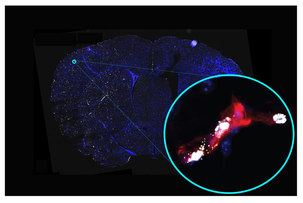
The ability of SARS-CoV-2 to infect human pancreatic cells—which produce insulin and are harmed and eventually destroyed in diabetics—was proven later that year in a study published in the journal Cell Metabolism. The loss of these cells could potentially be caused by infection, suggesting a potential underlying mechanism to explain the relationship.

Since then, cohort studies have indicated a greater incidence rate of type 2 diabetes, but not type 1 diabetes, after infection. One such study was reported in Diabetologia. Another retrospective cohort study employing Veterans Health Administration data, which was published in Diabetes Care, revealed that males, but not women, saw a rise in the incidence of all diabetes diagnoses following SARS-CoV-2 infection.

Children are more likely to be diagnosed with type 1 diabetes than adults are, according to a cohort study that was published in PLOS One. The risk of type 1 diabetes diagnosis after infection was also found to be higher in American Indian/Alaskan Native, Asian/Pacific Islander, and Black populations.

Diabetes is 22% more likely to develop

Now, a study involving 629,935 persons, with an average age of 32, has found that men who tested positive for SARS-COV-2 between January 1, 2020, and December 31, 2021, had a 22% higher risk of developing diabetes in the eight months after infection than men who hadn’t been exposed.

Based on age, sex, and date of infection, researchers matched pairings of individuals with a confirmed case of COVID-19 and those who hadn’t, using data from the British Columbia COVID-19 Cohort, a database of SARS-CoV-2 infection in British Columbia, Canada.

When the results were stratified by the severity of the disease, researchers discovered that those who had COVID-19 when they were admitted to the hospital had a 2.4-fold increased risk of developing diabetes compared to those who hadn’t been infected, and those who were admitted to intensive care had a 3.29-fold increased risk.

When these cases were taken into account, the data revealed that women were also more likely to acquire diabetes following infection with SARS-CoV-2, albeit this tendency was not significant when only moderate cases were taken into account.

The scientists were unable to differentiate between type 1 and type 2 diabetes using the data they had access to since this link was only discovered for non-insulin-dependent diabetes.

Risk of diabetes with viral infections

It is unclear precisely how SARS-CoV-2 infection causes these long-term consequences, as it is with other long-term side effects. It is not the first time that a viral infection has been connected to a higher chance of acquiring diabetes, but the mechanisms underlying the association are still unknown.

The effect of Coxsackievirus B infection on the risk of type 1 diabetes has been extensively investigated, along with the effects of mumps, rubella, and cytomegalovirus, according to Dr. Fares Qeadan, associate professor of biostatistics at Loyola University Chicago who was not involved in the study.

Researchers have also looked into the potential roles of inflammation, insulin resistance, and impacts on pancreatic cells in the relationship between hepatitis C virus infection and the risk of type 2 diabetes.

In conclusion, viral infections have been linked to a higher risk of developing both type 1 and type 2 diabetes. The evidence for type 1 diabetes is stronger and includes a wider range of viruses, but the data for type 2 diabetes is more limited and mostly concentrates on particular viral diseases like the hepatitis C virus. Dr. Fares Qeadan stated that more study is required to pinpoint the precise processes by which viral infections influence the onset of diabetes and to create preventative measures.

Diabetes or long COVID, which is it?

As the clinical characterisation of long-COVID is still being developed, experts cautioned that it was a complicated topic to determine whether the onset of diabetes following infection with SARS-CoV-2 might be regarded a symptom of long-COVID.

Dr. Morgan Birabaharan, a physician and virus researcher from the University of California, San Diego’s Division of Infectious Diseases and Global Public Health who was not involved in the study, stated:

The onset of diabetes may fall within the category of protracted COVID, which is used to characterise a variety of symptoms and illnesses that appear after the acute phase of SARS-CoV-2 infection (>30 days).

To classify what side effects of SARS-CoV-2 infection are ‘long COVID’ vs. some other process, he said, “is difficult because we are still trying to understand the pathophysiology of long COVID, whether it be persistent viremia, dysregulated immune response, or some other phenomenon.”

This most recent article backed suggestions for aggressive management of this, saying that the population-level effects of a rise in diabetes cases caused by the COVID-19 pandemic could also be considerable.

In any case, Dr. Qeadan said, “Recognising the potential link between SARS-CoV-2 infection and the onset of diabetes is important for healthcare professionals as it highlights the need for careful monitoring of blood glucose levels and early intervention in people who have had COVID-19.”

“This can help lessen the long-term effects of diabetes on the affected individuals and reduce the overall burden on healthcare systems,” he continued.

After COVID, diabetic symptoms

Increased thirst and hunger, frequent urination, unexplained weight loss, exhaustion, and hazy eyesight are all typical early indicators of diabetes, according to Ricordi.

If you had COVID-19 and any of these symptoms, it would be worthwhile to request a diabetic screening from your doctor, especially if you have risk factors or a family history of the disease.

One should see their primary care physician if any of these symptoms or indicators are present, according to Ricordi.

The conclusion

An increased risk of diabetes has been linked with COVID-19, according to recent research. Diabetes may be another factor contributing to extended COVID, according to the study. Endocrinologists think COVID-19 may harm the pancreas and affect how it releases insulin, though additional research is required to fully understand the association.

REFERENCES:

For Diabetes medications that have been suggested by doctors worldwide are available here https://mygenericpharmacy.com/index.php?therapy=13

Detect the Cognitive ability by a test before its arrival.

Detect the Cognitive ability by a test before its arrival.

According to researchers, a quick card memory test can identify cognitive decline years before symptoms appear. According to experts, the test would make it possible for those who are more likely to develop dementia to receive treatment and preventative measures sooner.

One specialist advocates administering the test to everyone over 45. Researchers claim to have created an easy test that can forecast a person’s future risk of developing cognitive impairment.

The test only applies to persons without pre-existing cognitive and memory impairments, the researchers write in their study, which was just published in the journal Neurology.

The study’s lead author and clinical professor at the Albert Einstein College of Medicine in New York City, Ellen Grober, Ph.D., said in a statement that there is “increasing evidence” that some people who appear to be healthy and have no cognitive or memory issues may fact be displaying very mild indicators of early cognitive impairment. In our study, a quick and accurate memory test indicated the likelihood that individualsnormally thought to have normal cognition will experience cognitive impairment.

Cognitive impairment study

969 participants in the study, whose average age was 69, took a basic memory test and then underwent follow-up exams over ten years.

There were two phases of the test.

The participants were first instructed to examine four cards, each of which had four drawings of various objects.

Then, the respondents were required to identify each item as a member of a specific category. For instance, when asked to name a fruit, participants might respond “grape.”

In the following stage, participants were required to recall the things to gauge their memory capacity. They were given category cues, which calculated the amount of memory storage, for the items they couldn’t recall.

Result of the tests for cognitive impairment

Using the Stages of Objective Memory Impairment (SOMI) approach, researchers categorized the participants into five groups with stages zero through four based on their test results.

Stage 0 was for patients with no memory issues (47% of cases).

Memory retrieval problems, which researchers noted can occur five to eight years before dementia, were more difficult to recall in stages one (35%) and two (13%) of the disease. When given indications, these participants could recall information.

The individuals in the third and fourth stages (5% overall) had trouble remembering all the objects, even with hints. These stages, according to the researchers, occur 1 to 3 years before dementia.

234 of the 969 subjects experienced cognitive impairment in the end.

Subjects at stages one and two were twice as likely to experience cognitive impairment compared to those at SOMI stage zero, even after accounting for factors such as age, gender, education level, and the APOE4 gene, which affects a person’s chance of developing Alzheimer’s disease.

Cognitive impairment was three times as likely to develop in those in stages three and four.

The significance of testing for cognitive impairment

The SOMI method continues to forecast an elevated risk of cognitive impairment even after accounting for indicators of Alzheimer’s disease including amyloid plaques and tau protein tangles.

According to research, 72% of people in the third and fourth stages will have cognitive impairment after ten years, compared to 57% of people in the second stage, 35% of people in the first stage, and 21% of people in stage zero.

Our findings confirm the SOMI system’s application in locating those most at risk for cognitive decline, according to Grober. “Researchers looking for remedies can benefit from spotting cognitive impairment early on. By working with their doctor and implementing strategies to support healthy brain aging, those persons who are discovered to be at elevated risk may also benefit.

Neura Health’s virtual headache and migraine clinic’s medical director, Dr. Thomas Berk, a neurologist, pointed out that present testing only reflect the brain’s current condition.

“Predicting neurological change years later is very difficult,” said Berk. When someone has neurological testing, “we are getting a snapshot of their current brain function, not what their brain will look like years later,”

“This does give some evidence for being able to assess the future risks of developing memory issues,” he continued.

There is unquestionably a need for “a simple and fairly rapid test,” particularly in light of the growing body of evidence that early intervention can have a positive impact, according to Dr. Dale Bredesen, head of the University of California Los Angeles’ Easton Centre for Alzheimer’s Disease Research.

Standard neurocognitive tests can take hours, making them impractical for screening, and common quick tests like the MMSE aren’t sensitive enough to catch these early alterations, according to Bredesen.

Using fresh memory tests

According to Bredesen, the subjects of present testing are those who already have cognitive problems.

“Simple tests like the one described in this report should be included for everyone over the age of 45, to identify those who should be evaluated further, and potentially treated,” he advised.

One physician claimed that the SOMI system made him think of a well-known kid’s game.

According to Dr. Clifford Segil, a neurologist at Providence Saint John’s Health Centre in California, “I advise my patients to make a mental image of a scene with all three words to help in their recall when they have to recall the three words.” “I would suggest the same for this proposed card cognition exam. Currently used cognitive exams include additional memory tests.

“Go Fish,” which is utilised as a learning tool for children rather than a cognitive exam for senior people, is similar to the suggested test to be employed in my elderly population, which interests me as a parent and a practising adult neurologist, Segil added.

“When you start to worry that you might be losing your memory, you should be evaluated by a neurologist to see if your complaints are generally age-appropriate normal or something else,” he said.

REFERENCES:

For Cognitive disease medications that have been suggested by doctors worldwide are available here https://mygenericpharmacy.com/index.php?cPath=77_478

Atrial fibrillation catheter therapy and dementia risk.

Atrial fibrillation catheter therapy and dementia risk.

Researchers looked into how medications and catheter ablation for atrial fibrillation affected people’s risk of developing dementia.

They discovered that catheter ablation therapy lowered the risk of dementia more effectively than medicine alone.

For the correlation to be verified and the cause of it to be understood, more research is required.

Atrial fibrillation (Afib)

A cardiac disorder called atrial fibrillation (Afib) is characterised by erratic beating in the upper chambers of the heart, which restricts blood flow to the lower chambers. Patients who have afib may experience discomfort and have a five-fold increased risk of stroke.

183,321 death certificates in the US in 2019 listed AFib as a cause of death. By 2030, 12.1 million persons in the United States are predicted to be affected by the illness.

AFib has been linked in studies to dementia and cognitive decline. Additionally, studies have shown that patients with dementia and cognitive impairment who also have AFib suffer from a greater rate of cognitive decline than those who do not.

Understanding whether treating Afib patients lowers their likelihood of developing dementia may improve patient outcomes.

Recent studies evaluated the effects of Afib medication versus catheter-based therapy on the risk of dementia. They discovered that compared to medicine alone, catheter-based Afib therapy was associated with improved cognitive function.

“Despite having negative brain imaging results, I have encountered multiple patients with long-term persistent atrial fibrillation who had cognitive impairment, decreased swallowing capacity, and slower motor performance.

Based on this, I do not find [the results] surprising,” Dr. Vicken Zeitjian, a San Antonio, Texas-based cardiologist board-certified in nuclear cardiology and echocardiography who was not involved in the study, told.

He went on to say that “these results further demonstrate that catheter ablation is a superior method of atrial fibrillation management than medical management alone.” The research will be presented at the 75th Annual Meeting of the American Academy of Neurology.

Medicine versus catheter ablation

887 Afib patients with an average age of 75 were included in the study by the researcher. Before enrollment, 193, or 21.8%, of the participants got catheter ablation, whereas the remaining patients just received Afib medication.

Catheter ablation is the process of utilising radiofrequency to remove tiny patches of cardiac tissue that may be the source of an irregular heartbeat.

The individuals’ cognitive function was evaluated at baseline, one year later, and two years later. Out of a possible 30, a 23 or below score indicated cognitive impairment.

The study’s findings showed that those who underwent catheter ablation had an average cognitive score of 25, compared to those who did not, who had a score of 23.

The researchers discovered that those who received catheter ablation were 36% less likely to experience cognitive impairment than those who were just given medication after controlling for conditions like heart disease, kidney disease, and sleep apnea.

However, they pointed out that there were no appreciable variations in the incidence of heart attacks between patients who underwent catheter ablation or medication alone.

They also discovered that using warfarin and other anticoagulants had no appreciable impact on cognitive deterioration.

The risk of dementia and catheter ablation

We enquired about how catheter ablation might lower the risk of dementia from Dr. Aaron Ritter, director of the Memory & Cognitive Disorders Programme at Hoag Hospital in Newport Beach, California, who was not involved in the study. Although the study doesn’t say how, he pointed out that there could be a number of causes.

“For me, ablation may be a more conclusive or long-lasting therapy than pharmaceutical management, which necessitates a commitment to a daily prescription schedule, perhaps twice daily. We have to wonder if the compliance issue is important to the outcome in those with memory issues, he said.

“Furthermore, we may also hypothesise that ablation may be more a successful treatment for atrial fibrillation, and as a result, individuals may have fewer blood clots or better consistent delivery of blood and oxygen to the brain,” he added.

Future research should, he hoped, incorporate measurements of blood flow, which would help researchers understand why ablation performed better in this trial.

less cognitive impairment overall

In the recent study, data for 887 older persons with AFib were analysed. Participants were 75 years old on average, about half were women, and more than 87% were white.

Approximately 22% of patients underwent catheter ablation. Compared to those who just received medicine for their AFib, these individuals were more likely to have both a persistent AFib and an implantable cardiac device.

Before the study began, participants performed cognitive function tests that evaluated their short-term memory, attention, concentration, and language skills. One and two years later, they conducted similar tests once more.

These tests merely evaluated whether a person had problems completing particular mental activities; they did not determine whether a person had dementia.

In contrast to individuals who only received pharmacological treatment during the two-year research, those who underwent catheter ablation had a 36% lower chance of experiencing cognitive impairment.

Dementia is caused by many things.

The results of the current study should not be taken too seriously because they have not yet undergone peer review, said Dr. Keith Vossel, a neurologist and the director of UCLA’s Mary S. Easton Centre for Alzheimer’s Research and Care in Los Angeles.

Additionally, the study must be published before its influence on clinical practise can be evaluated, the researcher added.

However, “it does this add to other research supporting the possible use of certain treatments for reducing dementia risk,” he said to Healthline.

The fact that blood flow to the brain was not measured in the present study’s limitations to determine if patients treated with catheter ablation or medication had different blood flow patterns to the brain.

Limitations

Additionally not engaged in the study, Dr. Fanny Elahi, a physician-scientist who is an assistant professor of neurology, neuroscience, pathology, molecular, and cellular-based medicine at Mount Sinai, stated:

“Although these findings are intriguing, more investigation is required to determine the connection between catheter ablation (CA) and dementia. With cognition as a co-primary endpoint and the study appearing to be observational rather than randomised, I wonder if the study is tainted by the baseline health of individuals who receive CA.

The main drawback is that cerebral blood flow measurements were not provided to go along with the cognitive testing. The authors’ use of a 30-point cognitive test, a somewhat constrained measure of cognition, is another drawback, says Dr. Ritter added.

Implications

cardiologist at Staten Island University Hospital, Dr. Rina Shah, who was not involved in the study, stated:

“It is significant to note that dementia and the prevalence of AFib both rise with ageing. AFib, however, can result in a variety of other medical issues, including hemodynamic deterioration and cardiomyopathies if rates are difficult to manage. We can assist prevent or delay the harmful effects of AFib by administering catheter ablation to patients earlier.

The study, according to Dr. Elahi, emphasises the significance of connections between the brain and the body.

“We must care for the whole person if we want to increase brain health. Implementing molecular and imaging biomarkers may help to further define the impact if there is one, she said.

“I am a strong believer in knowing the biological impacts of such therapies since we probably need several shots on goal to battle brain ageing and cognitive loss. Finding out why something works allows us to use synergistic methods to enhance the intervention, she added.

REFERENCES:

For Cardiovascular medications that have been suggested by doctors worldwide are available here https://mygenericpharmacy.com/index.php?therapy=11

Researchers identify potential new stroke treatment targets

Researchers identify potential new stroke treatment targets

When blood flow to a portion of the brain is stopped or reduced by a hemorrhage or obstruction, a stroke results. Although some stroke survivors recover completely, many still struggle with long-term repercussions and are at increased risk of having another stroke.

Brain damage following a stroke is thought to be a result of changes in small blood arteries that exist in addition to the blockage.

In the injured small blood arteries in the brain, a recent study identified multiple changes in gene activity that may serve as targets for pharmacological therapy to enhance stroke recovery.

An artery in the brain becomes clogged or bursts, resulting in a stroke. Beyond the blockage or bleeding, the brain cells are starved of oxygen and nutrients and suffer damage or degeneration. Researchers have been looking for strategies to lessen damage after a stroke and hasten recovery.

Researchers from Weill Cornell Medicine have now discovered alterations in gene activity in small blood arteries after a stroke. The results imply that these alterations could be targeted with current or upcoming medications to lessen brain damage or enhance stroke recovery.

The research was released in PNAS. Weill Cornell Medicine assistant professor of pathology and laboratory medicine and the lead author, Dr. Teresa Sanchez, told:

“By providing a knowledge platform of the molecular alterations in the cerebral microvasculature, our study has improved our understanding of the pathophysiology of stroke. This is critical to developing novel therapeutic strategies for this devastating condition.”

Stroke symptoms

The majority of strokes are ischemic strokes, in which a blood clot obstructs a blood vessel leading to the brain. This prevents nutrition and oxygen from reaching brain cells.

Immediate signs could be:

  • bewilderment and difficulty speaking
  • Headache, maybe accompanied by dizziness or nausea
  • numbness or a lack of movement in certain body areas, especially on one side
  • vision issues
  • Walking difficulties, a loss of coordination, and vertigo.

It’s crucial to get therapy and a diagnosis right away to reduce long-term damage. Many stroke victims, however, continue to experience physical and psychological after effects.

Over 795,000 people experience a stroke each year in the United States. Also, the condition is one of the main causes of long-term disability. This is according to the Centres for Disease Control and Prevention (CDC).

Long-term damage from a stroke

Despite the fact that only 10% of stroke survivors experience a near-complete recovery, survivors frequently experience a variety of symptoms, such as:

  • weakness or paralysis on one side of the body.
  • thinking, memory, and speaking issues.
  • difficulty swallowing and chewing.
  • issues controlling one’s bowels and bladder.
  • Depression.

Inflammation and long-term alterations in the brain’s small blood arteries, result in constrained blood flow to brain cells and leaking over the blood-brain barrier. They are the root of many of these symptoms.

In a recent study, gene activity in mouse cerebral microvasculature after a stroke was observed to vary. Similar modifications were found in stroke patients from people as well.

Gene activity changes following a stroke

The researchers discovered 541 genes whose activity was altered similarly in mice and people after stroke, and they also discovered many clusters of genes with various roles.

According to Dr. Teresa Sanchez, “Our work has also clarified the shared transcript alterations between human and mouse stroke and identified common changes in pathways associated with vascular/endothelial dysfunction, sphingolipid metabolism, and signalling.

They discovered genes linked to vascular disease, general inflammation, brain inflammation, and the specific form of vascular malfunction. This results in leaky cerebral microvessels. The blood-brain barrier, which controls the flow of chemicals between the blood and brain cells, is weakened as a result of these leaky arteries.

The activity of molecules that regulate the blood-brain barrier was shown to vary after stroke, according to the researchers.

Dr. Sanchez stated that a stroke causes “robust changes in the genes regulating the blood-brain barrier and endothelial activation, i.e. upregulation of genes causing blood-brain barrier leakage and downregulation of genes protecting the blood-brain barrier.

Additionally, they found that after a stroke, there was a disruption in the activity of genes that regulate sphingolipid levels, which are fat molecules involved in a broad variety of biological activities, including inflammation.

Potential therapeutic routes

Some of these molecular alterations, according to the researchers, might make for fresh medication therapy targets. They draw attention to the elevated sphingolipid concentrations in the cerebral microvasculature and hypothesize that sphingolipid targeting may be therapeutic after stroke.

We questioned Dr. Sanchez on the possibility of using medicines to either prevent these changes or undo the harm already done.

Since endothelial dysfunction is a major contributor to stroke and, concurrently, stroke-induced cerebral ischemia causes additional injury to the endothelium, which further compromises cerebral blood flow and exacerbates brain injury,” she said.

According to Dr. Allder, the discoveries could influence research into other neurological conditions:

I can see how it might make post-stroke treatments more accessible, but I also see how it might open up new treatment options for dementia and post-brain injuries, particularly repetitive brain injuries.”

More research is required

Therefore, the results may possibly suggest novel therapeutic avenues, but Dr. Iyer emphasised the need for more study:

The key drawback of this research is that human genome and transcriptome models aren’t usually transferable from mouse models. However, this study reveals a hitherto unrecognised cellular signalling route that is unquestionably ready for further study.

Preclinical research is currently being conducted by Dr. Sanchez and her team to see whether it would be advantageous for stroke patients to reverse some of the specific microvascular abnormalities that were observed in their study. They are doing this by employing candidate medications or genetic techniques.

REFERENCES:

For Stroke medications that have been suggested by doctors worldwide are available here https://mygenericpharmacy.com/index.php?cPath=77_99

Possible link between migraine and carpal tunnel syndrome.

Possible link between migraine and carpal tunnel syndrome.

Researchers looked into the prevalence of migraine headaches in patients who have nerve decompression surgery.

They discovered that people who have surgery to decompress a nerve at particular points on their bodies may be up to 70% more likely to get migraines than others who have the procedure elsewhere.

To determine whether nerve decompression can treat migraines, more research is required.

There may be pain and a loss of function when the nerves in the hands and arms contract around muscles and soft tissues. Between 5% and roughly 9% of the population are affected by various types of nerve compression in these places.

Surgery is frequently used to treat the illness and might result in full or partial symptom relief.

The muscles, blood arteries, and bone in the vicinity of the head’s surrounding nerves can also compress those nerves. Improvement or alleviation from migraine and headaches may result by decompressing these nerves.

What is Carpal tunnel syndrome?

Carpal tunnel syndrome is caused by compression of the median nerve. On the hand’s palm side, the carpal tunnel is a small opening encircled by bones and ligaments. Numbness, tingling, and weakness in the hand and arm are signs of median nerve compression.

Carpal tunnel syndrome can be caused by repetitive hand motions, health issues, and wrist morphology.

The tingling and numbness are typically reduced with appropriate care, and wrist and hand function is recovered.

Symptoms

The following list of signs and symptoms of carpal tunnel syndrome includes:

Feeling tingly or numb. Numbness and tingling in the fingers or hand may be apparent. Normal afflicted fingers include the thumb, index, middle, and ring fingers, but not the little finger. In certain fingers, you might experience something like to an electric jolt.

The wrist may feel the sensation before it moves up the arm. These symptoms may awaken you from sleep and frequently happen while you are holding the phone, newspaper, or steering wheel.

To try to alleviate their symptoms, many people “shake out” their hands. Over time, the numb sensation could persist continuously.

Weakness. You can feel weak in your hands and drop things. This can be because the thumb’s pinching muscles, which are similarly regulated by the median nerve, are weak or because the hand is numb.

Migraine and carpal tunnel syndrome

When the nerve that travels from the forearm to the palm of the hand is squeezed at the wrist, carpal tunnel syndrome develops.

In comparison to 16% of those without carpal tunnel syndrome, 34% of those with the condition get migraines, according to a cross-sectional study with 25,880 participants. It might be more effective to screen patients for the disorders if it is known whether nerve compression around the head is related to nerve compression in the hands and arms.

Researchers looked at how frequently people who had nerve decompression surgery for the hands and arms were diagnosed with migraines.

According to their findings, people who have particular types of nerve compression are more prone to suffer from migraine headaches. Not a part of the study, Dr. Chantel Strachan is an internist at ColumbiaDoctors and an assistant professor of medicine at Columbia University Irving Medical Centre in New York. She said.

“I wouldn’t jump to advise carpal tunnel release in every migraine patient. The choice to proceed with surgical treatment for nerve compression is specific to the patient and should be carefully considered with the patient’s medical care team.

Journal of Plastic and Reconstructive Surgery published the findings.

Most likely to experience migraine

Data from 9,558 patients who underwent nerve decompression surgery of the hands and arms between 2009 and 2019 were analysed for the study.

Participants were also evaluated by the researchers for the presence of migraine.

Of the subjects, the median nerve was decompressed in about 71% of cases. Surgery is done on the wrist to release pressure on the nerve, which lessens carpal tunnel syndrome symptoms.

A decompression of the ulnar nerve was done on about 14% of subjects. That is an elbow nerve decompression. 6.5% of patients underwent decompression procedures at various body locations.

In the end, the researchers discovered that people with multiple nerve decompression and median nerve decompression were respectively 30% and 70% more likely to experience migraines than people with ulnar nerve decompression.

Nerve compression and migraine

Dr. Sean Ormond, a specialist in anesthesiology and interventional pain management, did a study to learn more about the potential connection between nerve decompression and migraine.

He mentioned that there are a number of possibilities, but that the causes of nerve compression in the arms and hands and migraine are not entirely known.

“Both upper extremity nerve compression syndromes and migraine may share common risk factors, such as obesity, sedentary lifestyle, poor posture, or repetitive stress injuries,” stated Dr. Ormond.

The affected area may experience inflammation as a result of nerve compression. It is also recognised that inflammation contributes to the pathophysiology of migraines. The presence of inflammation in one place of the body may cause inflammation to spread throughout the body, potentially aggravating migraines, the doctor added.

Ormond observed that although further research is need to establish this, some people may be more prone to higher nerve compression and migraine due to a hereditary tendency.

According to Dr. Strachan, migraine sufferers may become more sensitive to pain due to nerve damage.

Limitations of the migraine study

Dr. Strachan pointed out that because the study was retrospective in nature, the results suggest association rather than causality.

She stated that different providers and their subspecialties, such as primary care, neurology, and pain, may have utilised different criteria to diagnose migraine.

The association between migraine and pain from nerve compression may be the consequence of other variables, as the researchers stated in their report that there is a general overlap across chronic pain disorders.

REFERENCES:

For Migraine disease medications that have been suggested by doctors worldwide are available here https://mygenericpharmacy.com/index.php?therapy=17

Analyze the links between BMI, obesity & cognitive ability.

Analyze the links between BMI, obesity & cognitive ability.

According to the World Health Organisation (WHO), there were more than 650 million obese adults in the world as of 2016. Obesity has been linked in the past to an increased risk of cognitive deterioration.

Evidence from University College London researchers challenges the idea that fat and cognitive capacity are causally related.

Around the world, more than 1.9 billion adults were obese in 2016, with more than 650 million of those adults suffering from obesity, a disease in which a person’s weight is over normal ranges and may lead to various health issues.

According to current estimates, 167 million adults and children will be overweight or obese by 2025. A multitude of disorders, including diabetes, cardiovascular disease, hypertension, osteoporosis, rheumatoid arthritis, and cancer, have been linked to obesity in previous studies, including these.

Furthermore, previous research has connected obesity to a higher risk of cognitive deterioration.

The causal relationship between obesity and cognitive performance has now been called into question by University College London academics. They contend that common family variables have tainted the research linking cognitive aptitude and BMI.

Obesity

If a person’s present weight is excessive for their height, they are considered obese. The body mass index (BMI) is the most popular metric for determining a person’s level of obesity.

This tool determines if an adult is obese or not based on their height and current weight:

  • BMI less than 18.5 indicates underweight.
  • Suitable BMI range: 18.5-24.9
  • BMI of 25 to 29.9 indicates obesity
  • obesity: a BMI of 30 or above

Children and teenagers need a different BMI calculator, which considers height, age, and gender to evaluate obesity because they are still developing.

The BMI measurement is not without problems, though. It is unable to distinguish between muscle and fat when weighing someone. Additionally, it disregards a person’s race, overall body composition, or bone density.

Cognitive function and obesity

Lead author of this study and senior research fellow at the Centre for Longitudinal Studies at University College London in the U.K., Dr. Liam Wright, Ph.D., states that there are several reasons why the research team decided to investigate the causal relationship between cognitive capacity and obesity:

“Over the past forty years, there has been a significant rise in the prevalence of obesity, but BMI hasn’t increased uniformly throughout the population. Therefore, it is crucial to understand why some people are more predisposed to obesity than others.

Additionally, there is a substantial body of research in the field of cognitive epidemiology that demonstrates a connection between cognitive function and practically every measure of health and health behaviour, including obesity.

Unfortunately, the majority of the cognitive epidemiology literature employs observational research designs that may be biassed and fail to show causal effects, according to Dr. Wright. “There are some compelling theoretical arguments for why cognitive ability might have a causal effect on health, but regrettably, these arguments are based on observational research designs,” she said. Because a sibling design could take into account some of the variables that can skew relationships found in previous research, we felt it was crucial to investigate for a relationship between cognitive capacity and BMI.

Examining siblings to reduce bias

Dr. Wright and his research group evaluated data from four distinct young population cohort studies carried out in the United States that included 12,250 siblings from 5,602 homes. Each participant’s data were tracked from youth to age 62.

The scientists were able to take into consideration unobserved characteristics associated with family background by analysing the relationship between cognitive capacity and BMI among families.

“Sibling designs account for factors that are shared between siblings by design,” Dr. Wright said. They don’t require the measurement of these factors, which is both a benefit and a drawback because it is difficult to determine which common factors actually contribute.

With this qualification, he continued, “There are four main factors that we thought might be significant: genetics (siblings share 50% of DNA), parental socioeconomic class (wealth, location, etc.), parenting styles (particularly regarding dietary choices), and parental cognitive ability (cognitive ability could operate indirectly!). “Once more, we didn’t directly examine these.”

According to Dr. Wright, they predicted that these variables would make general population studies more biassed and lead to weaker relationships than in earlier studies, which is exactly what they found.

However, he cautioned, “remember that sibling designs have their own flaws, including the ability for siblings to influence one another, for example, by modelling one another’s behaviour. This may imply that our findings are also skewed, albeit downwardly and smaller than the actual causal effect.

Association between BMI and cognitive ability

When the researchers evaluated the data from study participants who were not related, they discovered that, after accounting for family socioeconomic status, the change in teenage cognitive capacity from the 25th to the 75th percentile was associated with an estimated 0.61 kg/m drop in BMI.

And when the researchers analyzed the information from siblings, they discovered that the change in BMI from the 25th to the 75th percentile of teenage cognitive ability was only correlated with a 0.06 kg/m drop in cognitive capacity.

The relationship between cognitive capacity and BMI was less pronounced when siblings were compared than when the entire population was, according to Dr. Wright, but he was not surprised by this given the overall characteristics he mentioned.

However, he noted, “I was shocked at how little of an association there was when comparing siblings. As said, there are strong arguments to support the idea that cognitive ability has an impact on health and health-related decision-making“.

“Two possibilities for this small association are that one, our results were biassed towards finding smaller associations (e.g., by siblings influencing each other), and two, reflective decision-making isn’t as important in determining BMI as other factors like satiety, etc.,” Dr. Wright continued. Both of these are hypothetical.

Unproven causality

As a parent and a neurologist, Dr. Segil claimed that he has never observed a connection between obesity or a healthy weight and cognitive aptitude in people.obese

The purpose of this study, according to Dr. Segil, “is to argue that people with higher cognitive abilities, who have a higher socioeconomic position, have made healthier decisions.” Additionally, it’s possible that people’s cognitive function increases as their BMI decreases when they make healthier decisions.

He continued, “I do not believe that there is any evidence linking obesity to cognitive function. And I believe that their research’s use of siblings or other family members who are in a similar social economic situation to real-life situations such as brothers or sisters or siblings is realistic.”

After reading this study, Dr. Segil stated that he would be curious to know whether maternal or paternal obesity had a greater impact on adolescent cognitive development.

As a result of reading this, they claimed that adolescent cognition is linked to a lower adult BMI, he continued. So I’m keen to know if stronger adolescent cognitive abilities are related to the maternal and paternal BMI. Does having a thin or fat parent, using the same dataset, alter their children’s cognitive ability?I was shocked, though, by how little of an association there was when comparing siblings. As mentioned, there are strong arguments to support the idea that cognitive ability has an impact on one’s health and decision-making in relation to their health.

RFERENCES:

For Cognitive disease medications that have been suggested by doctors worldwide are available here https://mygenericpharmacy.com/index.php?cPath=77_478

Can hormones predict the possibility of weight loss?

Can hormones predict the possibility of weight loss?

In addition to being one of the world’s greatest killers, obesity is linked to several medical disorders. The majority of people who do succeed in losing excess weight do so only to gain it back, which frequently frustrates attempts to shed excess weight through lifestyle changes.

Understanding the complicated function that hormones play in this process may aid in the creation of effective long-term treatments for obesity.

Now that higher levels of the hormone neurotensin have been connected to greater weight loss maintenance in obese individuals, this information provides another hint as to how to rebound weight gain might be controlled.

It has long been understood that while many people who struggle with overweight or obese can lose excess weight through lifestyle changes like diet and exercise, it can be difficult for many of them to keep it off.

The World Health Organisation (WHO) views obesity as an epidemic since it causes more than 4 million deaths annually. Many of these fatalities are thought to be preventable with weight loss. For instance, it has been demonstrated that a weight loss of 5–7% can halt or delay the onset of type 2 diabetes.

Sadly, up to 80% of people who lose weight through diet and exercise gain back every bit of the weight they previously did. In the past, this weight increase has been attributed to a lack of self-control or a failure to follow weight maintenance plans.

A higher level of the hormone neurotensin may be associated with a superior capacity to maintain weight loss in obese people who have recently lost weight, according to the most recent research, which gives a fresh explanation.

Do we regain weight after weight loss?

The assumption that the body would return to a predetermined weight despite weight loss or growth has been proposed as one theory to explain why people experience rebound weight gain.

Bariatric surgeon Dr. Mir Ali of Orange Coast Medical Centre in Fountain Valley, California, who also serves as the center’s medical director, explained:

According to the set point theory, there is a weight that your body prefers to maintain at all times (within 5 to 10 pounds). This set point can be influenced by a variety of factors, including age, heredity, sex, health issues, and degree of activity. But it’s unclear what mechanisms might be at work in this observation.

The importance of hormones in hunger regulation has just recently come to light, despite the WHO reporting a rise in the number of persons with obesity starting in the 1970s. Only in 1999 was ghrelin, a hormone that promotes the desire to eat, separated and identified. Its ability to control hunger, fat storage, and energy regulation is now well documented. It also has interactions with other hormones like insulin which are important in regulating energy and storing fat.

Researchers have postulated that the fact that ghrelin levels tend to increase after weight reduction brought on by diet and exercise may be the cause of “rebound weight gain.” Contrarily, after bariatric surgery, which has a lower rate of rebound weight gain than diet and exercise-based therapies, levels of this hormone drop.

Less than 20 years ago, in 2004, researchers found that ghrelin and leptin, a hormone that controls satiety and long-term energy balance, interact. Since then, it has been shown that obese individuals have leptin resistance, which makes them less susceptible to its appetite-suppressing benefits.

looking into the neurotensin hormone

Recently, the focus has shifted to a different hormone termed neurotensin and its potential contribution to weight gain following weight reduction. The brain and intestines both generate this hormone. Previous studies have demonstrated that neurotensin levels rise following bariatric surgery, much like other hormones that control hunger and energy expenditure.

According to Dr. Gina Leinninger, an associate professor of physiology at Michigan State University who studies how the body manages its energy and how it affects obesity in people.

The possibility that neurotensin could modify body weight has drawn increasing attention. We were aware from earlier studies that neurotensin could cause animal models to eat less and move around more, two behaviors that might help them lose weight.

Higher neurotensin levels following meals may be associated with an individual’s likelihood of maintaining weight loss, according to a group of Danish researchers. In mice and obese adults, they also demonstrated a drop in neurotensin levels following weight loss.

Effects of neurotensin on preserving weight loss

8 obese mice were kept on a regular diet as controls, while 9 obese mice were placed on a calorie-restricted diet for 8 days. After the animals were put to sleep, the scientists collected samples from the duodenum, jejunum, ileum, and proximal colon to examine the levels of neurotensin expressed in those regions.

The goal of the calorie-restricted diet for mice was to promote weight loss that was comparable to what was shown in the 8-week human research.

The amounts of neurotensin in the jejunum region of the small intestine were much lower in the mice whose access to food was restricted, the researchers discovered.

The next step was to examine how a group of 42 obese patients responded to an extremely low-calorie diet plan of roughly 800 calories per day for eight weeks. Following this regimen, participants lost 12.3 kg on average. Participants underwent testing to see how their leptin, insulin, ghrelin, and neurotensin levels changed in the three hours following a meal after these 8 weeks.

For a further 52 weeks, researchers gave this group of participants instructions to eat a diet with a calorie restriction (600 fewer calories than their daily requirements).

Researchers examined data for a subcohort of participants from both groups—those who had lost over 3% weight during the maintenance period and those who had gained at least 5% of their initial weight loss—in order to examine the effect of hormones on weight loss maintenance.

After the initial 8-week weight loss phase, they discovered that those who had initially displayed a higher level of neurotensin in the three hours following a meal were more likely to have continued to lose weight during the maintenance phase.

What does this indicate for the management of obesity?

The Metabolism research is the first to link variations in circulating neurotensin to better/more sustained weight loss results in humans, and it suggests that (in the future), manipulating the neurotensin system might be helpful to support and/or optimise weight reduction. Dr. Leinninger acknowledged that there is still much to learn before that objective can be met.

In her own research, Dr. Leinninger examined the areas of the brain that neurotensin affects and if these areas are involved in the management of energy and obesity. That will be the focus of the following research steps.

How neurotensin can regulate weight and, more particularly, where in the body it acts, are the next great unanswered mysteries. We have some promising results in my lab’s investigation into how and where in the brain neurotensin can affect body weight, she said.

REFERENCES:

For Weight loss medications that have been suggested by doctors worldwide are available here https://mygenericpharmacy.com/index.php?therapy=20

Can Bariatric surgery treat people with obesity & diabetes?

Can Bariatric surgery treat people with obesity & diabetes?

Diabetes is a chronic disease that can cause nerve damage among other long-term problems. One method of weight loss is bariatric surgery, which may be necessary for some obese people.

According to a recent study, bariatric surgery may benefit those who have type 2 diabetes by reducing peripheral neuropathy. To assist patients in maintaining a healthy body weight, doctors employ a range of strategies. Bariatric surgery may be advantageous for those who are obese.

To fully comprehend the advantages of bariatric surgery, researchers are still working. How bariatric surgery might affect particular diabetic problems is one topic of investigation.

An investigation of the effects of bariatric surgery on diabetic complications in obese patients was recently published in Diabetologia.

Researchers saw stabilisation of retinopathy and cardiac autonomic neuropathy as well as improved peripheral neuropathy.

long-term impact of diabetes

Diabetes is a persistent disease. It has to do with how well the body can use glucose (sugar) for energy and the hormone insulin.

Diabetes has a number of long-term problems that might develop if it is not well managed. For instance, peripheral neuropathy, which is nerve damage that affects the feet, legs, arms, and hands, is one problem.

Another is retinopathy, which eventually harms the eyes and impairs vision. The nerves that govern the heart are also damaged in cardiac autonomic neuropathy. These damage areas may result in poor health and a lower quality of life.

Dr. Denise Pate, a board-certified medical professional and Medical Director at Medical Offices of Manhattan who was not engaged in the current study, provided additional details regarding the harm peripheral neuropathy causes:

Peripheral nerves are responsible for detecting sensations of touch, pain, and warmth. When these nerves are harmed by high blood sugar levels, the body is no longer able to perceive these stimuli. This can cause discomfort and, even worse, put a diabetic at danger of failing to notice minor damage to their extremities. These minor wounds carry the risk of skin and bone infection and, ultimately, amputation.

Bariatric surgery is recommended?

Obesity is a condition that some type 2 diabetics may also have. Some surgical treatments may be suggested by doctors as a result of these contributing variables.

There are several different bariatric surgery choices, and they can help in maintaining weight loss. For those who have had trouble reducing weight through other methods including diet and exercise, these solutions might be helpful.

Without participating in the study, Dr. Mir Ali, a bariatric surgeon and medical director of MemorialCare Surgical Weight Loss Centre at Orange Coast Medical Centre in Fountain Valley, California, told that “the indications for bariatric surgery currently accepted by most insurance carriers is a BMI greater than 40 or 35 with additional comorbid conditions, such as diabetes, hypertension, or sleep apnea.”

“The goal with surgery, which has been demonstrated in many studies, is to resolve or improve comorbid conditions, increase life expectancy, and improve the quality of life,” he continued.

Bariatric surgery benefits for diabetes

This prospective cohort study examined the effects of bariatric surgery on metabolic risk variables and the reduction of diabetic complications. The components of the analysed metabolic risk factors included weight, cholesterol, and blood sugar levels.

79 patients who underwent bariatric surgery and finished the 2-year follow-up were included in the study. All subjects had obesity of class 2 or 3.

The data revealed a decrease in peripheral neuropathy after two years. Cardiovascular autonomic neuropathy and retinopathy remained constant. Additionally, participants’ quality of life and pain levels improved, according to researchers.

The majority of the metabolic risk indicators, including weight loss, showed improvement. They also discovered a link between improvement in retinopathy and improvement in fasting glucose levels.

Benefits of weight-loss surgery

In the 18 to 24 months following surgery, the majority of patients lose 50 to 80% of the extra weight. But following surgery, the impacts on blood sugar start happening right away (within days). Patients who have this procedure can decrease or stop taking their diabetes medications.

Particularly helpful at managing diabetes is Roux-en-Y surgery. After surgery, about 33% of these patients do not require diabetes medication. 85% of patients are medication-free within two years of surgery. Their diabetes is no longer an issue.

People with a kind of diabetes that does not require medication and those with diabetes for less than five years have a higher likelihood of experiencing complete remission from the disease.

The dangers of weight loss surgery

The following are possible risks linked to these procedures:

  • Anaesthesia doesn’t work well.
  • surgery-related harm to neighbouring organs.
  • Bleeding.
  • production of blood clots.
  • Infection.
  • The tissue that surrounds and supports the internal organs in the abdomen experiences peritoneal inflammation, or peritonitis.

Additional dangers comprise:

  • the intestines are blocked.
  • development of kidney and gallstones.
  • Anastomotic stenosis is the medical term for narrowing of the stomach-to-intestine opening.
  • Early and late dumping syndromes include nausea, abdominal pain, and vomiting after eating.
  • Malnutrition.

limitations of the study and further research

There were certain restrictions on the study. First of all, there were not enough volunteers and there was no control group. Additionally, some of the patients who underwent bariatric surgery made it challenging for the researchers to follow up with them.

The fact that they were then constrained by the follow-up period shows the need for research that focus more on the immediate impacts and have even longer follow-up periods. There is a need for future studies with a wider range of participants because more than 98% of the participants were non-Hispanic and more than 73% were women.

The results of the study do not prove a causal connection between the variables they looked at. Further research into this topic may shed more light on the subject because researchers only employed particular assessments of peripheral neuropathy.

When reviewing the study’s findings, D.R. Pate issued the following warnings: The type of bariatric surgery was also not thoroughly analysed in that of the 79 patients, 71 received sleeve gastrectomy and 8 got gastric bypass surgery. We may conclude that weight loss in general, and not necessarily the method through which the weight loss occurred, was crucial to the outcomes as the type of technique was not further evaluated for the outcome.

Dr. Callaghan stated that he and his associates “We are currently conducting a study to see which treatment for peripheral neuropathy, exercise, bariatric surgery, or both, helps the most.”

He explained, “This is a randomised trial that will offer even more reliable information on the effects of exercise and bariatric surgery on peripheral neuropathy.”

REFERENCES:

For Diabetes medications that have been suggested by doctors worldwide are available here https://mygenericpharmacy.com/index.php?therapy=13

Could eating on a time schedule affect my fertility?

Could eating on a time schedule affect my fertility?

An increasingly well-liked weight-control strategy is time-restricted eating. This entails eating all of your meals and snacks during that time and fasting outside of it.

Evidence suggests that it may also reduce the risk of metabolic illnesses like diabetes. Some people find that it aids them in losing weight or maintaining a healthy weight.

An unfavorable effect of time-restricted eating on zebrafish fertility was discovered in a recent study. More study is required to find out similar occurrences in humans.

A type of intermittent fasting called time-restricted eating (TRE) emphasizes meal timing rather than calorie intake. It entails sticking to a rigid schedule of eating all of your meals and snacks — often between six and twelve hours each day. And only consuming water and calorie-free beverages outside of that window.

When following a TRE plan, a participant will select the eating window that best fits their lifestyle. By limiting their eating window, many people discover that they tend to eat less. This may make it an easy way to manage their weight. In recent years, the practice has grown in acceptance.

According to studies, TRE has various advantages. People with obesity who followed a 10-hour eating 14-hour fasting schedule for 8 weeks experienced clinically significant weight loss. Also, improvements in fasting blood glucose levels were observed. When obese women followed an 8:16 fasting schedule for three months, similar weight loss was observed.

Results haven’t always been favorable, either. According to a meta-analysis of 43 trials, calorie restriction was the best weight loss strategy, while intermittent fasting had a smaller impact.

TRE was found to have deleterious impacts on the quality of sperm and eggs in zebrafish. These effects persisted even when normal feeding was resumed.

Little impact on physical growth

For the study, the researchers employed zebrafish (Danio rerio), a little tropical fish that shares more than 70% of its genome with people. Zebrafish are tiny, thrive in big shoals in tanks, and reproduce quickly, making them a popular choice for research.

All of the fish had been fed an unrestricted diet before the experiment and were sexually mature. They were then randomly split into two groups by the researchers. One kept up the unrestricted diet, while the other went on a fast. The entire fish was added back to the diet after 15 days, according to the researchers.

The researchers measured the tail fin to determine somatic (body) growth during the 15-day experimental phase and after the animals were allowed to resume unrestricted feeding. They also evaluated reproductive performance, including the quality of the eggs and sperm produced.

The study’s authors discovered no distinction in somatic growth between the fish that had been fed normally and those that had been starved. Female fish, however, exhibited quicker fin growth than male fish after the fasting fish were put back on their regular diet.

Decline in egg and sperm quality

Females that were fasting had fewer offspring overall than those who were eating normally during the fast. However, the distinctions between fed and fasting fish vanished once they started re-feeding.

The quality of the offspring did differ before and after fasting, according to the researchers. Females produced fewer, but higher-quality children when fasting. The number of progeny increased once the fasting females resumed feeding, but their survival rate decreased.

Similarly, there was a decline in the quality of male sperm both during the fast and when feeding resumed.

Thus, fasting appeared to have a deleterious impact on gamete quality in both sexes, and the effects persisted even when normal food was resumed. According to the researchers, when food was scarce, fish focused more of their energy on maintaining their bodies and surviving rather than reproducing.

Similar effect on people?

UEA’s School of Biological Sciences professor and study’s corresponding author, Alexei Maklakov, stated: “Time-restricted fasting is a well-liked fitness and health trend that people follow to get in shape and lose weight,”

Prof. Alexei Maklakov stated, “But the way organisms adapt to food scarcity can affect the quality of eggs and sperm, and such effects could possibly persist after the end of the fasting period.”

Few research on the effects of TRE on fertility and reproduction have been conducted thus far, and the majority of these have involved rodents. The few human investigations, the majority of which had modest sample sizes, created more questions than they did answers.

Studies on humans

An extremely limited window (4-6 hours) for eating was discovered in a recent study to result in lower DHEA levels in obese women. DHEA is a steroid hormone that is crucial for the production of both estrogen and testosterone. Although this study was modest, experts emphasized the necessity for comparative studies in adults who are of a healthy weight.

Although the authors highlighted that there was little data in this area, another evaluation of papers revealed that intermittent fasting may lower androgen indicators in both men and women. This effect might help women with the polycystic ovarian syndrome (PCOS), but it could also have negative effects on men, like a loss of muscular mass.

The University of Illinois at Chicago’s Dr. Krista Varady, a professor of nutrition who was not involved in the study, has written extensively about TRE, She uttered:

Overall, I don’t believe that humans can benefit from these zebrafish research findings. According to findings from TRE studies conducted on humans, fasting has almost no negative effects on either a woman’s or a man’s reproductive hormones.

More study is required

Even though this study was done on fish, the authors claim that the results demonstrate how crucial it is to take into account how fasting may affect human fertility.

The study’s corresponding author, Dr. Edward Ivimey-Cook of the School of Biological Sciences at the University of East Anglia, said:

These findings emphasise how crucial it is to take into account not just how fasting affects body maintenance but also how it affects egg and sperm production.

He continued, “More research is needed to determine how long it takes for sperm and egg quality to get back to normal following the fasting period“.

Every year, hundreds of TRE articles in humans are published. I believe that rather than worrying about what is occurring in other non-mammalian species, we should concentrate on human discoveries. Humans and fish have radically different reproductive processes, making them quite different creatures. If this study had been conducted on people, it would have had a considerably greater impact.

REFERENCES:

For Fertility medications that have been suggested by doctors worldwide are available here https://mygenericpharmacy.com/index.php?therapy=50

Higher risk of heart failure: Urban vs Rural area.

Higher risk of heart failure: Urban vs Rural area.

According to an observational study, American people who live in rural locations are 19% more likely overall to get heart failure than those who reside in metropolitan areas.

American black men who live in rural areas are 34% more likely than their urban counterparts to experience heart failure. Compared to Black women in rural regions, Black women in urban areas had an 18% higher risk.

Also, compared to white women living in urban settings, white women in rural regions had a 22% higher risk of heart failure.

According to the U.S. Department of Agriculture, approximately 46 million Americans, or 14% of the population, resided in rural regions in 2020.

In the 1980s, the number of deaths per 100,000 people in rural and urban areas was roughly similar, but by 2016, there were 135 more deaths per 100,000 people in rural areas than in urban ones, according to a 2019 study.

According to the US Centres for Disease Control and Prevention, people who live in rural areas have a higher risk of dying from heart disease, cancer, accidental injury, chronic lower respiratory disease, and stroke than people who live in urban areas.

As per a sizable observational study conducted by scientists at the National Heart, Lung, and Blood Institute (NHLBI), a division of the National Institutes of Health, and the Vanderbilt University Medical Centre in Tennessee, Americans who live in rural areas are 19% more likely overall to develop heart failure than those who live in urban areas.

Researchers acknowledged that this study is the first to examine the relationship between rural American life and heart failure cases that are newly diagnosed.

Study of heart failure in rural vs. urban areas

More than 2,700 persons in 12 states (Alabama, Arkansas, Florida, Georgia, Kentucky, Louisiana, Mississippi, North Carolina, South Carolina, Tennessee, Virginia, and West Virginia) provided data that was collected over a 13-year period.

The information was obtained from The Southern Community Cohort Study, a National Cancer Institute-funded long-term health study of persons in the Southeast of the United States.

At the end of the study period, the researchers found that living in rural America was associated with an increased risk of heart failure among both women and Black men, even after adjustment for other cardiovascular risk factors and socioeconomic status,” a news release for the study stated.

The National Heart, Lung, and Blood Institute (NHLBI), a division of the National Institutes of Health (NIH), provided the majority of the funding for the study. The research results, which were developed in association with Nashville, Tennessee’s Vanderbilt University Medical Centre, were released in JAMA Cardiology.

Conduction of Rural heart disease study

The Southern Community Cohort Study (SCCS) included 27,115 persons who were the subject of the study.

Participants from Alabama, Arkansas, Florida, Georgia, Kentucky, Louisiana, Mississippi, North Carolina, South Carolina, Tennessee, Virginia, and West Virginia were among the first to be enrolled in the study by its researchers, which got underway in 2002.

Approximately 86% of those people were chosen from community health centers that offer medical care to underserved groups.

A little over 20% of the SCCS participants who were chosen for the study were residents of rural areas. This study only included participants who identified as Black or non-Hispanic White because, as the researchers explain in their publication, there were insufficient numbers of participants from other racial and ethnic groups.

Approximately 69% of the chosen individuals were Black. Participants’ ages ranged from 24 to 54. Only those participants were chosen for the trial who did not initially report having heart failure.

According to research, participants from rural areas had somewhat higher body mass indices and rates of hypertension, diabetes, cardiovascular disease, and hyperlipidemia than those from urban areas.

Both stroke and depression rates were lower among rural inhabitants than among their urban counterparts. Diet and exercise habits were comparable between the two populations. In addition to being more likely to be married and have less formal schooling, individuals from rural areas were also less likely to now smoke.

Greatest risk is for Rural Black men

Participants experienced 7,542 occurrences of heart failure between the study’s beginning and a median 13-year follow-up.

When the heart is unable to adequately pump blood throughout the body, heart failure results. Shortness of breath when performing daily tasks or difficulty breathing while lying down are symptoms.

1,865 of the heart failure incidents involved rural individuals, whereas 5,677 were city dwellers.

After accounting for variables like age, sex, and race as well as cardiovascular risk factors, health behaviors, and socioeconomic factors in their analysis, the researchers concluded that adults who live in rural areas have an overall higher risk of developing heart failure than their counterparts in urban areas of 19%.

Black men from rural areas had the highest risk of heart failure. Compared to their urban counterparts, this group had a 34% higher chance of developing heart failure.

Women were likewise more vulnerable. Rural Black and White women had an 18% and 22% higher risk of heart failure than women who lived in metropolitan areas.

“We addressed, as much as we could, things that we thought could be playing a role in our observation,” Roger stated. We anticipated that we would discover such a difference, but we did not anticipate the size of the difference we discovered.

Racial and gender biases play?

The fact that Black men and women had a higher chance of developing heart failure in rural locations wasn’t surprising to Keisha Ray, an assistant professor of bioethics and medical humanities at McGovern Medical School at UTHealth Houston in Texas.

“It is consistent with what health disparities scholars have consistently maintained—discrimination like racism and sexism touches all parts of Black people’s and women’s lives,” she said. “Racism also affects people’s access to the resources they need for good heart health, such as access to wholesome foods, leisure activities, adequate housing and income, and access to preventive healthcare.”

Because of chauvinism, health professionals frequently fail to take women seriously, Ray continued.

She stated that “women are frequently not believed when they complain of illness or their poor health is downplayed and dismissed.” This might delay the delivery of care that could save their lives.

REFERENCES:

For Heart failure medications that have been suggested by doctors worldwide are available here https://mygenericpharmacy.com/index.php?cPath=77_99