Browsed by
Month: February 2023

Explore each types of Colitis and its numerous symptoms.

Explore each types of Colitis and its numerous symptoms.

When your big intestine becomes inflamed, colitis develops. It can result in discomfort as well as other symptoms like ulcers, bloating, and diarrhoea. Different therapies are needed for various forms of colitis.

Your colon, sometimes referred to as your large intestine, is inflamed when you have colitis. You will have abdominal discomfort and agony if you have colitis. This discomfort could be modest and recurrent over time, or severe and striking out of nowhere.

On the colon’s lining, ulcers can develop in serious situations. These ulcers may bleed, resulting in the production of pus and mucous. There are many different drug options, and doctors can customise the course of treatment to match specific needs.

Types and causes of colitis

Ulcerative colitis

One of two disorders categorised as inflammatory bowel disease is ulcerative colitis (UC). The second is Crohn’s illness. The inner lining of your large intestine becomes inflamed and develops bleeding ulcers as a result of the chronic condition known as UC. Typically, it starts in the rectum and progresses to the colon.

The most typical colitis diagnosis is UC. Experts don’t know why the immune system overreacts to bacteria and other chemicals in the digestive tract, which causes it to happen.

Typical forms of UC include:

  • Proctosigmoiditis, a condition that affects the rectum and lower colon,
  • Left-sided ulcerative colitis, which starts from the rectum and affects the left side of the colon.
  • Having an effect on the entire large intestine, pancolitis

Pseudomembranous colitis

The expansion of the bacterium Clostridium difficile causes pseudomembranous colitis (PC) (C. diff). In the intestine, this type of bacteria is typically present, but because “good” bacteria exist to balance it out, it doesn’t create any issues.

Antibiotics in particular are known to kill beneficial bacteria. This enables C. diff to take control and release inflammatory toxin-producing toxins.

Ischemic colitis

When blood supply to the colon is abruptly cut off or reduced, ischemic colitis (IC) develops. A abrupt blockage may be brought on by blood clots. The most common cause of recurrent IC is atherosclerosis, or the accumulation of fatty deposits in the blood arteries supplying the colon.

This kind of colitis frequently results from underlying diseases. These may consist of:

  • An inflammatory condition of the blood vessels is called vasculitis.
  • diabetes
  • stomach cancer
  • dehydration
  • losing blood
  • heart disease
  • Blockage or hindrance
  • injury or trauma

Although it’s uncommon, taking certain medications, including fibrates and nonsteroidal anti-inflammatory drugs, can cause IC (NSAIDs). To fully comprehend all drugs that might be a contributing component, more research is required.

Microscopic colitis

A physician can only diagnose microscopic colitis by using a microscope to examine a sample of colonic tissue. A doctor will search for inflammation-related indicators, such as white blood cells called lymphocytes.

Occasionally, doctors would divide microscopic colitis into lymphocytic and collagenous colitis. When a large number of lymphocytes are found, a doctor says the patient has lymphhocytic colitis. The tissues and lining of the colon, however, are not abnormally thickened.

Collagenous colitis is a condition in which the outermost layer of tissue of the colon’s lining thickens abnormally as a result of an accumulation of collagen. Doctors are unsure of the specific cause of microscopic colitis. They are aware that certain persons are more susceptible to the disease than others.

People who are more vulnerable are:

  • habitual smokers
  • individuals who were born as females
  • persons who have had an autoimmune disease in the past
  • those over the age of 50
  • those who take specific medications, such as some varieties of:
  • proton pump blockers (PPIs)
  • inhibitors of selective serotonin reuptake (SSRIs)
  • Aspirin and other NSAIDs (nonsteroidal anti-inflammatory medications)

The following are the signs and symptoms of microscopic colitis:

  • persistent diarrhoea
  • stomach bloating
  • abdominal pain

Babies with allergic colitis

Infants are susceptible to the disorder known as allergic colitis, which typically appears in the first few months following birth. Infants with the syndrome may have the following symptoms:

  • reflux
  • uncontrollable spitting up
  • fussiness
  • potential blood specks in a baby’s poop

Doctors are unsure of the specific cause of allergic colitis. One of the most widely accepted explanations is that some components in breast milk cause allergic or hypersensitive reactions in newborns with allergic colitis. A review of papers published in 2020 suggested that formula, cow’s milk, or breast milk protein allergies may be at blame.

Infants with similar symptoms may also develop an allergic colitis called eosinophilic colitis. Similar to that, its causes are unknown, but they are probably connected to a protein allergy.

Symptoms of Colitis

Typically, diarrhoea is ulcerative colitis’s initial sign. Stools becoming gradually looser, and some people may get cramping in their stomachs and have a strong urge to use the restroom.

The onset of diarrhoea may be gradual or abrupt. The degree and spread of inflammation affects the symptoms.

The following list of ulcerative colitis symptoms is common:

  • abdomen ache
  • crimson and mucus-filled diarrhoea

Some folks might also go through:

  • drowsiness or tiredness
  • slim down
  • reduced appetite
  • anaemia
  • an increased temperature
  • dehydration
  • a persistent urge to urinate

Early in the morning is usually when symptoms are worse. Mild or nonexistent symptoms may last for months or years at a time. However, depending on the area of the colon that is afflicted, they frequently come back without therapy.

Risk factors of colitis

Following are a few well-known risk factors for ulcerative colitis:

  • Age: Although ulcerative colitis can affect anybody, it is more prevalent in people between the ages of 15 and 30.
  • Ethnicity: White people and persons of Ashkenazi Jewish origin are more likely to develop the illness than other ethnic groups.
  • Genetics: Although new research has discovered particular genes that may contribute to ulcerative colitis, the relationship is unclear because environmental variables also play a part.

Diagnosis of Colitis

Your symptoms may be frequently asked about by your doctor, along with when they first appeared. The doctor will do a complete physical examination and use diagnostic procedures like:

  • A flexible tube with a camera is inserted through the anus to see the rectum and colon during a colonoscopy.
  • Similar to a colonoscopy but only displaying the rectum and lower colon, sigmoidoscopy
  • stool specimens
  • scans of the abdomen using MRI or CT technology
  • ultrasonography, which, depending on the area being examined, can be useful
  • An X-ray of the colon taken after it has been injected with barium, which improves image clarity, is known as a barium enema.

Treatment of colitis

Treatments, which attempt to lessen symptoms, can differ depending on things like:

  • a kind of colitis
  • age
  • overall state of health

Bowel rest

Limiting your oral intake can be beneficial, especially if you have IC. It could be required to administer fluids and other nutrients intravenously at this time.

Medication

To help you control the symptoms of colitis, your doctor may recommend a number of drugs. These medicines may consist of:

  • Corticosteroids or 5-aminosalicylates are examples of anti-inflammatory drugs that can be used to alleviate pain and swelling.
  • immune system suppressants such cyclosporine, azathioprine, or tofacitinib (Xeljanz) (Gengraf, Neoral, Sandimmune)
  • Biologics like ustekinumab, adalimumab, and infliximab (Remicade) (Stelara)
  • antibiotics for infection treatment
  • taking painkillers
  • antibiotics for diarrhoea
  • spasmolytic medications
  • nutritional deficiency supplements

Surgery

Your colon or rectum may be completely or partially removed during surgery for colitis. In the event that other therapies fail, this might be required.

Prevention of colitis

Surgery is the only surefire method of avoiding a flare-up of colitis. You can lessen the likelihood of flare-ups by doing the following if you want to avoid surgery:

  • To keep note of the things that can make symptoms worse, keep a food journal.
  • Find out from your doctor whether you should alter how much fibre you consume.
  • Ask your doctor if eating more frequent, smaller meals will benefit you.
    If you can, up your level of activity.
  • Learn stress-reduction techniques like yoga, meditation, and mindfulness practises.
  • Always follow the directions on your prescriptions, and let your doctor know if you haven’t.
  • Make sure your doctor is aware of all of your other prescription and over-the-counter medications, as well as vitamins.

REFERENCES:

  • https://www.healthline.com/health/colitis
  • https://www.medicalnewstoday.com/articles/163772
  • https://my.clevelandclinic.org/health/diseases/10351-ulcerative-colitis
  • https://www.mayoclinic.org/diseases-conditions/ulcerative-colitis/symptoms-causes/syc-20353326
  • https://www.webmd.com/ibd-crohns-disease/ulcerative-colitis/what-is-ulcerative-colitis

For more details, kindly visit below
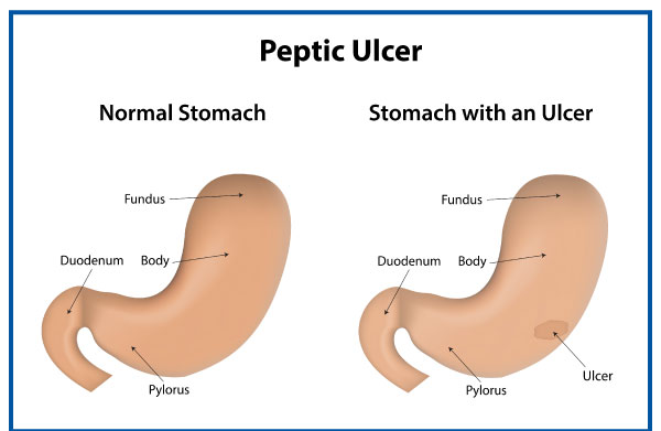
Characteristics of peptic ulcers and its complications.

Characteristics of peptic ulcers and its complications.

What are peptic ulcers?

If you have open sores in your stomach or upper small intestine, you may have a peptic ulcer. That occurs when the mucus lining your digestive tract’s protective layer is removed by stomach acids. There can be no symptoms, or there might be discomfort or a sharp ache. Internal bleeding brought on by peptic ulcers can occasionally need receiving blood transfusions in a medical facility.

Researchers predict that 8.09% of people worldwide experienced at least one peptic ulcer in 2019; which is a 25% increase since 1990. According to the same study, fewer occurrences have been documented in the US since the first half of the 20th century. Between 1990 and 2019, they started to rise once more.

Peptic ulcers come in three different varieties:

  • Internal stomach ulcers are referred to as gastric ulcers.
  • Esophageal ulcers are so named because they appear inside the oesophagus.
  • Duodenal ulcers are ulcers that form in the duodenum, the upper portion of the small intestine.

Symptoms of peptic ulcers

Peptic ulcers may not show any symptoms at all or they may just produce minor discomfort. Others can feel a stomach ache that is both dull and searing.

Additional signs can include:

  • meal is tough to swallow
  • nausea
  • vomiting
  • feeling full or bloated
  • sticky, dark stool (if bleeding)

Ulcers can result in severe symptoms, bleeding, and indications. These are symptoms of an ulcer that is bleeding:

  • throwing up blood
  • faeces that are dark crimson in colour or that are black and tarry
  • nausea and vomiting that is particularly intense and continuous

A medical emergency is indicated by these symptoms. The patient has to see a doctor right away or dial 911.

Causes of peptic ulcers

There are numerous potential causes of peptic ulcers, however the two most frequent ones are as follows:

H. pylori cause ulcers

It is unclear why H. pylori bacteria only cause ulcers in some people, despite the fact that many people naturally carry them. H. pylori spread via water and food. They reside in the mucus that covers the lining of the stomach and duodenum and generate the enzyme urease, which reduces the acidity of stomach acid, neutralising it.

The stomach creates more acid to make up for this, which irritates the stomach lining. Additionally, the germs irritate the stomach and impair its immune system.

Nonsteroidal anti-inflammatory drugs cause ulcers

NSAIDS are a class of drugs frequently used for headaches, menstrual cramps, and other symptoms. They can also aid in lowering inflammation and fever. Ibuprofen and aspirin are two examples. Many NSAIDs are accessible over-the-counter.

They lessen the stomach’s capacity to produce a thick layer of mucus for protection. As a result, stomach acid has a greater potential to harm Trusted Source. NSAIDs may also hinder blood flow to the stomach, which would hinder the body’s capacity to repair cells.

Other causes of peptic ulcers

Among the additional probable causes of peptic ulcers are:

  • Genetics: Many people who have peptic ulcers have relatives who also have the condition, indicating that hereditary factors could be at play.
  • Smoking: It’s possible that people who smoke tobacco frequently have a higher risk of developing peptic ulcers than people who don’t smoke, though it’s unknown how smoking affects peptic ulcers specifically.
  • Alcohol consumption: Alcohol consumption can aggravate the lining of the stomach and raise acidity.
  • Use of corticosteroids: Studies have shown that those who use high or ongoing dosages of corticosteroids are also at higher risk.

Studies on the link between stress and an increased risk of ulcers are contradictory, but one study revealed that psychological stress may make peptic ulcers more common in a way that cannot be explained by other factors like NSAID use.

Complications of a peptic ulcer

Ulcers that are left untreated may worsen over time. They may trigger additional, more severe health issues like:

  • Perforation: An infection results from a hole that forms in the stomach or small intestine lining. A perforated ulcer can cause abrupt, excruciating stomach pain.
  • Internal bleeding: Because bleeding ulcers can cause severe blood loss, hospitalisation is often necessary. Lightheadedness, wooziness, and dark stools are symptoms of a bleeding ulcer.
  • Scar tissue: After an accident, thick tissue known as scar tissue forms. Food has a tough time moving through your digestive tract because of this tissue. Vomiting and weight loss are indications that there is scar tissue.

All three issues are significant and might necessitate surgery. If you suffer any of the following symptoms, get emergency medical help right away:

  • unexpectedly acute abdominal ache
  • extreme perspiration, fainting, or confusion, as these could be symptoms of shock
  • blood in the stool or vomit
  • a difficult-to-touch abdomen
  • stomach ache that gets better when lying perfectly motionless but gets worse when you move around

Diagnosis of peptic ulcers

During a regular exam, a doctor will frequently become suspicious of a peptic ulcer due to a patient’s description of their symptoms.

The following tests can support a diagnosis:

  • an H. pylori blood test, while a positive result does not usually indicate a current infection
  • a breath test that detects H. pylori using a radioactive carbon atom.
  • a stool antigen examination to find H. pylori in the stools
  • using a GI (upper gastrointestinal) X-ray) to look for ulcers

The most popular device used by doctors to make diagnoses is Upper endoscopy or esophagogastroduodenoscopy. In order to check for the presence of an ulcer in the stomach and upper intestine, a tube must be inserted through the mouth.

The doctor may take a biopsy (a small sample of tissue) to be examined under a microscope if the test reveals an ulcer. A biopsy can check for cancerous tissue and test for H. pylori. A second endoscopy may be required a few months later to check on the ulcer’s progress.

Treatment of peptic ulcers

The goal of treatment will be to either completely eradicate the H. pylori infection or reduce stomach acid levels to allow the ulcer to heal. The following are typical treatments that a doctor might suggest:

  • Antisecretory substances. To help reduce the production of stomach acid, these include proton pump inhibitors and H2 receptor antagonists.
  • Triple treatment. To get rid of H. pylori, two antibiotics plus a proton pump inhibitor are used.
  • Staying away from NSAIDs. If someone regularly uses NSAIDs, doctors advise against using them.
  • Surgery. A doctor may advise surgery in cases of bleeding, with an average recovery time of 8 to 12 weeks.

REFERENCES:

For more details, kindly visit below.

Lets Explore the types of Cardiomyopathy and its treatment.

Lets Explore the types of Cardiomyopathy and its treatment.

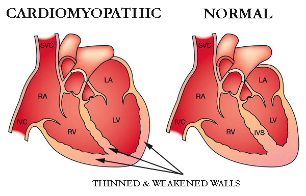
Cardiomyopathy is a condition that affects the heart muscle that gets worse with time and can be fatal. As the heart muscle ages, it becomes less effective in pumping blood.

Cardiomyopathy can be brought on by a genetic trait that is inherited or it can develop as a result of a number of different medical disorders, including heart disease, hyperthyroidism, or alcohol abuse.

Weakness, lightheadedness, shortness of breath, elevated blood pressure, and fluid retention, or edoema, are among the symptoms. Cardiomyopathy can be treated with lifestyle modifications, medicines, and surgery.

Cardiomyopathy is a condition that progressively affects the heart muscle, or myocardium. The heart muscle typically weakens and becomes less capable of properly pumping blood to the rest of the body.

Cardiomyopathy comes in a variety of forms and is brought on by a number of conditions and medications, including coronary heart disease. Heart failure, an irregular pulse, and other consequences can result from cardiomyopathy.

It’s crucial to receive medical care and continue receiving it. They can lessen the risk of heart disease or heart failure.

Types of Cardiomyopathy

Cardiomyopathy comes in a variety of forms, including the following:

Dilated

Heart palpitations and shortness of breath are two symptoms of cardiomyopathy. The most prevalent type of the condition is dilated cardiomyopathy. Adults between the ages of 20 and 60 are the typical victims.

Although the disease frequently begins in the left ventricle, it can later spread to the right ventricle as well. Additionally, the atria’s structure and operation might be impacted by dilated cardiomyopathy.

Hypertrophic

A hereditary disorder known as hypertrophic cardiomyopathy causes the heart muscle fibres to develop abnormally, thickening or “hypertrophying” as a result. The thickening restricts blood flow and stiffens the heart’s chambers. Additionally, it may raise the chance of electrical disturbances, called arrhythmias.

It is the second most prevalent kind of cardiomyopathy in children, according to the Children’s Cardiomyopathy Foundation. Before the age of one year, a diagnosis is made in around one-third of the affected children.

Restrictive

When the tissues in the ventricles stiffen and cannot properly fill with blood, restrictive cardiomyopathy develops. It might eventually result in cardiac failure. It is more prevalent in elderly persons and may be brought on by infiltrative disorders, such as amyloidosis, which involve the buildup of aberrant substances in physiological tissues.

Arrhythmogenic

In arrhythmogenic cardiomyopathy, the healthy tissues of the right ventricle are replaced by fibrotic and fatty tissue, which may lead to irregular heart beats. This procedure may occasionally take place in the left ventricle as well.

Arrhythmogenic cardiomyopathy increases the risk of sudden cardiac mortality, particularly in young individuals and sports, according to study published in the journal Circulation Research. It is a genetic inherited disorder.

Other types of cardiomyopathy

The majority of the following forms of cardiomyopathy fall under one of the previous four categories, however each has different causes or side effects:

Peripartum cardiomyopathy. Cardiomyopathy peripartum can happen before, during, or after pregnancy. This uncommon kind happens in the final month of pregnancy or within five months after delivery when the heart weakens. Postpartum cardiomyopathy is the term sometimes used to describe it after delivery. This dilated cardiomyopathy variant poses a serious risk to life. One instance raises your chances for subsequent pregnancies.

Alcoholic Cardiomyopathy. Alcoholic cardiomyopathy is caused by consuming excessive amounts of alcohol over time, which can weaken your heart and make it less effective at pumping blood. Additionally, your heart may grow. It’s a type of dilated .cardiomyopathy

Ischemic cardiomyopathy. Ischemic cardiomyopathy happens when coronary artery disease prevents your heart from pumping blood to the rest of your body. Heart muscle blood vessels constrict and become clogged. The cardiac muscle cannot receive oxygen as a result. The most common cause of heart failure is ischemic cardiomyopathy. Alternatively, any form of cardiomyopathy that is unrelated to coronary artery disease is referred to as nonischemic cardiomyopathy.

Noncompaction cardiomyopathy. The medical term for this is spongiform cardiomyopathy. It is a rare condition that manifests at birth and is brought on by faulty heart muscle development during pregnancy. Any stage of life may experience a diagnosis.

Pediatric cardiomyopathy. Pediatric cardiomyopathy is the term used to describe cardiomyopathy that affects children.

Idiopathic cardiomyopathy. Idiopathic cardiomyopathy indicates the reason is unknown.

Symptoms of cardiomyopathy

Early on in the development of cardiomyopathy, there may be neither indications nor symptoms. However, when the illness worsens, several indications and symptoms, such as:

  • a lack of breath during exercise or even while at rest
  • Legs, ankles, and feet swelling
  • abdominal bloating brought on by a fluid buildup
  • Cough when seated
  • difficulty falling asleep flat
  • Fatigue
  • fast, hammering, or fluttering heartbeats
  • chest pressure or discomfort
  • Unsteadiness, fainting, and dizziness

If not treated, symptoms and signs frequently worsen. It depends on the individual whether the illness becomes worse swiftly or gradually.

Causes and risk factors

There are certain well-established risk factors for cardiomyopathy, albeit its exact aetiology is not always known. For instance, disorders that cause the heart to inflame or sustain damage can raise a person’s risk of cardiomyopathy.

Cardiomyopathy can also be brought on by heart failure, which can happen as a result of a heart attack or other medical issues.

Additional risk elements consist of:

  • a history of cardiomyopathy, sudden cardiac death, or heart illness in the family
  • blood pressure is high.
  • cardiovascular disease
  • The diseases sarcoidosis and amyloidosis, which can harm the heart
  • viruses that affect the heart
  • diabetes
  • drinking disorder

After giving birth, certain women may be at an increased risk for cardiomyopathy.

Research from 2016 indicates, cardiomyopathy risk is also increased by HIV and HIV therapies. Heart failure and dilated cardiomyopathy in particular are risks that are increased by HIV.

Talk to your doctor if you have HIV about obtaining frequent testing to monitor the condition of your heart. Additionally, you ought to make an effort to maintain an active and heart-healthy diet.

Diagnosis of cardiomyopathy

To confirm cardiomyopathy, doctors will do a physical examination and diagnostic procedures. They may employ one or more of the diagnostic tests listed below:

  • Chest X-ray: A chest X-ray can assist diagnose some medical disorders by revealing whether the heart has enlarged.
  • Electrocardiogram (EKG): A heart’s electrical activity, including how quickly it beats, is measured by an electrocardiogram (EKG). Additionally, it demonstrates if the heart rhythm is normal or irregular.
  • Echocardiogram: An echocardiogram produces a moving image of the heart using sound waves. It displays the heart’s size and shape.
  • Cardiac catheterization: This procedure examines the blood flow through the heart’s chambers.

How is cardiomyopathy treated?

Treatment for cardiomyopathy focuses on controlling your symptoms. The course of the disease is also slowed by treatment. You will get routine examinations to monitor the condition of your heart.

Your healthcare provider might advise:

Medication: Heart drugs can increase blood flow, address underlying issues, or manage symptoms. You may use beta blockers like propranolol (Inderal), blood thinners like warfarin (Coumadin), or drugs to decrease cholesterol.

Arrhythmia-treating equipment: Pacemakers and implantable cardioverter defibrillators (ICDs) are used to treat erratic heartbeats. Your heartbeat is monitored by these gadgets. When an arrhythmia begins, they send electrical impulses to your heart.

Devices that improve blood flow: Some gadgets make your heart’s blood-pumping process more effective. Devices used in cardiac resynchronization therapy (CRT) regulate the heart’s left and right side contractions. Your heart is assisted in pumping blood with a left ventricular assist device (LVAD).

Surgery: Your doctor might advise heart surgery if you have serious symptoms or underlying heart issues. Open heart surgery or a heart transplant are typically only suggested by healthcare professionals after all other options have failed to provide relief.

REFERENCES:

For more details, kindy visit below.