Browsed by
Month: July 2023

When is a temperature too high for a human body?

When is a temperature too high for a human body?

According to research, the safest temperature range for humans is probably between 40°C and 50°C, or 104°F and 122°F, respectively.

Your body has to exert more effort to function in hotter environments. Heat-related illnesses and even mortality can result from extreme temperatures. Those who are elderly or have a chronic ailment are at higher risk, for example.

When temperatures are high, it’s crucial to take precautions to stay cool.

The reduction of ice sheets and glaciers altered geographic ranges for animals and plants, and changing seasons are just a few of the severe repercussions that the Earth is now experiencing as a result of climate change.

And this past week, scientists reported that the Earth had the warmest day ever on July 4.

What is the highest temperature that people can safely accept as a result of climate change, which is causing temperatures to rise?

Professor Lewis Halsey and a group of scientists from the University of Roehampton in London claim to have discovered what this temperature range is now.

The upper critical temperature (UCT) is most likely to range between 40°C and 50°C (104°F and 122°F), according to research that Halsey will present at the SEB Centenary Conference in Edinburgh, Scotland, from July 4–7, 2023.

The authors of the study claim that this is crucial since it has broad ramifications for workers, athletes, travelers, and medical professionals to understand the temperatures that cause our metabolic rate to increase as well as how this temperature differs for various people.

How the body is impacted by temperature and humidity

The researchers enlisted 13 healthy participants between the ages of 23 and 58 for the study. There were seven female contestants.

Each subject spent an hour relaxing while being exposed to five different temperatures. The circumstances included:

  • 50% relative air humidity (RAH) and 28°C (82.4F)
  • 40% RAH and 104°C
  • 50% RAH and 40°C (104F)
  • 50 °C (122 °F), 25% RAH
  • 50% RAH and 50°C (122F)

The researchers kept track of a number of measures during each condition and at baseline, including:

  • skin and internal temperatures
  • systolic pressure
  • perspiration rate
  • a heartbeat
  • respiration rate
  • the amount of air that is breathed in and out every minute
  • levels of movement

At 40°C (104F) and 25% RAH, the participants’ metabolic rate increased by 35%. However, at 40°C (104F) and 50% RAH, it climbed by 48%.

Although the metabolic rate was not greater in the 50°C and 25% RAH condition compared to the 40°C and 25% RAH condition, it was 56% higher than baseline in the 50°C (122F) and 50% RAH condition.

At the 40°C-25% RAH condition, the increase in metabolic rate was not followed by a rise in body temperature. Participants in the 50°C-50% RAH condition, however, noticed a 1°C (1.8 Fahrenheit) increase in core body temperature.

These results, according to the researchers, imply that the body can expel heat at 40 °C (104 °F), but not at 50 °C (122 °F).

Dr. Mark Guido, an endocrinologist of Novant Health Forsyth Endocrine Consultants in Winston Salem, North Carolina, who was not involved in the study, told us that “the findings do seem likely to vary by humidity.”

“The study found some indication that, even at the same temperature, the resting metabolic rate was higher at higher humidities. The metabolic rate appears to be significantly influenced by dampness as well, he continued.

How are metabolic rate and health impacted by climate?

Thermoneutral zone and metabolic rate may be impacted by living in various climates, according to Dr. John P. Higgins, a sports cardiologist at McGovern Medical School at The University of Texas Health Science Centre at Houston (UTHealth), who was not involved in the study.

People who live in warm climates typically acclimatise and don’t raise their body temperature or metabolic rate as much. Similarly, Dr. Higgins noted that those who live in cool or frigid climates may react to heat exposure more strongly because they have less heat tolerance.

Dr. Ulm was also contacted, and he provided the following insight: “The body, in general, will find ways to activate the various feedback loops needed to achieve homeostasis, i.e., the painstaking regulation of physiological processes that allow for the complex biochemistry of organs and tissues to be carried out efficiently and properly.”

“Body temperature and metabolic rate are essential elements of this delicate dance, and it may be more likely for such opposing feedback loops to be active and functional in people who live in hotter areas year-round. This could be attributed to both heritable factors for populations that have endured these conditions over an extended period and, more generally, short-term adaptations.

It’s comparable to how people who live permanently in high-altitude locations will adjust through compensatory mechanisms, such as in their red blood cell physiology and other characteristics of oxygen-carrying ability, both acutely as through iron turnover rates and chronically.

What are the study’s constraints and key findings?

Dr. Ulm and I discussed its limits.

As is typically the case with these kinds of studies, the issue of how representative the cohort sample of participants is of both the general and the targeted populations is raised in relation to the physiological traits and reactions being assessed.

“The studies, in this case, were also particularly challenging given the ambient conditions, and there is also the perennial issues of the applicability of the experimental environment to real-world correlates,” he continued.

The main finding, according to Dr. Guido, is that greater heat stress does appear to increase the resting metabolic rate by increasing how hard the body must work to stay cool, especially by causing a significant increase in heart rate. It is difficult to draw practical conclusions from a small laboratory study, he added. This could very well result in an increase in cardiovascular disease by placing extra strain on the heart if it remains true under real-world circumstances, he said.

Further research is required, Dr. Higgins continued, “Also, might it be advantageous for weight management to perform exercise in warmer temperatures indoors or outdoors to boost metabolic rate and thus burn more calories.”

How to safeguard oneself?

According to Atkinson and Ali, some methods for avoiding excessive heat include the following:

  • Take in plenty of water to stay hydrated. Ali also advised against drinking alcohol and coffee because they can dehydrate you.
  • Wear lightweight, loose-fitting clothing in lighter colors. According to Ali, this enables sweat to dissipate and cool your body.
  • When it’s hot outside, try to stay inside. According to Atkinson, the hottest part of the day is often from 11 a.m. to 3 p.m.
  • Keep the air in your house or office well-ventilated. Ali recommended using fans or air conditioners to stay cool.
  • To keep the sun out, close your curtains. For windows that face the sun, you should do this especially, according to Atkinson.

On warm days, stay away from strenuous exertion. This can quickly boost your core temperature, increasing your risk of heat exhaustion or heat stroke, according to the Academy of Nutrition and Dietetics.

Observe heat advisories and local weather forecasts. When extreme weather events are expected, the National Weather Service issues heat alerts.

Ali said that it’s crucial to keep an eye on those who are most susceptible, such as the elderly and those who are suffering from chronic illnesses, to make sure they can maintain their composure.

“If necessary, seek medical attention for severe symptoms or [seek] shelter in designated cooling centers during heatwaves,” he advised.

REFERENCES:

For Fever treatments that have been suggested by doctors worldwide are available here https://mygenericpharmacy.com/index.php?therapy=77

Epilepsy risk is 2.5 times higher in hypertensive people.

Epilepsy risk is 2.5 times higher in hypertensive people.

Researchers looked into the connection between epilepsy and high blood pressure. They discovered that taking antihypertensive drugs lowers the risk of epilepsy. This is increased by high blood pressure by about 2.5 times.

They come to the conclusion that while high blood pressure is a risk factor for epilepsy, further research is required to fully understand the mechanisms that underlie the association.

Ablestock

After stroke and dementia, epilepsy is the third most prevalent neurological illness that affects elderly individuals.

According to research, late-onset epilepsy has increased in prevalence over the past 20 years. As the population ages, the prevalence of the disorder will undoubtedly continue to climb. This makes epilepsy a substantial public health concern.

Despite this, 32-48% of cases of epilepsy have unknown underlying causes. According to several studies, vascular risk factors may raise the chance of developing late-onset epilepsy. According to another study, epilepsy may be caused by vascular risk factors starting in a person’s 30s.

Policymakers may be better able to develop public health initiatives and preventive actions to lower and manage rates of the condition if they have a better understanding of the involvement of vascular risk factors in late-onset epilepsy.

A recent investigation of the relationship between vascular risk factors and the start of epilepsy was conducted by researchers in the United States under the direction of the Boston University School of Medicine.

They discovered a connection between hypertension and a nearly 2-fold increased incidence of late-onset epilepsy. For those who did not use medication to control their blood pressure, this risk was significantly larger.

Data evaluation

Data from the Framingham Heart Study (FHS), an ongoing, community-based study that started in 1948, was used by the researchers. Through health exams every four years, the Offspring Cohort follows the health outcomes of 5,124 of the children of the original participants.

The researchers combined data from 2,986 individuals who underwent their fifth checkup between 1991 and 1995, were at least 45 years old at the time, and whose health records contained information on vascular risk factors.

Systolic and diastolic blood pressure were measured as vascular risk factors. The researchers defined high blood pressure as having a systolic pressure of at least 140 millimeters of mercury (mm Hg) and a diastolic pressure of at least 90 mm Hg, as well as taking antihypertensive drugs.

Additionally, the researchers looked for:

  • diabetes
  • cholesterin amounts
  • smoking history
  • a cardiovascular condition
  • stroke
  • BMI, or body mass index

The ICD-9 codes associated with epilepsy or seizures, self-reported seizures, routine chart reviews for neurological diseases, and antiepileptic medication use were utilised by the researchers to screen participants for epilepsy or seizures.

To identify cases of epilepsy, the researchers also analysed electroencephalography (EEG), cardiac, and other pertinent data, as well as brain imaging.

The study found 55 cases of epilepsy in the group, of which 26 were confirmed, 15 were likely, and 14 were suspected. The average age of the subjects at the time of a possible diagnosis was 73.8 years old.

The study’s findings revealed a nearly 2-fold increased risk of epilepsy in people with hypertension. The other risk variables, however, were unrelated to epilepsy.

They also discovered that the probability of having epilepsy increased by 17% for every 10 mm Hg change in systolic blood pressure.

Underlying processes

The connection between epilepsy and hypertension is explained by several theories. The renin-angiotensin system (RAS), which controls blood pressure, may be one mechanism.

According to research, rats who have had repeated seizures had 2.6–8.2 times the RAS components of seizure-free mice. Antihypertensive medications that decreased RAS component levels postponed the start of seizures and decreased their frequency.

More investigation is required, though, as other studies indicate the system may only have a modest impact on the relationship between epilepsy and hypertension.

Small vascular disease (SVD), a disorder in which the walls of small arteries and capillaries are damaged and do not adequately convey oxygen-rich blood to numerous organs, is another potential underlying mechanism, claim the researchers. According to a recent study, the duration of high blood pressure is a reliable indicator of developing SVD in later life.

The researchers also mention that temporal lobe epilepsy and SVD are related. Cortical microinfarcts, tiny lesions in the cortical tissue, and the disruption of U fibers are a few potential explanations. These conditions may cause excessive excitability and hence seizures. U fibers link adjacent regions of the cerebral cortex.

The researchers draw the conclusion that hypertension is a standalone predictor of late-onset epilepsy and carries a 2-fold chance of getting seizures after 45 years.

They do acknowledge that their study has some limitations. It might not accurately represent all races and ethnicities because it was mostly made up of white people. The study’s observational design, according to the researchers, precludes the ability to establish causality.

Dr. Jason Hauptman, a neurosurgeon at Seattle Children’s Hospital, told us that these findings were particularly intriguing because there has been debate over whether elevated blood pressure (hypertension) is a standalone risk factor for stroke.

living with high blood pressure and epilepsy

A healthy blood pressure reading is less than 120/80 mmHg; if you are unsure of your current blood pressure, ask your doctor. High blood pressure is closely linked to a variety of health risks, including cardiac arrest, so it is important to try and maintain this level. The good news is that there are lots of easy lifestyle adjustments you can make to lower your blood pressure, like:

  • healthy eating
  • not a smoker
  • controlling stress
  • lowering the consumption of alcohol
  • Exercising
  • consuming less salt

Speak to your doctor if you have concerns about your epilepsy and high blood pressure. They can assist you in creating a management strategy to lower your blood pressure through dietary and activity modifications. while also making sure that you are controlling your seizures.

REFERENCES:

For Epilepsy disease treatments that have been suggested by doctors worldwide are available here https://mygenericpharmacy.com/index.php?therapy=49

New brain imaging techniques for the treatment of epilepsy.

New brain imaging techniques for the treatment of epilepsy.

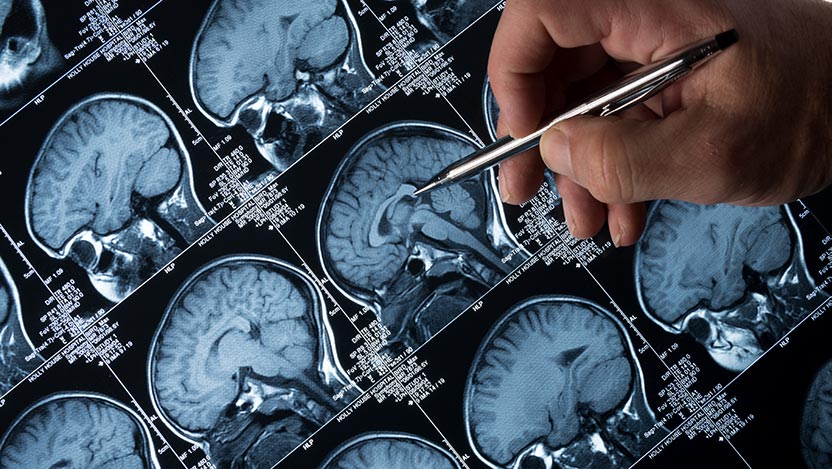
In a recent study, researchers identified a brain circuit that can be targeted with brain stimulation by mapping abnormalities in the brain linked to epilepsy.

They stated that they hoped their discoveries could lessen the symptoms that come along with seizures.

They stated that the brain mapping method might also aid in predicting whether stroke survivors will experience seizures.

According to a recent study published in the journal JAMA Neurology, deep brain circuit stimulation may be able to identify whether people who have had a stroke may eventually acquire the disease and assist treat epilepsy.

Scientists from Brigham and Women’s Hospital in Massachusetts examined five datasets that had more than 1,500 individuals with brain injuries for their study.

The lesions have several diverse causes, such as tumors, trauma, and stroke.

The ability to explore across many brain regions and forms of brain injury for common network connections associated with epilepsy as a result allowed researchers to do so.

Brain mapping: What is it?

There are specific functions for each region of the brain. The surgeon wants to comprehend how the brain regions close to the seizure onset operate before doing any type of brain surgery, including epilepsy surgery. This enables your team to determine how much of the seizure focus can be safely removed.

The process of brain mapping can be used to pinpoint the functions of various brain areas.

Different people have different locations for different bodily processes (such as movement, voice, vision, and more). Tumours, seizures, or other brain abnormalities may alter which regions of the brain are in charge of a certain function. Sometimes general laws don’t apply.

By activating particular brain regions, one can create a “map” of each person’s brain. The map reveals to medical professionals which regions of the brain are in charge of vital processes like speech, sensation, or movement.

Brain mapping for epilepsy

The sites of brain damage in epilepsy patients and those without the condition were compared by the researchers.

According to the researchers, the brain was filled with lesions connected to epilepsy. They did, however, have a common network.

The researchers pointed out that epilepsy may be brought on by disruption of brain connections rather than the site of the damage. The basal ganglia and cerebellum, two deep-brain regions, were the locations of the linkages.

According to the researchers, identifying lesions in a brain network may aid in determining whether or not a person may experience epilepsy following a stroke. They claimed that common brain pathways could connect various damages and result in epilepsy.

The researchers point out that earlier studies have linked deep brain regions to modifying and regulating seizures in epilepsy-prone animals. They might have a braking effect on the brain.

How have scientists used deep brain stimulation?

The researchers examined the deep brain stimulation results in 30 patients with drug-resistant epilepsy.

If the stimulation was linked to the same brain network they discovered when mapping brain lesions, they claimed that the benefit would be greater.

Dr. Frederic Schaper, an assistant scientist at the Centre for Brain Circuit Therapeutics at Brigham and Women’s Hospital and an instructor of neurology at Harvard Medical School in Massachusetts, said, “In our study, we analysed existing data from patients that received deep brain stimulation for drug-resistant focal epilepsy.”

Although all patients had electrodes for deep brain stimulation implanted in the anterior thalamus, Schaper informed us that each patient’s precise electrode placement and stimulation sites varied slightly. “We found that patients with deeper brain stimulation sites that were more connected to deep brain regions in the cerebellum and basal ganglia had better seizure control than patients who were less connected to these regions.”

“This finding suggests an important role for brain networks distant from the anterior thalamic deep brain stimulation site in the mechanism of action of deep brain stimulation for epilepsy and seizure control,” he continued.

Deep brain stimulation principles

The American Association of Neurological Surgeons defines deep brain stimulation as a surgical procedure in which electrodes are placed in particular parts of the brain. Then, in order to assist manage aberrant brain activity, these electrodes transmit electrical impulses.

The amount of stimulation is managed via an implanted programmable device that resembles a pacemaker. The device is connected to the brain’s electrodes by a wire.

The full mechanism through which deep brain stimulation reduces seizure frequency is unknown, according to Schaper. “Previous research in people and animal models indicates that deep brain stimulation disturbs the brain networks responsible for seizures. It is uncertain, nevertheless, whose brain networks are in charge of [deep brain stimulation]-induced seizure control.”

Schaper mentioned that deep brain stimulation is a safe and efficient treatment for drug-resistant focal epilepsy and has received approval from federal regulators.

Improving epilepsy symptoms

In this investigation, brain networks were sought after. They claimed that deep brain stimulation can lessen epilepsy symptoms if it activates just one node in the network.

“This study is quite exciting,” said Dr. Jean-Philippe Langevin, a neurosurgeon and the director of the Restorative Neurosurgery and Deep Brain Stimulation Programme at the Pacific Neuroscience Institute at Providence Saint John’s Health Centre in California. He was not involved in the research.

“The scientists discovered that networks were more associated with epilepsy than brain lesions. “They could influence epilepsy symptoms if they could concentrate stimulation within the networks,” Langevin told us.

The roadways in the brain are called brain networks. The roadside stops are called lesions. The researchers discovered that the entire network was influenced when electrical currents were applied anywhere along a network of streets.

According to Langevin, “Deep brain stimulation works for other diseases.” These include essential tremors, Parkinson’s condition, dystonia, obsessive-compulsive disorder, and dystonia. For certain conditions, “working within a single network would also hold true.”

“This is exciting because, in the future, when patients come to us with seizures, a scan can look at how the network is connected, making it easier to use [deep brain stimulation],” continued Langevin. “The scans do exist, but we don’t typically use them in the study.”

Symptoms of a seizure

Different people experience different pre-seizure warning symptoms.

But there are a few widespread indications:

  • a sense of impending disaster
  • For every seizures, the same tone or sound is produced.
  • trouble generating ideas
  • having trouble finding the right words
  • Having underwater-like audio perception
  • experiencing déjà vu or believing that nothing is familiar
  • feeling queasy in the stomach
  • having the impression that everything is deformed, either larger or smaller than it should be.

It is suggested that you lay on your side if you are experiencing a seizure. Someone else should roll a seizure victim over if they are unable to move.

Additionally, a person experiencing a seizure ought to be relocated to a location where they won’t damage themselves. For instance, a space free of any furnishings.

Ensure that they are not wearing anything tight around their neck, such as a necktie, scarf, or button-up shirt. If so, you ought to take these things off.

Never abandon a person experiencing a seizure. Until the seizure is finished, be at their side.

REFERENCES:

For Epilepsy medications that have been suggested by doctors worldwide are available here https://mygenericpharmacy.com/index.php?therapy=49

Is high BMI linked with an increased risk of death?

Is high BMI linked with an increased risk of death?

The validity of body mass index (BMI) as the only predictor of all-cause mortality is further questioned by a recent study.

The majority of earlier research, according to the study’s authors, rely on more dated data that isn’t sufficiently diverse, so they’re hoping the current study may remedy that.

A BMI that indicates overweight or obesity can increase the chance of developing several chronic, fatal diseases, but it may not be a reliable predictor of mortality as a whole.

According to a recent study, it is advisable to take into account a person’s body mass index, or BMI, together with other risk variables when forecasting all-cause death. As an independent variable, BMI might not be a reliable predictor of premature death.

There was no variation in the risk of death from all causes among persons in the healthy and overweight BMI categories, from a BMI of 22.5 to 27.4, according to the research.

However, the study found that in persons with a BMI greater than 30, the risk of all-cause death rose by 21% to 108%.

No appreciable increase in mortality was observed in older persons between BMIs of 22.5 to 34.9, the higher range indicating obesity.

Older statistics on BMI and early death are displaced by new data.

Data from the 1970s that concentrated on non-Hispanic white adults formed the basis of the majority of research on BMI and mortality.

The new study examined more recent, comprehensive data while keeping in mind the changes in lifestyles since that time, including the rise in overweight and obesity, and sought a more varied study population sample.

Self-reported BMI data from 554,332 American individuals who took part in the National Health Interview Survey from 1999 to 2018 and data from the 2019 US National Death Index were used in the analysis.

The average age was 46, there were equal numbers of males and women, and 69% of the population identified as non-Hispanic white, while 12% identified as non-Hispanic Black.

Among the individuals, 35 percent had a BMI between 25 and 30, which is normally regarded as overweight, and 27.2 percent had a BMI of 30 or more, which is categorised as obesity.

A total of 75,807 fatalities were reported throughout the average follow-up period of 9 years and the maximum follow-up period of 20 years.

Why BMI is a poor indicator of health?

We spoke with Dr. Pedro J. Caraballo, medical director of the Mayo Clinic Clinical Decision Support Programme, who was not engaged in this investigation.

“It is extremely debatable whether or not BMI alone should be used to define obesity or health. Different types of obesity that may have an impact on health have clearer definitions. BMI may be easily calculated and found in any medical records, though.

A person’s BMI is calculated by dividing their weight in kilogrammes by the square of their height in metres.

However, BMI ignores other aspects of the body, such as the ratio of fat to muscle, how fat is distributed throughout the body, and metabolic health. For instance, having excess fat around the waist raises your risk of getting sick.

“[BMI] does not distinguish between muscle mass and fat mass, and some individuals, like bodybuilders, may have a high BMI because of more muscle mass,” said Dr. Dagfinn Aune, a research associate in the Faculty of Medicine, the School of Public Health at Imperial College London in the United Kingdom, who was not involved in this study.

Despite these drawbacks, according to Dr. Aune, BMI performs a good job of capturing the elevated risk of chronic disease and mortality that is connected to obesity at the population level.

BMI as a measure of obesity is “not a suitable tool”

Dr. Aune provided a lengthy list of chronic diseases linked to an oversized BMI. These included kidney stones, gallstones, diverticular disease, coronary heart disease, stroke, heart failure, sudden cardiac death, atrial fibrillation, hypertension, type 2 diabetes, and a dozen distinct cancers.

Additionally, preeclampsia, gestational diabetes, gestational hypertension, stillbirth, and infant death are just a few of the pregnancy issues that are linked to being overweight or obese while pregnant, according to Dr. Aune.

The results of this study are outdated in Dr. Caraballo’s eyes. He referenced his own research, which “showed that BMI is an independent risk factor only in extreme values, very low (20) or very high (>40), with risk stratification based on comorbidities.”

Multiple studies, according to Dr. Caraballo, have found that mild to moderate obesity “may help survival when considering a specific subpopulation that is under stress.”

He noted various papers on this subject for “heart disease, kidney disease, cancer, stroke, and rheumatoid arthritis, etc.” and concluded that maintaining energy reserves may be beneficial for people.

According to Dr. Visaria, “the United States has undergone a significant transformation since the 20th century in terms of racial/ethnic makeup, age distribution, healthcare access and treatments, and sociocultural behaviours.”

It is crucial to comprehend the relationship in a more modern population since all of these can affect the association between BMI and all-cause mortality, he said.

Dr. Visaria further emphasized the importance of utilizing the most rigorous techniques to eliminate bias and ensure that observational data is as nationally representative as feasible.

Lower risk of older adults having greater BMI

Dr. Visaria proposed theories as to why this would be the case, given that older adults did not exhibit an increased mortality risk up to a BMI of 35.

We believe that the decline in bone mineral density and sarcopenia that occurs as people age have a role in this. Despite having large quantities of fat, losing these two types of weight can cause you to have excessively normal BMIs, he warned us.

Because of their maintained bone and muscle mass, those with higher BMIs may actually be in better health.

What factors predict total mortality more accurately?

According to Dr. Caraballo, the link between fat and mortality is extremely convoluted.

“Obesity by itself, in the range of mild to moderate, may not be an independent risk factor,” he said. “However, obesity is a significant risk factor for the emergence of numerous metabolic disorders that, over time, raise the mortality risk (diabetes, heart disease, etc.). People may also put on weight when they have a chronic illness because they do less exercise and eat poorly.

In his recommendation, Dr. Visaria said that “physicians should consider supplementing BMI with other measures such as waist circumference, waist-to-height ratio, and waist-to-hip ratio.”

According to Dr. Visaria’s study, “We show that waist circumference significantly modifies the association between BMI and all-cause mortality.”

Dr. Visaria stated, “Bioimpedance scales are another alternative to determine total body fat percent, but they still need to be verified and are known to have some mistakes. Additionally, doctors should consider patients’ cardio-metabolic health factors like blood pressure, blood sugar, and cholesterol levels when interpreting adiposity measurements.

REFERENCES:

For Obesity medications that have been suggested by doctors worldwide are available here https://mygenericpharmacy.com/index.php?therapy=20

Higher doses of Ozempic improve blood sugar, weight loss?

Higher doses of Ozempic improve blood sugar, weight loss?

According to experts, glucagon-like peptide 1 (GLP-1) medications like Ozempic, often referred to as semaglutide, can aid in weight loss in those who are obese.

For weight loss, Ozempic is not FDA-approved. There is also the brand Wegovy, which is semaglutide.

Wegovy and Ozempic should not be used by persons who are not diabetic or obese for any reason, including to shed minor amounts of weight, according to experts.

In the latter part of 2017, the FDA approved the use of the GLP-1 medication Ozempic in people with type 2 diabetes. But lately, it’s made news for other reasons as well: Benefits of purported weight loss and scarcity.

“People are talking about them because there is a trend where celebrities and influencers are increasingly turning to off-label use of GLP-1 medications like Ozempic for weight loss by people who are not obese or diabetic,” claims Dr. Rekha Kumar, an endocrinologist in New York City and the head of medical affairs at the weight management program Found.

This, according to Kumar, is very troubling.

“The trend of medispas, boutique weight-loss clinics, and illegal telehealth businesses liberally prescribing to people who don’t meet criteria is not only irresponsible prescribing, but it may also prevent the medication from reaching those who need it most,” Kumar claims.

By responding to the following frequently asked questions regarding Ozempic, Kumar and other professionals distinguished fact from fantasy.

What is Ozempic?

Dr. Angela Fitch, FACP, FOMA, president of the Obesity Medicine Association and chief medical officer of knownwell, a weight-inclusive healthcare firm, says that Ozempic is a brand name for the medication recognised as semaglutide.

Ozempic is an injectable medicine for persons with type 2 diabetes, according to Kumar. The FDA first approved it for 0.5 mg or 1 mg dosages. The FDA authorized a higher dose of 2 mg in 2022.

According to Kumar, it helps the pancreas produce insulin, which decreases blood sugar levels.

Adverse effects of semaglutide

All GLP-1 medications, as pharmacological agents, have the potential to have side effects, according to Dr. Jay Shubrook, professor of the Primary Care Department at Touro University in California.

According to Dr. Shubrook, these “are frequently dose-dependent and can be more obvious during dose changes.”

In any case, typical adverse effects of semaglutide “include an excessive loss of appetite, nausea, and less frequently, vomiting or diarrhoea. Most patients only experience temporary adverse effects, he noted.

He pointed out that teaching patients to eat slowly and mindfully, as well as how to control their portions, can lessen the negative effects of semaglutide.

Is Ozempic an FDA-approved weight-loss product?

No. “Ozempic is only approved for diabetes,” claims Dr. Charlie Seltzer, a Philadelphia-based medical professional who is board-certified in both internal medicine and obesity.

But here’s where some of the ambiguity arises. “The active ingredient, semaglutide, is approved for weight loss under the trade name Wegovy,” claims Seltzer.

Elon Musk tweeted about Wegovy’s assistance with his weight loss in October 2022.

Distinction between Wegovy and Olympic

Semaglutide and injectables are both sold under the trade names Ozempic and Wegovy. They aren’t precisely the same, though.

“Wegovy is FDA-approved for the treatment of overweight and obesity,” claims Kumar. “Wegovy was developed specifically for the treatment of overweight and obesity,” according to the manufacturer. “It contains a higher dose of semaglutide, [2.4 mg], than Ozempic.”

Does Ozempic aid in shedding pounds?

Kumar points out that Wegovy’s dosing was employed in the studies on semaglutide and weight loss, including one from 2021 that showed that once-weekly doses of 2.4 mg of semaglutide could lower body weight when paired with dietary and lifestyle modifications.

“[In the] study,] those who took the medication and made lifestyle changes lost almost 15% of their body weight, on average, compared to 3% in the placebo group,” Kumar claims.

So certainly, semaglutide may aid in weight loss, at least at a greater dose of 2.4 mg. Although Seltzer observes that the two medications function similarly, it is uncertain whether the 0.4 mg dosage difference between Ozempic and Wegovy is significant.

As food takes longer to leave the stomach and suppresses hunger, ozempic prolongs satiety, according to Seltzer. “It does nothing magical to the metabolism.”

In addition, Kumar points out that despite what some celebrities and social media influencers may say, these medications are not intended for those who just want to drop a few pounds.

“Normal-weight patients without diabetes might lose weight if they take GLP-1s, but the risks of the medication outweigh the benefit of weight loss just to be thin versus treating a disease,” says Kumar. “GLP-1s have not been studied in this population, and with this type of inappropriate use, we probably will see more side effects.”

Is Ozempic safe?

For adults with type 2 diabetes, ozempic is typically regarded as safe in doses up to 2 mg, however doctors agree that some people shouldn’t take it.

“It should be avoided in many populations, including but not limited to people with a history of pancreatitis, people who have had medullary thyroid cancer, or who are at increased risk for medullary thyroid cancer,” says Seltzer.

If you are a good candidate for Ozempic, your doctor can help you decide. Furthermore, some persons might suffer negative effects. According to Fitch, typical ones include:

  • nausea
  • constipation
  • dizziness
  • reduction in appetite
  • diarrhea

Can you regain weight after using semaglutide?

Patients who quit taking 2.4 mg dosages of semaglutide had gained back two-thirds of the weight they had lost one year after stopping, according to a trial of nearly 2,000 patients published in 2022.

The same problems that got the people into difficulty in the first place will still exist once the drug is stopped or loses its effectiveness, according to Seltzer, and the weight will quickly regain.

Fitch concurs,

Whatever you do personally to aid in weight loss, Fitch advises, “You have to keep doing it, or the weight will come back.” “The human body was created in this manner. It is constructed to safeguard its weight at all costs. Care for the elderly is crucial.

“Since obesity is a chronic disease, you must treat it chronically, ongoingly, and in a coordinated, comprehensive way,” adds Fitch. For a comprehensive approach to metabolic health, weight control, and primary care, patients must collaborate with their doctor.

What other therapies are there for obesity?

First, Fitch emphasises the need of being nonjudgmental and emphasising joint decision-making in all obesity treatments.

According to Fitch, “obesity is a lifelong chronic disease and should be treated in a compassionate and thorough patient-centered way, such as shared decision making around taking medication or having surgery with the risks and benefits in mind.”

Although diet and exercise are frequently suggested as first-line therapy, they are not always effective.

“Obesity is a complex disease with many factors,” explains Fitch. “We add in other treatments to help patients live longer, healthier, better quality lives when lifestyle changes are not enough.”

REFERENCES:

For Weight loss medications that have been suggested by doctors worldwide are available here https://mygenericpharmacy.com/index.php?therapy=20

Experimental drugs could improve several cancer therapies.

Experimental drugs could improve several cancer therapies.

In order to combine medication with diagnostics, researchers have found a novel radioactive compound named CB-2PA-NT as a possible possibility.

The chemical exhibits strong uptake and retention in tumors while maintaining a distinct difference from surrounding tissues. It works by targeting neurotensin receptors, which are prevalent in numerous malignancies.

Shortly, researchers intend to carry out human imaging investigations utilizing CB-2PA-NT, which may have an impact on personalized cancer treatment for patients. Regulatory approval is still waiting.

The University of Wisconsin and the University of North Carolina worked together to create the anti-cancer medication candidate CB-2PA-NT, which has the potential to be used widely.

This study lays the framework for future investigations that will use CB-2PA-NT in human imaging, albeit these studies still require regulatory approval in order to start.

New research positions CB-2PA-NT as a promising candidate for an original theranostics method, according to data presented at the Society of Nuclear Medicine and Molecular Imaging Annual Meeting (SNMMI 2023).

Therapy and diagnostics are combined in theranostics.

Correct diagnosis is a prerequisite to choosing the best course of action. A precise diagnosis is even more crucial in the era of personalized medicine. This is where treatments can be tailored to a person’s unique biomarkers.

This is especially true in the field of cancer, where it is crucial to get a precise diagnosis.

Theranostics offers a potent method for battling cancer by fusing two crucial components. It entails locating cancer cells throughout the body and removing them with specialized radiation.

Positron emission tomography (PET) is used to localise the malignancy, and then medicine is given to kill it. Theranostics’ astounding accuracy greatly reduces the possibility of damaging nearby healthy tissues.

CB-2PA-NT has the potential to significantly advance the field of precision medicine by particularly targeting neurotensin receptors (NTSRs) present in diverse cancer types.

Researchers pursue the receptors on cancer cells

When it comes to many malignancies, including lung, colorectal, breast, pancreatic, and prostate cancers, NTSRs are more prevalent receptors.

A radioactive chemical that can precisely bind to NTSR1, one of these receptors, has been developed by scientists.

In terms of how well they are absorbed and retained by tumors, earlier attempts to synthesize these compounds have had mixed results.

One of the study’s authors and doctorate candidate at the University of North Carolina in Chapel Hill, Xinrui Ma, gave us an explanation of the main conclusions.

“This [study] abstract reports] a novel theranostic agent targeting neurotensin receptor 1 (NTSR1) and its application in cancer imaging and therapy,” said Ma.

The optimization of NTSR1-related compounds has already been attempted by numerous organizations, including our own. In fact, we have been working on this topic since 2012. Challenges include limited serum stability, significant liver absorption, some ligands’ agonistic character, and/or quick washout, she added.

The cross-linked propylamine moiety, Ma continued, “can significantly improve tumor uptake and retention, building on previous research and experience.”

“The tumor absorption increased 10-fold as compared to the peptide-based ligand while still maintaining the high contrast. The high uptake was also sustained at 24 and 48 hours following injection, which is more significant, the researcher said.

We have a rare chance to create theranostic drugs for patient treatment because of the much better tumor absorption and retention. Indeed, to investigate the theranostic potential of these novel compounds in a variety of cancer types, such as lung cancer, colorectal cancer, and PSMA-negative prostate cancer, we have forged a close partnership with Prof. Jonathan Engle’s team at the University of Wisconsin, said Xinrui Ma.

Which NTSR1 inhibitor is most effective?

The best NTSR1 antagonist for imaging and therapy was determined by the researchers’ investigation, which looked at a variety of NTSR1 antagonists.

They then carried out studies to radioactively designate these compounds. They verified that the NTSR1 receptor was in fact present in the lung cancer cells (H1299 cells) using a procedure known as western blot.

The compounds were also examined for stability in test tubes and on living lung cancer cells. Also, for their capacity to bind to lung cancer cells in laboratory settings and on animals.

Finally, to examine how the chemicals were dispersed throughout the body, they used PET and CT imaging on tiny animals.

Western blot analysis of the results revealed that the NTSR1 receptor was substantially expressed in the H1299 lung cancer cells. The chemical known as CB-2PA-NT has shown a potent ability to bind to the H1299 lung cancer cells among the NTSR1 antagonists.

Small animal imaging provided proof that CB-2PA-NT was taken up by the tumor in large numbers. Clearly contrasted with the surrounding tissues, and stayed in the tumor for a considerable amount of time.

CB-2PA-NT was chosen for additional research because it stood out as the most promising compound when compared to the other NTSR1 antagonists.

There is a “need to confirm application in humans”

If this theranostic strategy is successful, it may provide a reliable method for imaging to detect the presence of NTSR1 in many cancer types.

This would be helpful for making diagnoses, selecting patients, and keeping track of how well treatments were working. It might also function as a radioactive material used in therapy.

We were informed by Yale resident doctor Dr. Tejasav Sehrawat. He was unrelated to the experiment, that “theranostics is a young discipline for diagnosing and treating malignancies. The growth of the field overall is quite intriguing and has a lot of potential. The competent execution of this investigation and the good preclinical findings are encouraging.

“While the authors have already demonstrated their findings in animal models, application in people still has to be verified. We should all be eagerly awaiting the findings of the authors’ next human investigations. Because there is significant inter-species heterogeneity in these studies, according to Dr. Tejasav Sehrawat.

Possibly negative future effects for cancer patients

This finding, according to Ma, “is important because it could offer personalised medicine for cancer patients,”

She informed us that, in terms of the disease, NTSR was discovered to be overexpressed in prostate cancer tissues. Specifically in PSMA-negative prostate cancer tissues.

This shows that NTSR1-targeted theranostics may be a prostate cancer treatment option in populations that are ineligible for PSMA-based methods. Lung, colorectal, breast, and pancreatic cancers are only a few of the tumors that could potentially benefit from NTSR-targeted theranostics, says Ma, Xinrui

“A broad spectrum of patients may benefit from the newly developed agents,” Ma noted. The researchers are still awaiting regulatory authorization to carry out the first-ever human imaging tests with CB-2PA-NT, and more study is required.

REFERENCES:

For Cancer disease medications that have been suggested by doctors worldwide are available here https://mygenericpharmacy.com/index.php?therapy=10

High levels of lean muscle may help prevent Alzheimer.

High levels of lean muscle may help prevent Alzheimer.

High quantities of lean muscle may help prevent Alzheimer’s disease, according to recent studies. To determine whether this connection is causal, more study is required.

Resistance training and a sufficient intake of protein in the diet are advised by experts as ways to build lean muscle mass. Previous studies have shown a link between obesity and an elevated risk of Alzheimer’s.

A recent study that was published in BMJ Medicine suggests that having a lot of lean muscle may prevent Alzheimer’s disease. The authors of the study pointed out that additional study is required to comprehend the biological mechanisms underlying it.

Researchers gathered data on 450,243 participants’ genetics, lean muscle mass, cognition, and health for this study from the U.K. Biobank. They next used a method known as Mendelian randomization to search for genetic relationships between lean muscle mass and genetic variants.

The quantity of lean muscle and fat tissue in the arms and legs was measured using bioimpedance, an electric current that moves through the body at varying speeds. The researchers next discovered 584 genetic variations related to lean muscle mass, but none of these were located in an area of the genome known to include genes connected to an elevated risk of Alzheimer’s disease.

The chance of developing Alzheimer’s disease did, however, appear to be reduced in individuals who had high amounts of lean muscle mass and associated genetic variations.

These results were confirmed by measuring the quantity of lean muscle mass and body fat tissue in a second cohort of 252,879 individuals without Alzheimer’s disease and 7,329 individuals with the condition.

The findings indicated that lean mass was associated with enhanced cognitive performance, but this association did not account for lean mass’s protective effect against the onset of Alzheimer’s disease.

The study’s objectives

The majority of the data utilized by the researchers came from the UK Biobank, a continuous database that collects health and genetic data on 500,000 people. The researchers used data from hundreds of thousands of people.

Mendelian randomization, which employs gene variations as a proxy for specific risk variables, was used to analyze the data.

Additionally, bioimpedance measurements which determine the speed at which an electrical current travels through the body based on its composition in terms of muscle and fat were used to calculate each person’s lean mass.

A total of 584 genetic variations were connected to lean muscle mass, but none of them were discovered in the region of the brain linked to Alzheimer’s susceptibility.

Participants’ chance of developing the disease was statistically significantly (12%) lower on average for those with higher amounts of (genetically proxied) lean muscle.

The analysis was performed using data from an additional 260,208 individuals, of whom 7,329 were identified as having Alzheimer’s disease, in order to confirm these findings. They measured lean muscle mass over the entire body, not only in the arms and legs.

Again, they discovered that having more lean muscle was linked to a lower risk of Alzheimer’s.

Unexpectedly, the analysis did not reveal a correlation between body fat and the probability of developing Alzheimer’s disease once lean mass was taken into account. Body fat was associated with inferior performance in cognitive activities.

The significant contrast between the protective effect of lean mass on dementia risk and the lack of an effect of fat mass on dementia risk, according to Daghlas, startled him.

How Alzheimer’s disease and lean muscle are related?

The fact that the processes underlying the association between lean muscle and Alzheimer’s disease are still unknown is another drawback of the study.

The researchers made brief speculations about possible connections. For instance, Alzheimer’s has long been linked to cardiovascular illness, though Daghlas cautioned that the connection is “complicated.”

According to Daghlas, heart disease problems like stroke and hypertension are what fuel vascular dementia. Though this is a contentious idea, he added, “the most recent causal evidence suggests weaker evidence for an effect of vascular risk factors on Alzheimer’s disease risk.”

Lean mass may very possibly lower the incidence of vascular dementia through lowering the risk of cardiovascular disease, according to Daghlas, however more research is needed in this area. “This can be looked into in upcoming studies.”

In the publication, the researchers also made the speculative claim that “new” processes, such as centrally acting myokines, may be at work.

Myokines are proteins that muscles produce that have an impact on other tissues, according to Daghlas. “Experimental studies have demonstrated that they are induced by exercise and have a positive impact on brain function.”

Other muscle-related issues, in addition to these, “may explain the larger picture,” according to Dr. Anna Nordvig, a neurologist at NewYork-Presbyterian and Weill Cornell Medicine who was not involved in the study. Examples include “bone hormones, cardio vs. strength training differences, sex hormones, glymphatic drainage depending on exercise, and sleep, to name a few.”

In the end, further clinical intervention studies are required to demonstrate the link between lean muscle and Alzheimer’s disease and the mechanisms underlying it.

The best way to build lean muscle mass

Having lean muscle mass has several health benefits in addition to possible advantages for the brain.

Resistance training using weights, bands, and pleiomorphic activities are advised by Dr. Joseph C. Maroon, clinical professor, vice chairman, and Heindl scholar in neuroscience at the Department of Neurosurgery at the University of Pittsburgh.

Additionally, he recommends supplementing with B-hydroxy B-methylbutyrate (myHMB), a good source of dietary protein.

This naturally occurring compound aids in the maintenance of a healthy weight and the development of lean muscle mass in humans. B-hydroxy helps muscles recover from hard activity, improves athletic performance, and enhances muscle and strength, according to him.

According to Dr. Sullivan, the best food, the best kind and frequency of exercise, the optimum amount of rest, and stress management are the primary factors that influence muscle growth.

These are the recommendations she makes:

Exercise: four to five quick strength-training sessions each week. In comparison to two or three longer aerobic exercises each week, this will produce greater lean muscle mass.

Diet: Put your attention on lowering insulin resistance by eating fewer carbohydrates and more protein, the building block of muscle.

Sleep: 8 to 9 hours of sleep per night are recommended if you want to recuperate from this kind of workout completely.

Stress management: With the rise in inflammation and blood sugar that stress hormones like cortisol induce, chronically high stress can sabotage any self-improvement effort. Long-term high cortisol levels can contribute to persistent muscle tension and lactic acid buildup, which can inhibit muscle growth. Moving your body more, going outside, eating more good foods, being an aggressive communicator, and finding your purpose are the simplest ways to relieve chronic stress.

How to lower your risk of developing Alzheimer’s?

Although there is no cure for Alzheimer’s, doctors think there are a number of steps you may take to lower your risk.

“Protect” and “stimulate” are the two categories that these fall under, according to Nordvig, and “physical activity falls into both of these.”

“Protect” includes monitoring factors like blood pressure, sugar levels, weight, nutrition, and sleep, she said. These are topics covered at a yearly checkup.

We should also work to safeguard ourselves from environmental risk factors associated to Alzheimer’s, according to Dr. Rena Sukhdeo Singh, a vascular neurologist at the University of Maryland Shore Regional Health.

The incidence of dementia has also been connected to fine particulate matter in air pollution.

According to Sukhdeo Singh, systemic inflammation also contributes to the development of Alzheimer’s disease. Numerous factors, including medications, a diet high in sugar and processed foods, smoking, and binge drinking, can contribute to chronic inflammation.

Optimising additional daily inputs that influence cognition is what “stimulate” entails. For instance, Sukhdeo Singh proposed, “learning a new skill, hobby, language, or instrument,” or taking part in shorter activities like “sudokus, puzzles, and number games.”

Unfortunately, there are some factors that we have no control over. Age, genetics, and sex are non-modifiable risk variables, she added.

Restrictions of this research

Researchers solely considered lean muscle mass for this investigation. However, there are other things to think about.

The protein amyloid, which is damaging to the functioning of the brain, is found in higher amounts in adipose tissue, but the researchers neglected to test these signs of inflammation and insulin resistance, according to Maroon. “This probably diminishes the significance of their findings.”

Furthermore, “while their positive finding was statistically significant, the effect size was modest in lean muscle mass reducing the risk of dementia and only explained 10% of the variance,” added Dr. Sullivan.

The link between more lean muscle mass and a lower incidence of Alzheimer’s disease has to be further investigated.

According to Nancy Mitchell, a registered nurse, “For now, people with lower muscle mass tend to be obese, which is a risk factor for type 2 diabetes.”

We refer to Alzheimer’s disease as “diabetes of the brain” because it has been hypothesized that high blood sugar harms the nerve endings in the regions of the brain that are most impacted by cognitive decline. Therefore, the link may actually be between a reduced risk of obesity and diabetes. This can be a study drawback in and of itself because greater specificity is still needed. Not all correlations indicate causation. Nancy Mitchell is a nurse practitioner.

REFERENCES:

For Alzheimer’s disease medications that have been suggested by doctors worldwide are available here https://mygenericpharmacy.com/index.php?therapy=31

Can Hormone therapy for menopause increase Dementia risk?

Can Hormone therapy for menopause increase Dementia risk?

Menopausal hormone therapy is used by about 45% of all women to lessen menopause symptoms.

According to prior studies, some types of hormone replacement treatment may make women more susceptible to developing serious illnesses.

Menopausal hormone therapy is linked to an increased risk of dementia and Alzheimer’s disease, according to researchers from Copenhagen University Hospital, says Rigshospitalet.

These results go against earlier research that suggested HRT might reduce a woman’s risk of dementia.

Menopausal hormone therapy, often known as hormone replacement therapy (HRT), is used by about 45% of all women worldwide to cope with menopause symptoms.

HRT can cause adverse effects like nausea and migraines. According to earlier studies, women who use specific forms of HRT may be more susceptible to strokes, gallbladder problems, and malignancies including breast and endometrial.

Menopausal hormone therapy is now linked to a higher risk of dementia and Alzheimer’s disease, according to study from Copenhagen University Hospital, as per Rigshospitalet.

These results go against earlier research that suggested HRT might reduce a woman’s risk of dementia. The BMJ recently published an article based on this study.

What occurs throughout menopause?

Every woman experiences menopause, which is the end of the monthly cycle and the last time the ovaries release eggs.

Menopause usually begins in a person between the ages of 45 and 55. Perimenopause, often known as the menopausal transition, can persist between seven to fourteen years.

A woman who is beginning menopause may experience symptoms like:

  • a hot flash
  • morning sweats
  • irregular or absent
  • vulvar aridity
  • difficulty sleeping
  • mood swings like anxiousness and depression

Menopause is a natural part of ageing, but it comes with some changes that some people may desire to minimise. Menopause-related symptoms may be treated with the following methods:

  • HRT
  • hormonal birth control at a low dose
  • low-dose mood stabilisers
  • prescription or over-the-counter drugs for vaginal dryness

Additionally, several lifestyle modifications can assist in relieving some symptoms:

  • routine exercise
  • wholesome diet
  • meditation techniques
  • restricting alcohol
  • giving up smoking
  • counselling for mood changes
  • maintaining a healthy sleep routine

What is hormone therapy for menopause?

The purpose of HRT is to enhance and balance the levels of the female hormones progesterone and oestrogen in the body.

Although the body’s ovaries naturally produce both of these hormones, their production declines after menopause, leading to menopausal symptoms.

There are two primary types of menopausal hormone treatment that a doctor could recommend, depending on a woman’s situation and requirements:

  • treatment with just oestrogen
  • combined treatment utilising progesterone and oestrogen

HRT can be applied topically or vaginally, and comes in tablet, nasal spray, skin patch, and vaginal cream or suppositories forms.

The following are possible HRT adverse effects:

  • bloating
  • headaches
  • breast discomfort
  • nausea
  • acne
  • mood changes
  • uterine bleeding

How Does HRT Affect the Risk of Dementia?

Dr. Nelsan Pourhadi, the study’s lead author and a researcher at the Danish Cancer Society and the Danish Dementia Research Centre in the Department of Neurology at Copenhagen University Hospital – Rigshospitalet in Copenhagen, Denmark, claims that the study’s objectives were dual and based on understudied facets of the subject matter.

“First, we sought to look into whether menopausal hormone therapy use, as advised by guidelines, increased the incidence of dementia.” Second, he told us, “we were looking into continuous versus cyclic therapy regimes.”

Dr. Pourhadi and his team used data from a national registry database for this investigation. The study’s controls were about 56,000 age-matched women without a dementia diagnosis and approximately 5,600 women with dementia. Danish women between the ages of 50 and 60 in 2000 who had no history of dementia or any conditions that would exclude the use of HRT were included in the data, which covered the years 2000 to 2018.

The subjects’ average age at the time of dementia diagnosis was 70. In comparison to controls, 32% of women with dementia and 29% of controls had used estrogen-progestin therapy starting at an average age of 53 before receiving a diagnosis. For dementia-stricken women, therapy lasted an average of 3.8 years, compared to 3.6 years for males.

Analysis revealed that women who got estrogen-progestin therapy had a 24% higher incidence of Alzheimer’s disease and all-cause dementia. Even ladies who started the treatment at age 55 or younger experienced this.

The Women’s Health Initiative Memory Study (WHIMS), the largest clinical experiment in the field, found similar results, according to Dr. Pourhadi.

Does HRT alter the risk of dementia?

Researchers have previously searched for a link between HRT and the risk of dementia.

Menopausal hormone therapy may aid in lowering a woman’s risk of dementia, according to earlier studies. Menopausal hormone therapy use was associated with a lower chance of developing all neurological disorders, including Alzheimer’s disease and dementia, according to a study released in May 2021.

Additionally, a study published in June 2022 discovered that women with depression who used HRT after menopause had a lower risk of developing Alzheimer’s disease and vascular dementia.

Several research have shown a connection between HRT use and an elevated risk of dementia. HRT was linked to a higher incidence of dementia, according to research released in September 2022.

And according to a study that was just released in April 2023, women who had HRT more than five years after the onset of menopause or who started menopause early had greater levels of tau protein in their brains, which is thought to be one of the main causes of Alzheimer’s disease.

When questioned why prior and current studies may have conflicting results, Dr. Pourhadi responded, “It is crucial to emphasise that our findings are in line with those of the largest clinical trial on the topic, WHIMS. The majority of prior clinical trials were hindered by issues like poor selection, limited sample sizes, brief follow-up periods, and results that were purely dependent on cognitive testing rather than a clinical evaluation of dementia.

Furthermore, earlier observational studies, particularly short-term ones, were unable to evaluate the use of menopausal hormone therapy close to menopause, the author continued. The discrepancies between the findings of those studies and those of our study “may be explained by these differences.”

Can HRT lead to dementia?

Dr. Pourhadi explained that because this study is an observational one and not a causal one, it is impossible to establish a link between menopause hormone therapy and dementia.

Therefore, more investigation is required to determine whether or not the observed link may be assumed to be causal. Additionally, it is important to distinguish between the various menopausal hormone therapy delivery methods such as tablets, patches, and gels,” he continued.

Dr. Mindy Pelz, a specialist in holistic medicine who specialises in women’s and hormonal health but was not part in this study, concurred.

It’s vital not to overestimate the results of this new observational study. Correlation does not imply causality, and prior research has shown that menopausal hormone therapy lowers the incidence of dementia, so it’s conceivable there’s a variable missing that we haven’t thought of yet, the researcher added.

She told us that many women go for hormone replacement medication to deal with their symptoms when they have cognitive deficiencies after menopause, which could be a sign of dementia in the future.

Dr. Jewel Kling, assistant director of women’s health internal medicine at the Mayo Clinic in Arizona and a non-participant in this study, informed us after reviewing the findings that because this was an observational study using data from a national registry, we could not draw any conclusions about the cause-and-effect relationship between menopausal hormone therapy and dementia risk.

The only way to conclude causation is through a randomised control design, which this wasn’t. “(We) can only claim that there was a relationship identified between the two in their data. According to the study’s design, there are numerous additional factors that could potentially explain this association, the researcher said.

REFERENCES:

For Mental disease medications that have been suggested by doctors worldwide are available here https://mygenericpharmacy.com/index.php?cPath=77_478

Can you prevent heart attack with monthly Vitamin D dose?

Can you prevent heart attack with monthly Vitamin D dose?

A crucial fat-soluble vitamin for supporting the immune system and bone health is vitamin D. Australian researchers monitored a group of elderly individuals. This is to determine whether vitamin D supplements could lower the incidence of serious heart disease events.

For five years, the test group received a monthly vitamin D supplement from the researchers.

The participants who took vitamin D supplements did have a slight risk decrease for several major cardiovascular events, even though it was not as significant as the researchers had hoped.

According to a study that was recently published in the BMJ, an Australian research team conducted a clinical trial. This was to see if vitamin D may help prevent major heart disease events like heart attacks and strokes.

The older persons were tracked by the researchers between the ages of 60 and 84. Heart disease is thought to be more likely to strike people in this age bracket.

When comparing the control and test groups, the researchers found that vitamin D had no effect on strokes. However, they did discover that the rate of major cardiovascular events was 9% lower in the vitamin D supplement group.

What is Vitamin D?

In addition to supporting the immune system and other processes, vitamin D is crucial for strong bones. The human body responds to sun exposure by producing vitamin D. A person can increase their vitamin D intake by eating particular foods or taking supplements.

For strong bones and teeth, vitamin D is necessary. In addition, it performs a variety of other crucial functions in the body, including controlling immunological response and inflammation.

Despite its name, vitamin D is actually a hormone or prohormone rather than a vitamin.

Detailed research on vitamin D

The leading cause of death in the United States is cardiovascular disease (CVD). Although CVD can affect adults of any age, those 65 and beyond have the highest illness rates.

CVD can be dangerous and may be and the potential strain it may have on the healthcare system. Researchers have been exploring strategies to both treat and prevent the illness.

The authors of the study noted that earlier studies had limitations and did not demonstrate a link between vitamin D and lowering the risk of CVD. Because of the author’s observation, vitamin D has biological effects which suggest it could influence cardiovascular disease. Therefore, they conducted a more thorough investigation.

21,315 participants in the study ranged in age from 60 to 84. Participants who were already taking vitamin D supplements or had a history of diseases like sarcoidosis and hypercalcemia were excluded from the study.

A 60,000 IU vitamin D-3 pill was given to the test group once a month for five years. The control group consumed a sugar pill.

In order to understand the individuals’ socioeconomic level, way of life, and eating habits, the researchers gathered baseline data. They collected surveys, tested blood samples, and kept an eye out for adverse events throughout the trial. This is to ensure sure the participants were taking their supplements as prescribed.

The individuals also allowed researchers access to their medical records. So that they could gather data on mortality, prescribed drugs, and cardiovascular events.

Does vitamin D aid the heart?

A few previous observational studies have hinted at a potential link between reduced incidence of CVD and higher blood levels of vitamin D.

This new study suggests that vitamin D supplementation may have some advantages, even if clinical studies have not yet conclusively shown that it improves heart health.

When compared to the placebo group, the number of heart attack events among people taking vitamin D was 19% lower. Additionally, the vitamin D group had decreased rates of coronary revascularization, which might involve treatments like a heart bypass or a coronary artery bypass graft.

Although the rate of major cardiovascular events was 9% lower overall in the groups receiving vitamin D, the study’s results did not indicate a lower rate among minor cardiovascular events.

The scientists cautioned about the 9% decline, saying it’s likely that users of statins or other cardiovascular medications may have contributed to it.

“For total major cardiovascular events, there was some indication of a stronger effect in those who were using statins or other cardiovascular drugs at baseline,” the authors wrote.

Because of this, the authors state that additional research is necessary before they can state with certainty that vitamin D alone prevents CVD.

The authors conclude that their research “indicates that supplementation with vitamin D may reduce the incidence of major cardiovascular events, particularly myocardial infarction, and coronary revascularization.”

“Those who were taking statins or other cardiovascular medications at the outset may have noticed this beneficial impact more clearly. The authors write, “Subgroup studies in other major trials might assist to explain this issue.

Does vitamin D suffice to lower the risk of CVD?

We had a discussion about the study with Dr. Yu-Ming Ni, a cardiologist from MemorialCare Heart and Vascular Institute at Orange Coast Medical Centre in Fountain Valley, California. Dr. Ni did not believe that the study’s findings were yet significant enough to demonstrate that vitamin D supplementation can lower rates of CVD.

“After reading this study, it is tempting to draw the conclusion that there may be a trend towards a benefit for vitamin D supplementation for the prevention of cardiovascular disease, especially as it relates to the prevention of heart attacks myocardial infarction,” he stated.

According to Dr. Ni, the new study “did not demonstrate a significant benefit of Vitamin D supplementation, even if there was a small benefit” when compared to previous studies on vitamin D and CVD.

Dr. Ni stated that vitamin D is still an essential supplement for bone health even though she did not believe the study offered hope for using it to lower the incidence of CVD.

When we discussed the report with Dr. Dmitriy Nevelev, associate director of cardiology at Staten Island University Hospital in New York, he had a somewhat different perspective on it.

Dr. Nevelev added that although earlier sizable studies on vitamin D and CVD had not revealed a “significant effect,” “many of these studies had limitations such as suboptimal adherence with daily therapy, an insufficient dose of vitamin D, or an overall lower risk population.”

REFERENCES:

For Heart disease medications that have been suggested by doctors worldwide are available here https://mygenericpharmacy.com/index.php?cPath=77_99

Once-weekly insulin vs daily injection: Which is better?

Once-weekly insulin vs daily injection: Which is better?

The effectiveness of once-weekly and once-daily insulin therapy for type 2 diabetes was compared by researchers.

They discovered that the once-weekly medication icodec reduced blood sugar levels more successfully than the conventional once-daily injections.

Further research is required, according to experts, to validate the findings. A novel, once-weekly insulin regimen may revolutionize care for type 2 diabetics, finds a recent study.

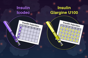
In a Phase 3 experiment, researchers compared the effectiveness and safety of once-weekly insulin termed “icodec” with the conventional once-daily injection degludec in adults with type 2 diabetes.

A long-acting insulin medication called Degludec aids in controlling blood sugar levels.

After 26 weeks, they discovered that once-weekly icodec therapy reduced blood sugar levels more than once-daily degludec. The research was released in JAMA.

Icodec may have similar glucose-lowering effects to daily insulin injections, according to a recent study.

Adherence issues with daily insulin injections

In the US, there are more than 37 million diabetics. These persons have type 2 diabetes in 90–95% of cases.

The hormone insulin, which is produced by the pancreas, enables cells to utilise glucose as fuel. When cells no longer react to insulin as they should, type 2 diabetes develops and elevated blood sugar levels follow.

The eyes, kidneys, and heart are just a few of the organs that elevated blood sugar can harm over time. Therefore, either lifestyle changes or the use of drugs that do not lower blood sugar with insulin is required for treatment.

When non-insulin treatments are ineffective, it is currently recommended by guidelines that persons with type 2 diabetes take insulin-based therapies to reduce blood sugar levels.

Currently, type 2 diabetes medications based on insulin necessitate daily injections. However, patients may find it difficult to administer daily injections, which lowers adherence rates.

According to research, weekly injections increase adherence. According to one study, individuals who receive insulin treatments once per week follow their treatment plans for an average of 333 days as opposed to 269 days for patients who receive daily injections.

Insulin therapy non-compliance might have serious repercussions. According to research, persons with diabetes who do not stick to their insulin medication have a higher risk of dying and being admitted to the hospital.

Thus, raising adherence rates is essential to enhancing diabetes patients’ quality of life and health outcomes.

Which is more effective? Once-weekly vs. daily insulin injection.

The researchers gathered 588 participants for the study, with an average age of 58, from 11 nations, including the USA, Argentina, and China.

Over a third of the participants were women, and every participant was on non-insulin glucose-lowering medication.

They were thereafter randomly assigned to receive one of the following treatment plans for a total of 26 weeks during the study:

  • once every week icodec
  • monthly placebo
  • every day degludec
  • a single-dose placebo

In the end, the scientists discovered that icodec more effectively lowered haemoglobin A1c (HBA1c) levels than degludec.

A measurement of the average blood sugar levels over the previous three months is called HBA1c. Those with diabetes are advised to maintain levels of 6.5% or lower. People without diabetes typically have HBA1c values of less than 5.7%.

Participants in the icodec group had HBA1c values that dropped from an average of 8.6% to 7% after 26 weeks. HBA1c values in the degludec group decreased from an average of 8.5% to 7.2% over this time.

The study’s authors found no discernible differences in participants’ fasting blood sugar levels or body weight between those taking icodec and those taking degludec.

We enquired about the potential causes of icodec’s superior results in lowering HBA1c readings from Dr. Absalon Gutierrez, associate professor of endocrinology at McGovern Medical School at UTHealth Houston who was not engaged in the study.

Although we can’t be certain, it probably has to do with the patient’s compliance with the drug. According to how the trial was set up, it was significantly simpler to forget to administer the degludec injections than the icodec injections. According to Dr. Gutierrez, this is most likely the case in real life as well.

Side effects of icodec weekly insulin

The researchers also reported that from the beginning of the study until week 31, 5.8% of those using Degludec and 8.9% of those taking icodec suffered hypoglycemia. This is characterized by blood sugar levels that are below the normal range.

Additionally, during the duration of the experiment, 167 patients receiving degludec and 177 patients getting icodec both had adverse effects. According to the researchers, 46 and 60 incidents, respectively, were in the degludec group and the icodec group. This may have been caused by the use of insulin.

However, they pointed out that the majority of the incidents were minor, and that these included COVID-19, influenza, and diabetic retinopathy, an eye disorder that can impair vision in people with diabetes.

What are the research’s constraints?

The study’s shortcomings were listed by the researchers in their paper. They pointed out that because the trial only lasted 26 weeks, longer-term consequences are still undetermined.

They also stated that they did not gather information on patient-reported outcomes or data from continuous glucose monitoring.

Dr. Gutierrez stated: “Icodec exhibited higher hypoglycemia even though it worked somewhat better in decreasing HBA1c. Given that it can’t be titrated as regularly, this is to be expected. Additionally, the degludec titrations were not ideal according to the study’s design.

Dr. Lushun Wang, Senior Consultant Orthopaedic Surgeon and Medical Director of Arete Orthopaedic Clinic in Singapore and a non-participant in the study, was also interviewed by us:

“The trials’ duration can be extended further in order to guarantee dependable long-term efficacy and safety. To comprehend Icodec more fully, rigorous and in-depth testing should be conducted.

Data from continuous glucose monitoring may ensure a more thorough understanding of blood glucose control and its impact on the quality of life of the patient. In addition, the trial’s design used more Icodec injections than would be necessary for a daily regimen, which does not adequately reflect real-world use or any potential advantages for treatment adherence.

Effects of once weekly injection on diabetes

The researchers observed that by lowering the number of injections from at least 365 to 52 annually, icodec may increase treatment adherence and convenience for individuals with type 2 diabetes.

They went on to say that the “small absolute risk of hypoglycemia” should be outweighed by the ease and little additional glycemic advantage of once-weekly dosing.

Icodec’s practical design enables daily injections to be replaced with this once-weekly alternative, according to Dr. Wang. Its main benefit is from its capacity to deliver an insulin release that is steady and continuous over the course of a week. Hence minimizing swings in blood glucose levels. The improved HbA1c reduction seen in the studies is evidence that Icodec’s ability can result in better overall blood glucose control.

Dr. Guitierrez concurred that icodec insulin would be a viable choice for patients who struggle to take once-daily basal insulin as prescribed. To better understand the risk of hypoglycemia associated with using icodec in comparison to once-daily insulin injections, he pointed out that more research is required.

REFERENCES:

For Diabetes medications that have been suggested by doctors worldwide are available here https://mygenericpharmacy.com/index.php?therapy=13