Browsed by
Category: Brain disease

Focused ultrasound can improve Parkinson’s symptoms?

Focused ultrasound can improve Parkinson’s symptoms?

In a recent study, a novel, non-invasive method of using focused ultrasound to lessen Parkinson’s disease symptoms was presented. In order to disrupt the neural network that causes uncontrolled movement and motor dysfunction, the approach entails deleting a tiny neuron cluster.

Twice as many research participants received the new treatment than those who received sham care, and these improvements in symptoms frequently sustained for at least a year.

An innovative, noninvasive strategy for lowering dyskinesia, or uncontrollable movements, and motor impairment in Parkinson’s disease patients is presented in a recent study. Focused ultrasound is used in the novel technique.

When compared to trial participants who underwent a sham, or placebo, therapy, twice as many people reported improvement in dyskinesia and motor impairment three months after undergoing the incision-less procedure.

In 77% of individuals who responded to the medication, the gains persisted for up to a year. Patients who receive focused ultrasound therapy often return home the same day.

Gait issues, speaking difficulty, and eye disruption were among the infrequently reported negative effects by the researchers. The group that received the treatment experienced more severe adverse effects than the group that received a placebo.

Focused ultrasound

Focused ultrasound is a minimally invasive, non-surgical technique that causes an ablation in a region that reduces Parkinson’s and tremor symptoms. To produce a clear thermal lesion deep into the brain without harming nearby structures, we use focused ultrasonic waves. The basal ganglia are what we are primarily aiming for. A set of brain regions are involved in the coordination and control of movement. We employ a thermal imaging equipment for magnetic radiofrequency imaging (MRI) during the procedure. This gauges alterations in the skull’s temperature and enables us to guarantee the patient’s security.

The treatment is fantastic because it allows us to work on really delicate places securely, effectively, and with very minimal adverse effects.

Dopamine and L-DOPA

Parkinson’s disease is caused by low levels of the neurotransmitter dopamine in the substantia nigra of the brain. This is brought on by the death of neurons that produce dopamine.

The predominant idea holds that autophagy, the brain’s housekeeping mechanism, has failed, causing an accumulation of waste that impairs brain function.

Dopamine synchronises various brain regions so that they are communicating with one another at the same frequency, according to Dr. Jean-Philippe Langevin, director of the Pacific Neuroscience Institute’s Restorative Neurosurgery and Deep Brain Stimulation Program, who was not involved in the study.

“Consider all these parts of the brain as walkie-talkies or phones trying to communicate, but the frequency at which they’re trying to communicate is off,” said Dr. Langevin. Movements become stiff, hesitant, and tremble as a result, and are no longer flowing.

Dr. Langevin stated that the absence of dopamine causes the motor system of the brain, which regulates movement, to be “quite noisy.” Researchers can actually recognise electrical noise as this “noise.” According to recent research, Parkinson’s disease actually causes some neuronal frequencies to rise.

Levodopa, also known as L-DOPA, which replenishes dopamine in the brain, is the most widely used medication for the illness.

Yet, for many people, the dyskinesia and motor dysfunction they encounter are genuine side effects of L-DOPA therapy. Over time, the medication’s effectiveness deteriorates as well.

New treatment

The novel method examined in the trial is based on an essential tremor medication that was previously authorised in 2016.

A small group of neurons in the globus pallidus area of the brain are removed or ablate during focused ultrasound therapy. By leaving scarring on the tissue, ablation causes tissue destruction.

Dr. Howard M. Eisenberg, co-author of the study and professor of neurosurgery at the University of Maryland Medical System, with the following explanation of how a little more than 1,000 sound energy beams ablate the target tissue:

“Like light, sound can be concentrated. Imagine you had a magnifying glass when you were a child. If you focused the sun’s rays on a piece of paper, you would receive a tiny dot of light that would then transform into heat and burn a hole in the paper. With sound, we may achieve the same results.

When the beams are concentrated and the intensity is strong enough, “you can burn a little hole in the brain” precisely at the target location without harming surrounding tissue, he told us.

In essence, Dr. Eisenberg said, “that’s how it works: Back to the future, similar to ablation that was done years ago for essential tremor, but using different technology.”

According to Dr. Eisenberg, the dyskinesia and motor dysfunction are not always brought on by the ablated neurons. Instead, “it’s a system of targets that are interconnected nuclei, and you’re interrupting that system,” he said.

Motives for excitement

If the procedure enables Parkinson’s patients to take less L-DOPA medication, then there are many reasons to be optimistic, according to Dr. Langevin.

According to Dr. Eisenberg, based on prior experience with essential tremor ablation, we can anticipate benefits in roughly 80% of patients, and they might last at least five years.

However, not every participant in the research benefited from this treatment. A few participants, according to Dr. Eisenberg, might have skulls that are less effective at transmitting acoustic energy.

The reason for this is that the skull is made up of two hard layers of bone sandwiched by a softer layer, “like an Oreo biscuit,” rather than being one solid piece of bone.

Focused ultrasound vs. DBS

Dr. Eisenberg pointed out that deep brain stimulation is thought to be more successful for treating essential tremors than ablation (DBS). DBS stimulates tissue rather than destroying it. Furthermore, Parkinson’s dyskinesia and motor disability are treatable by DBS.

DBS, however, necessitates creating one or more tiny holes in the skull through which a wire is placed and directed to the desired location in the brain. The wire is then attached to a tiny neurostimulator that has been inserted into the chest.

“They won’t consider it even though we always explain that deep brain stimulation has advantages over focused ultrasound,” Dr. Eisenberg said of individuals who would prefer it instead. Hence, it’s beneficial for individuals who simply don’t want deep brain stimulation, which is understandable.

Yet, he added, the advantages provided by the new focused ultrasound approach are still potent enough to be life-altering.

REFERENCES:

For more details, kindly visit below.

Quick survey on Paralysis and its different types.

Quick survey on Paralysis and its different types.

What is Paralysis?

Loss of muscle function in the body is referred to as paralysis. Sometimes paralysis is transitory, and occasionally it is permanent. While paralysis can affect any portion of the body, the majority of cases are seen in the limbs. Paralysis, both partial and total, can happen at any time. There is no immediate pain experienced by a patient who is paralysed.

The treatment plan is designed to either cure or treat the condition, depending on the nature of the underlying cause, in order to prevent the patient’s everyday lifestyle from being significantly impacted.

The most frequent underlying cause of partial or total paralysis in a patient is a stroke. Incomplete paralysis means the patient has no control over the afflicted muscle tissue while partial paralysis means the patient still has some control over the affected muscle.

Types and causes of Paralysis

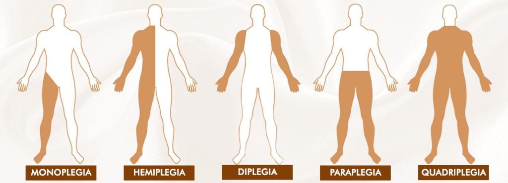
The paralysis types that are best known to the public are:

  • Monoplegia
  • Hemiplegia
  • Paraplegia
  • Quadriplegia
  • lock-in Syndrome

Monoplegia:

Monoplegia is a form of paralysis in which a person is unable to control one limb. The paralysis frequently affects just one arm, or occasionally only particular arm muscles. A common side effect of cerebral palsy is monoplegia. The brain’s capacity to control some body muscles is lost in cerebral palsy. The majority of cerebral palsy patients are young children and teenagers.

Since the patient can still do his everyday activities with only one limb paralysed, monoplegia is regarded as a positive indicator in the prognosis of this illness.

Hemiplegia:

Hemiplegia is a form of paralysis in which one side of the body loses control. This usually only has an impact on one arm or limb, though it might occasionally also have an impact on the torso. Hemiparesis is a condition in which a person’s functional use of their limbs is greatly diminished in terms of strength and endurance, but they retain some degree of functional use. In a few situations, hemiparesis progresses to hemiplegia.

When the corpus callosum between the left and right sides of the brain is damaged, hemiplegia can result. It can also result from spinal cord injury. A stroke, which impairs one side of the brain’s functionality, can also result in hemiplegia.

Left and right hemiplegia are additional divisions of hemiplegia. The diagnosis of right or left hemiplegia depends on where the injury is located.

Paraplegia:

In the case of paraplegia, the patient is unable to control their muscles below the waist. Each person experiences paraplegia differently. The fundamental cause of paraplegia typically arises in the brain or spinal cord, and people who experience it have completely normal legs. Sometimes a lower torso limb only has partial paraplegia, while other times it occurs completely. Regular physical therapy and medication are frequently responsible for partial paraplegia.

Paraplegia frequently results from a patient suffering from a brain, spinal cord, or both types of injuries. The impulses that the brain sends to the lower body are not returned to the brain through the spinal cord in paraplegic patients. Patients who suffer from this absence of brain communication not only lose their ability to move, but also their ability to feel.

A patient with partial or incomplete paraplegia may still be able to use one leg, whereas a patient with complete paraplegia loses sensation and use of both legs.

Quadriplegia

All four limbs of the body are paralysed in a quadriplegia. In this scenario, the patient’s hands and legs become impaled as a result of the brain’s signals to the areas below the neck not being returned. Sending and receiving signals from the brain is the responsibility of the spinal cord. This stops working in quadriplegia, resulting in the condition.

Quadriplegia may begin as a result of spinal cord or brain injuries. The likelihood of recovery from this condition increases as the wounds to the afflicted area heal or as the brain inflammation subsides. It is impossible to provide an accurate forecast, though.

In addition to exhaustion, sudden spasms, lack of sensation below the neck, trouble passing urine from the body, respiratory distress, bedsores, and depression, patients with quadriplegia also experience fatigue.

Locked-in Syndrome:

In a condition known as “Locked-in Syndrome,” the patient is unable to control any area of their body with their under-eye muscles. Locked-in syndrome is primarily a consequence of a serious brain damage, a stroke, or brain cancer. A person suffering from locked-in syndrome won’t be able to move any of his body’s limbs or his lips, jaw, or up and down or side to side movements in the neck. However, those who experience locked-in syndrome are still able to blink and move their eyes up and down.

Doctors rely on the moment of the eyes to make a precise diagnosis because locked-in syndrome frequently mimics the symptoms of a coma.

To ensure that the patient gets all the nutrients needed, adequate nutrition is essential. However, the patient must be fed through a stomach tube, which must either be inserted directly into the stomach through an incision made in the small intestines or through the nose.

Patients who spend a lot of time in bed can develop pressure sores, blood clots, damaged muscles, and damaged nerves. By moving the patient, rubbing the muscles, rotating the joints along their axes, and using physiotherapy, care should be given to prevent pressure sores.

Complications of paralysis

Other physiological processes including respiration and heart rate might be impacted by paralysis. Other body systems in the affected area may also be affected by the illness. Depending on the kind of paralysis you have, you could be vulnerable to:

  • breathing issues, coughing, and pneumonia risk.
  • Deep vein thrombosis (DVT) and clots in the blood.
  • issues with speech or swallowing (dysphagia).
  • both anxiety and depression.
  • Sexual difficulties and erectile dysfunction.
  • Excessively high blood pressure (autonomic dysreflexia) or low blood pressure (orthostatic hypotension) and heart problems.
  • bowel incontinence and urinary incontinence.
  • Sepsis and pressure wounds (bedsores).

How is paralysis diagnosed?

Paralysis is frequently simple to diagnose, particularly when your lack of muscle function is visible. Your doctor may employ X-rays, CT scans, MRI scans, or other imaging techniques to examine inside body parts where paralysis is more challenging to detect.

If you suffer a spinal cord injury, your doctor might perform a myelogram to determine how you are doing. In this surgery, a particular dye will be injected into the spinal cord’s nerves. This will make it easier for them to see your nerves on X-rays. In addition, they might conduct an electromyography. They will utilise sensors to assess the electrical activity of your muscles during this operation.

How is paralysis treated?

The underlying cause of the paralysis as well as the symptoms present will determine a therapy strategy. For illustration, a physician would advise:

  • operation or potential amputation
  • physical exercise
  • Workplace therapy
  • wheelchairs, braces, portable scooters, and other mobility aids
  • If you have spastic paralysis, you may take drugs like Botox or muscle relaxants.

Paralysis is frequently incurable. However, a medical team might suggest a range of medications, equipment, and tactics to assist manage symptoms.

REFERENCES:

For more details, kindly visit below.

Contrasting types of Brain aneurysm and their symptoms.

Contrasting types of Brain aneurysm and their symptoms.

An aneurysm is an artery that has enlarged due to weakened arterial wall. Even though an aneurysm rupture can result in catastrophic complications, it frequently has no symptoms. An aneurysm is a bulge or distention of the artery caused by a weakening of the artery wall.

The majority of aneurysms are not harmful and exhibit no symptoms. Some, however, have the potential to burst at their most serious state, resulting in potentially fatal internal haemorrhage.

Over 25,000 deaths in the United States (U.S.) are attributed to aortic aneurysms each year, according to the Centers for Disease Control and Prevention (CDC). Every year, some 30,000 brain aneurysms burst in the United States. About 40% of these cases result in death within 24 hours.

What is Brain aneurysm?

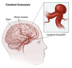
A brain aneurysm is a condition of the artery in which a region of the arterial wall bulges and swells with blood. It can also be referred to as a cerebral or intracranial aneurysm.

Any age can be affected by a brain aneurysm, which has the potential to be fatal. If a brain aneurysm ruptures, it is a medical emergency that, if left untreated, might result in a stroke, brain damage, and even death.

Types of Brain aneurysm

The location of an aneurysm within the body determines its classification. The two most typical locations for a severe aneurysm are the heart’s arteries and the brain’s arteries.

There are two primary shapes for the bulge:

  • Blood vessels with fusiform aneurysms bulge on all sides.
  • Saccular aneurysms only have a side that bulges.

The size of the bulge affects the chance of rupture.

Aortic aneurysm

The left ventricle of the heart is where the aorta originates. It then travels through the chest and abdominal cavities. The aorta’s diameter is from 2 to 3 centimetres (cm), but an aneurysm can cause it to swell to more than 5 cm.

Abdominal aortic aneurysms are the most prevalent aortic aneurysms (AAA). The area of the aorta that passes through the abdomen is where this happens. The annual survival rate for a AAA larger than 6 cm is 20% without surgery.

AAA can quickly turn fatal, but those who make it to the hospital have a 50% chance of surviving the rest of their lives.

Less frequently, the portion of the aorta that runs across the chest may be affected by a thoracic aortic aneurysm (TAA). Without treatment, TAA has a survival probability of 56% and an overall recovery rate of 85% after surgery. As only 25% of aortic aneurysms form in the chest, it is a rare disorder.

Cerebral aneurysm

Intracranial aneurysms are aneurysms in the blood vessels that supply the brain. They are frequently referred to as “cherry” aneurysms due to their appearance.

Within 24 hours, a brain aneurysm rupture can be fatal. Around 66 percent of people who survive brain aneurysms will have a neurological impairment or disability as a result. Brain aneurysms account for 40% of fatalities.

The most frequent cause of a form of stroke known as subarachnoid haemorrhage is ruptured brain aneurysms (SAH).

Peripheral aneurysm

An aneurysm in the popliteal region develops behind the knee. The most frequent peripheral aneurysm is this one.

  • Aneurysm of the splenic artery: This kind develops close to the spleen.
  • Splenic artery aneurysm: This type of aneurysm occurs near the spleen.
  • Mesenteric artery aneurysm: The artery that carries blood to the intestines is impacted by this.
  • Femoral artery aneurysm: The femoral artery is located in the groyne and can rupture.
  • Carotid artery aneurysm: This happens in the neck.
  • Visceral aneurysm: Aneurysm of the arteries supplying blood to the gut or kidneys is known as a visceral aneurysm.

Aortic aneurysms are more prone to burst than peripheral aneurysms.

Symptoms of Brain aneurysm

Unless it ruptures, a brain aneurysm rarely exhibits any symptoms (ruptures). If a brain aneurysm is unusually large or presses against brain tissues or nerves, it may occasionally induce symptoms even if it is unruptured.

Unruptured brain aneurysm symptoms can include:

If you have signs of a brain aneurysm that has not ruptured, you should visit a doctor as soon as possible. Although aneurysms typically do not burst, it is always crucial to have one evaluated in case medical attention is required.

An intense headache that strikes out of nowhere is typically the first sign of a ruptured brain aneurysm. It has been compared to getting hit on the head and causes blinding anguish unlike anything else ever felt.

Other signs of a ruptured brain aneurysm may also manifest suddenly and include the following:

  • an ill feeling or being
  • a sore neck or ache in the neck
  • responsiveness to light
  • double or blurry vision
  • abrupt confusion
  • consciousness is lost
  • fits (seizures)
  • weakness in any limbs or on one side of the body

Causes of Brain aneurysm

An artery in the brain experiences structural alterations that lead to brain aneurysms. The artery’s walls weaken and thin as a result of these alterations. The distortion may result from wall weakening, but it is also possible for inflammation or trauma to be the only cause of the deformity.

What specifically causes aneurysms to occur is still a mystery. But it’s thought that one or a few of the following elements might favour their development:

  • an elastic tissue breakdown inside the artery
  • stress brought on by the artery’s blood flow
  • because of an increase in inflammation, the artery’s tissue has changed.

Additionally, where an artery splits off into several directions, brain aneurysms are more likely to occur. This is due to the weaker arteries that are present there by nature.

Aneurysms can develop at any time after birth. But they typically come about throughout the course of your lifetime.

Complications of Brain aneurysm

Complications from a ruptured aneurysm could be one of its early warning indications. Instead of only the aneurysm, a rupture is more likely to cause symptoms.

The majority of persons who have an aneurysm do not have any complications. However, problems can also include the following in addition to thromboembolism and aortic rupture:

  • Severe chest or back pain: After an aortic aneurysm in the chest ruptures, severe chest or back pain may develop.
  • Angina: Another form of chest pain that can result from some aneurysms is angina. Myocardial ischemia and heart attacks can result from angina.
  • A sudden extreme headache: An unexpectedly strong headache is the primary sign of SAH caused by a brain aneurysm.

Any aneurysm rupture may result in pain, low blood pressure, a fast heartbeat, and dizziness. The majority of persons with an aneurysm won’t have any problems.

Prevention of Brain aneurysm

Since certain aneurysms are congenital—that is, existing from birth—it is not always possible to prevent them. But some lifestyle decisions can influence the risk:

  • Smoking increases the risk of developing aortic aneurysms and having an aneurysm rupture in any part of the body. Smoking cessation can lower the risk of developing a serious aneurysm.
  • Reduced aneurysm risk can also be achieved by controlling blood pressure. Dietary changes, regular exercise, and medication can all help lower blood pressure to a healthy level.
  • These actions are crucial for lowering pressure on the arterial walls since obesity can put the heart under additional strain.
  • Additionally, a healthy diet helps lower cholesterol and lower the risk of atherosclerosis. Atherosclerosis and fusiform aneurysms frequently go hand in hand.

Anyone who has been given a conservative treatment plan after being diagnosed with an aneurysm can work with a medical professional to address any risk factors.

REFERENCES:

For more details, kindly visit below.

Important note on Parkinson’s disease you need to know.

Important note on Parkinson’s disease you need to know.

Parkinson’s disease is a neurological condition that worsens over time. Movement issues are one of the earliest symptoms. Dopamine is a chemical that exists in the brain and is responsible for smooth and coordinated motions of the body’s muscles. The “substantia nigra” is a region of the brain where dopamine is made.

The substantia nigra cells begin to deteriorate in Parkinson’s disease. Dopamine levels drop as a result of this. Parkinson’s disease symptoms begin to show up when they have decreased by 60 to 80%.

Parkinson’s disease stages

Parkinson’s is a progressive illness, which means that over time, the condition’s symptoms usually get worse.

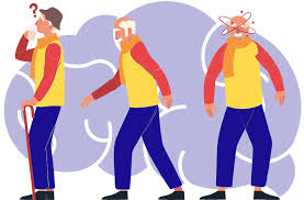
The Hoehn and Yahr scale is frequently used by doctors to categorise its stages. This scale categorises symptoms into five phases and aids medical practitioners in determining the severity of disease symptoms and signs.

Stage 1

Stage 1 is the mildest type is Parkinson’s. In fact, it’s so mild that you could not even notice any symptoms. Your regular activities and chores might not yet be affected by them.

Even if you do experience symptoms, they might just affect one side of your body.

Stage 2

It may take months or even years for stage 1 to move into stage 2. The experiences of each person will vary. You might feel symptoms at this stage, which is moderate such as:

Stiffness in your muscles can make regular tasks more difficult and take longer to complete. However, you probably won’t have any balance issues at this point.

The body’s two sides may experience the same symptoms. Posture, movement, and facial expression modifications may be more obvious.

Stage 3

The symptoms change course at this midpoint. You probably won’t develop any new symptoms, but they might become more obvious. They might also obstruct all of your daily activities.

Activities move more slowly as a result of the noticeable slower movements. Falls can grow more frequent as balance problems become more serious. However, people with Parkinson’s disease in stage 3 can typically keep their independence and carry out daily tasks without much help.

Stage 4

There are substantial modifications as the stages 3 and 4 proceed. Without a walker or other aid, it will be quite difficult for you to stand at this point.

Significantly slower reactions and muscle motions are also observed. It might be risky and unsafe to live alone.

Stage 5

In this most advanced stage, constant help is required due to the intense symptoms. Standing will be challenging, if not impossible. Most likely, a wheelchair will be needed.

Parkinson’s patients may also have disorientation, delusions, and hallucinations at this stage. The disease’s consequences can start developing in its latter stages.

Symptoms of Parkinson’s disease

Some of the early signs of Parkinson’s can show up years before there are any movement issues. These initial indications include:

  • reduced capacity to smell (anosmia)
  • constipation
  • tiny, squished handwriting
  • voice variations
  • hunched position

The four most prevalent motor issues include:

  • tremor (shaking that occurs at rest)
  • sluggish motions
  • rigidity in the arms, legs, and trunk
  • difficulties with balance and a propensity to tumble

Additional signs include:

  • blank look on the face
  • a propensity to become trapped while walking
  • low-pitched, muted speech
  • reduced swallowing and blinking
  • inclination to reverse direction
  • shortened arm walking while swinging
  • Parkinsonian gait, or the propensity to walk with shuffled steps

Additional signs and symptoms could be:

  • Seborrheic dermatitis is the condition when the skin develops flaky white or yellow scales on greasy areas.
  • greater likelihood of developing the deadly skin disease melanoma
  • vivid dreams, chatting, and movement while sleeping are all signs of disturbed sleep.
  • depression
  • anxiety
  • hallucinations
  • psychosis
  • issues with focus and memory
  • visual-spatial interactions are challenging

Parkinson’s disease’s early warning signals could go unnoticed. With these warning signals, your body may try to warn you of the movement issue years before any movement difficulties appear.

Causes of Parkinson’s disease

Parkinson’s disease is a neurological condition brought on by alterations in the brain. Although the exact reason why it occurs is unknown, scientists have found some varieties that do.

Low dopamine levels

Parkinson’s disease symptoms are mostly brought on by low or declining levels of the neurotransmitter dopamine. It occurs when brain cells that make dopamine die.

The area of the brain that regulates movement and coordination receives information from dopamine. Therefore, it may be more difficult for people to control their movement when dopamine levels are low.

The severity of the symptoms gradually worsens as dopamine levels continue to drop.

low levels of norepinephrine

Damage to the nerve terminals that create another neurotransmitter, norepinephrine, which supports blood circulation and other natural bodily activities, may also be a component of Parkinson’s disease.

Parkinson’s disease patients with low norepinephrine levels may have both motor and nonmotor symptoms like:

  • rigidity and stiffness
  • physical unsteadiness
  • tremor
  • anxiety
  • having trouble focusing
  • dementia
  • depression

This may help to explain why orthostatic hypotension frequently occurs in persons with Parkinson’s disease. When someone stands up, their blood pressure fluctuations, which can cause dizziness and a chance of falling, is referred to as this.

The Lewy body

The brain of a person with Parkinson’s disease may have Lewy bodies, or clusters of the protein alpha-synuclein. Changes in movement, thinking, behaviour, and mood can result from the buildup of Lewy bodies, which can also result in nerve cell death. Dementia may also result from it.

Lewy body dementia is not the same as Parkinson’s disease, although because the symptoms are similar, some people may have both.

Genetic factors

Although mutations in numerous genes have been found to be associated with Parkinson’s disease, experts do not believe this to be a hereditary ailment.

Only 10% of cases, particularly in those with early-onset disease, seem to be genetic in nature.

Autoimmune factors

According to a 2017 study, there may be a hereditary connection between inflammatory diseases like rheumatoid arthritis and Parkinson’s disease.

In 2018, Taiwanese health data were examined by researchers who discovered a 1.37-fold increased risk of Parkinson’s disease in patients with autoimmune rheumatic disorders.

Risk factors for parkinson’s disease

Parkinson’s disease risk may be increased by a number of environmental variables.

These consist of:

  • Past traumatic brain injury: For instance, head traumas sustained while playing contact sports may raise the likelihood of the illness.
  • Exposure to toxins: These include metals, pesticides, solvents, and other contaminants.
  • Males are 50% more likely than females to have the illness, while a 2016 study found that the risk for females may rise with age.
  • Age: The illness frequently manifests around the age of 60.
  • Some pharmaceuticals: Some drugs can cause Parkinsonism, a condition in which a person exhibits tremors and other symptoms but does not have Parkinson’s disease.

Symptoms typically start to show up around the age of 60. However, early-onset Parkinson’s, which manifests before the age of 50, affects 5–10% of those who have the condition.

Statistics in the past have indicated that Black Americans are less likely than people of other ethnicities to have Parkinson’s disease.

The lack of knowledge about how the disease can affect Black people and a higher likelihood of misdiagnosis as a result of unequal access to healthcare, according to experts, may be to blame for this.

REFERENCES:

  • https://www.healthline.com/health/parkinsons
  • https://www.medicalnewstoday.com/articles/323409
  • https://www.mayoclinic.org/diseases-conditions/parkinsons-disease/symptoms-causes/syc-20376055
  • https://my.clevelandclinic.org/health/diseases/8525-parkinsons-disease-an-overview
  • https://www.nia.nih.gov/health/parkinsons-disease

For more details, kindly visit below.

Important note on causes and treatment for brain cancer.

Important note on causes and treatment for brain cancer.

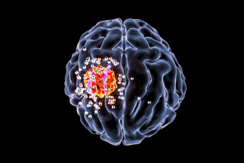
The development of malignant cells in the brain leads to brain cancer. Depending on the type of tumour, the cancer cells produce slow- or fast-growing tumours.

The goal of brain cancer treatment is to remove the tumour and then eradicate any cancer cells that may still be present. In particular for slow-growing tumours, new advancements in brain cancer therapies are increasing survival rates.

What is brain cancer?

Primary brain cancer, commonly referred to as brain cancer, is characterised by an excess of brain cells that develops into masses known as brain tumours. Cancer, on the other hand, begins in another place of the body and progresses to the brain. It is referred to as secondary or metastasized brain cancer when that occurs.

Some malignant brain tumours have a rapid rate of growth. These cancerous tumours may interfere with how your body functions. Brain tumours should be treated as soon as they are discovered since they can be fatal.

Brain tumours are rather rare. People have a less than 1% lifetime chance of getting a malignant brain tumour, according to estimates from the American Cancer Society.

Types of brain tumors

Names for brain tumours are determined by their location within the brain or upper spine. A grade is also assigned to tumours. You can determine the projected rate of growth of a tumour by looking at its grade. Grades range from one to four, with four being the fastest-growing grades and one being the slowest.

The following are some of the most prevalent kinds of brain tumours:

  • Glioma. About 3 out of 10 occurrences of brain cancer are gliomas, which are brain tumours that start in the glial cells.
  • Astrocytoma. Glioblastomas, the kind of quickly-expanding brain tumour, are a subtype of astrocytomas.
  • Meningioma. Meningioma tumours, the most prevalent kind of brain tumour in adults, develop in the tissue that surrounds your brain and spinal cord and are frequently benign and slow-growing.
  • Ganglioglioma. Surgery is typically used to treat the slow-growing tumours known as gangliogliomas that are present in glial and neuronal cells.
  • Craniopharyngiomas. Craniopharyngiomas are slow-growing tumours that develop between the pituitary gland and the brain. Because they frequently encroach on the optic nerves, they can impair eyesight.
  • Schwannomas. Almost typically benign, schwannomas are slow-growing tumours that develop around the cranial nerves.
  • Medulloblastoma. Children are more likely to develop medulloblastomas, which are rapidly developing tumours that develop on the nerve cells in the brain.

Symptoms of brain cancer

The size and location of the brain tumour affect the symptoms of brain cancer. Particularly in its early stages, brain cancer exhibits many of the same symptoms as a number of less dangerous diseases.

Numerous of these symptoms are extremely typical and are not likely to be signs of brain cancer. However, it’s a good idea to see a doctor if you’ve had any of these symptoms for longer than a week, if they came on suddenly, if they don’t go away with over-the-counter painkillers, or if any of them worry you.

Typical signs of brain cancer include:

  • morning headaches that are typically worse
  • nausea
  • vomiting
  • a miscommunication
  • a loss of equilibrium
  • having trouble walking
  • Memory problems
  • having trouble thinking
  • speech issues
  • vision issues
  • personality alters
  • inconsistent eye motions
  • body jerking
  • muscle jerking
  • unexpected fainting or syncope
  • drowsiness
  • tingling or numbness in the arms or legs
  • seizures

Early diagnosis significantly improves the prognosis for brain cancer. If you frequently suffer any of the aforementioned symptoms or suspect that your symptoms may be more serious, schedule an appointment with a doctor right away for a diagnosis.

Causes and risk factors

Primary brain cancer has an unidentified specific cause. However, studies have connected excessive ionising radiation exposure to a higher chance of developing brain cancer. The most frequent sources of ionising radiation include radiation therapy treatments, frequent medical imaging tests (CT scans and X-rays), and potential employment exposure.

Additional factors that could increase the risk of acquiring brain cancer include:

  • greater age
  • a history of brain cancer in the family
  • chronic smoking
  • being exposed to fertilisers, pesticides, and herbicides
  • working with substances like lead, plastic, rubber, petroleum, and some textiles that might cause cancer
  • having mononucleosis or Epstein-Barr virus infection

Secondary brain cancer Some types of cancer are more likely than others to cause brain cancer, which develops when cancer that started in another part of your body travels to your brain.

The following cancers frequently metastasis, or spread, to the brain:

  • lung disease
  • mammary cancer
  • renal cancer
  • urethral cancer
  • melanoma, a form of skin cancer

How is brain cancer treated?

Brain cancer can be treated in a number of ways. A primary brain cancer will be treated differently than a cancer that has spread to other organs.

The kind, size, and location of your brain tumour will determine whether you receive one therapy or more. There will also be considerations for your age and general health.

Treatments for brain tumours include:

Surgery.

The most frequent form of treatment for brain tumours is brain surgery. Depending on the tumor’s position, it might be possible to remove it whole, partially, or not at all.

Chemotherapy.

These medications can reduce your tumour and kill brain cancer cells. Chemotherapy can be administered orally or intravenously.

Radiation treatment. 

Using high-energy waves like X-rays, this procedure eliminates cancer cells and tumour tissue that can’t be removed surgically.

Combination treatment.

Combination therapy refers to the simultaneous administration of chemotherapy and radiation therapy.

Biologic medicines

These medications support, guide, or restore your body’s natural tumour defences. For instance, immunotherapy is a class of biologic medication that is frequently prescribed and increases your immune system’s capacity to recognise and combat cancer.

Other medicines.

To treat symptoms and adverse effects brought on by your brain tumour and brain cancer therapies, your doctor may prescribe drugs.

Clinical studies.

Clinical trial medicines and drugs may be employed in advanced brain cancer instances that don’t respond to treatment. These are medications that are still being tested. An immunotherapy trial and a CAR T cell therapy trial may be part of clinical trials for brain cancer.

Rehabilitation.

If your disease or treatment have made it difficult for you to speak, walk, or perform other daily tasks, you might need to go to rehabilitation sessions. Physical therapy, occupational therapy, and other types of therapies are all included in rehabilitation. These treatments can assist you in relearnng activities.

Various forms of treatment.

There isn’t much evidence to back up the use of complementary medicines to treat brain cancer. To make up for the nutrients lost during cancer treatment, some medical specialists do advise taking measures like following a bland diet and taking vitamin and mineral supplements. 

Before making any dietary changes, using any herbs or supplements, or pursuing any alternative treatments, see your doctor.

How to reduce your risk of brain cancer?

Although there is no known way to prevent brain cancer, you can lower your risk by staying away from:

  • pesticide and insecticide exposure
  • exposure to cancer-causing substances
  • smoking
  • radiation exposure that is not essential

REFERENCES:

  • https://www.healthline.com/health/brain-cancer
  • https://www.cancercenter.com/cancer-types/brain-cancer
  • https://www.mayoclinic.org/diseases-conditions/brain-tumor/symptoms-causes/syc-20350084

For more details, kindly visit below.

Important substancial factors to avoid Alzhiemer’s disease.

Important substancial factors to avoid Alzhiemer’s disease.

What is Alzhiemer’s disease?

A form of dementia that progresses is Alzheimer’s disease. A condition that adversely impacts memory, thinking, and behaviour is referred to as dementia. The modifications make daily life more difficult. There are numerous possible causes of dementia, including diseases and brain traumas. Sometimes there is no known cause.

The Alzheimer’s Association estimates that 60 to 80 percent of dementia cases are caused by Alzheimer’s disease. The condition is typically diagnosed in patients over the age of 65. Alzheimer’s disease is typically described as having a “early onset” or “younger onset” if it is discovered earlier. Alzheimer’s has no known cure, but there are medications that can halt the disease’s growth.

Importants facts about Alzhiemer’s disease

  • Alzheimer’s disease is a persistent, chronic (long-term) illness. It is not a normal ageing symptom.
  • Dementia and Alzheimer’s disease are not the same thing. A form of dementia is Alzheimer’s disease.
  • Its symptoms appear gradually, and its degenerative effects on the brain result in a steady decline.
  • Alzheimer’s disease can affect anyone, but some people are more susceptible to it than others. People over 65 and those with a family history of the illness are included in this.
  • Alzheimer’s patients cannot be predicted to have a particular outcome. While some persons experience a slower onset of symptoms and a faster rate of disease progression, others experience lengthy lifespans with minor cognitive impairment.

What does Alzheimer’s disease look like?

Even though the earliest symptoms of Alzheimer’s may differ from person to person, memory issues are often one of the first signs of the disease. The very early stages of Alzheimer’s disease may also be indicated by a deterioration in other cognitive abilities, including the ability to express oneself clearly, problems with vision or spatial awareness, and impaired reasoning or judgement. However, not everyone who has minor cognitive impairment (MCI) will go on to acquire Alzheimer’s. MCI is a condition that can be an early indicator of the disease.

Alzheimer’s patients struggle with simple tasks like driving a car, preparing food, and paying their bills. They might repeatedly ask the same questions, become disoriented quickly, misplace items or put them in strange places, and find even the most basic of tasks to be confusing.

Symptoms of Alzheimer’s disease

Everybody occasionally experiences moments of amnesia. However, those who have Alzheimer’s disease exhibit a number of persistent habits and symptoms that get worse with time. These may consist of:

  • Memory loss that interferes with regular tasks like remembering appointments
  • difficulty performing routine tasks, including using a microwave
  • inability to solve problems
  • difficulty speaking or writing
  • becoming uncertain of the time or location
  • reduced judgement
  • lower level of personal hygiene
  • changes in personality and mood
  • retreat from the community, family, and friends

These symptoms do not always indicate Alzheimer’s disease. To ascertain the cause, it’s crucial to visit a doctor.

As the condition progresses, the symptoms change. People with Alzheimer’s frequently experience substantial difficulty speaking, moving, or reacting to events around them in the later stages of the disease.

Causes of Alzhiemer’s disease(Factors to avoid)

According to current theories, the aberrant protein buildup in and around brain cells is what causes Alzheimer’s disease. Amyloid is one of the proteins involved, and deposits of it create plaques around brain cells.

The other protein is tau, which builds up inside brain cells to form tangles. Scientists now know that this process starts many years before symptoms manifest, even if the exact cause is unknown.

The chemical messengers (known as neurotransmitters) used to communicate or send signals between brain cells decline as brain cells are damaged. The brains of those who have Alzheimer’s disease have notably low levels of one neurotransmitter, acetylcholine.

Different parts of the brain diminish throughout time. Memory-related areas are frequently the first to be damaged. Different parts of the brain are affected in more uncommon forms of Alzheimer’s disease. Instead of memory issues, the earliest signs may be issues with vision or language.

Diagnosing Alzheimer’s disease

Examining brain tissue after death is the only surefire technique to determine if someone has Alzheimer’s disease. However, a doctor can measure your mental capacity, identify dementia, and rule out other disorders using different examinations and tests.

Taking a medical history will probably be the doctor’s first step. They might inquire as to:

  • symptoms
  • family’s history of illness
  • other health issues, present or previous
  • Medications taken now or in the past
  • alcohol consumption, nutrition, and other lifestyle choices

After that, your doctor will probably ask for a number of tests to see if you have Alzheimer’s disease.

What to do if you suspect Alzheimer’s disease

If you want to know if the symptoms you’re having are caused by Alzheimer’s disease or something more manageable like a vitamin deficiency or a drug side effect, make an appointment with your doctor.

A timely and correct diagnosis also gives you and your family the chance to think about financial preparation, create advance directives, sign up for clinical trials, and foresee care requirements.

REFERENCES:

For more details, kindly visit below.

What are the possible side effects of using Serpentina?

What are the possible side effects of using Serpentina?

A member of the milkweed family, rauwolfia (Rauwolfia serpentina), often spelled ravolphia, is a useful shrub. Its root is either marketed as pills or capsules, or it is ground into a powder and packaged in this way. It is a substance that is frequently utilised in Asian medicine, particularly the traditional Ayurvedic treatment that originated in India.

About 50 alkaloids, which are its active constituents, have been identified; however, reserpine, rescinnamine, and deserpidine seem to produce the strongest psychedelic effects. Indian snakeroot should not be confused with Yohimbe or Rauvolfia vomitoria. They are not equivalent.

Uses

Indian snakeroot is used to treat mental illnesses such agitated psychosis and insanity, as well as mild high blood pressure, anxiety, and sleeplessness. It is also used as a tonic for general debilities and for snake and reptile bites, fever, constipation, feverish intestinal problems, liver disorders, achy joints (rheumatism), fluid retention (edoema), and epilepsy.

Indian snakeroot has a substance that is similar to the prescription medication reserpine. Reserpine is used to treat schizophrenia, mild to severe hypertension, and a few signs of impaired circulation.

  • Difficulty sleeping (insomnia). Early research suggests that Indian snakeroot, when taken with two additional plants, may be able to treat insomnia.
  • Nervousness.
  • illnesses of the mind like schizophrenia.
  • Constipation.
  • Fever.
  • liver issues.
  • painful joints
  • due to inadequate circulation, leg spasms.
  • mildly elevated blood pressure
  • other circumstances

Side effects

When a standardised extract is administered under the guidance of a medical expert with experience using it, Indian snakeroot is POSSIBLY SAFE. Standardized Indian snakeroot has a predetermined dosage of medication. Indian snakeroot has a range of compounds, including reserpine, depending on the plant.

Indian snakeroot contains reserpine and other potentially hazardous compounds; therefore, a skilled healthcare expert must accurately calculate the dosage and monitor any side effects. Nasal congestion, stomach cramps, diarrhoea, nausea, vomiting, loss of appetite, drowsiness, seizures, Parkinson’s-like symptoms, and coma are just a few of the side effects that can range in severity from mild to serious. When driving or operating heavy machinery, Indian snakeroot should not be taken because it can impair reaction time.

SPECIAL PRECAUTIONS & WARNINGS

Pregnancy and breast-feeding: Indian snakeroot is NOT SAFE to take during pregnancy or when nursing a baby. Indian snakeroot has compounds that could lead to birth abnormalities. Additionally, it is NOT SAFE to utilise this Indian snakeroot when nursing. It contains toxins that can enter breast milk and damage a nursing infant.

Shock therapy (electroconvulsive therapy, ECT): Indian snakeroot should not be administered by patients undergoing electroconvulsive therapy (ECT), also known as shock therapy. Indian snakeroot should be stopped at least a week before starting ECT.

Gall stones: Indian snakeroot may exacerbate gallstones and gallbladder disorders.

Stomach ulcers, intestinal ulcers, or ulcerative colitis: If you have ever experienced stomach, intestinal, or ulcerative colitis, you should not use Indian snakeroot.

Allergic reaction to reserpine or other drugs classified as rauwolfia alkaloids: If you have an allergy to any of these medications, avoid taking Indian snakeroot.

Depression: If you have depression or suicidal thoughts, avoid using Indian snakeroot.

Pheochromocytoma: An adrenal gland tumour that raises blood pressure to hazardous levels: In this case, stay away from Indian snakeroot.

Surgery: Indian snakeroot may hasten the central nervous system during surgery. There is a worry that it can make surgery more difficult by raising blood pressure and heart rate. Before any scheduled surgery, stop using Indian snakeroot at least two weeks in advance.

REFERENCES:

For more details, kindly visit below.