Browsed by
Category: Mental health

Possible link between migraine and carpal tunnel syndrome.

Possible link between migraine and carpal tunnel syndrome.

Researchers looked into the prevalence of migraine headaches in patients who have nerve decompression surgery.

They discovered that people who have surgery to decompress a nerve at particular points on their bodies may be up to 70% more likely to get migraines than others who have the procedure elsewhere.

To determine whether nerve decompression can treat migraines, more research is required.

There may be pain and a loss of function when the nerves in the hands and arms contract around muscles and soft tissues. Between 5% and roughly 9% of the population are affected by various types of nerve compression in these places.

Surgery is frequently used to treat the illness and might result in full or partial symptom relief.

The muscles, blood arteries, and bone in the vicinity of the head’s surrounding nerves can also compress those nerves. Improvement or alleviation from migraine and headaches may result by decompressing these nerves.

What is Carpal tunnel syndrome?

Carpal tunnel syndrome is caused by compression of the median nerve. On the hand’s palm side, the carpal tunnel is a small opening encircled by bones and ligaments. Numbness, tingling, and weakness in the hand and arm are signs of median nerve compression.

Carpal tunnel syndrome can be caused by repetitive hand motions, health issues, and wrist morphology.

The tingling and numbness are typically reduced with appropriate care, and wrist and hand function is recovered.

Symptoms

The following list of signs and symptoms of carpal tunnel syndrome includes:

Feeling tingly or numb. Numbness and tingling in the fingers or hand may be apparent. Normal afflicted fingers include the thumb, index, middle, and ring fingers, but not the little finger. In certain fingers, you might experience something like to an electric jolt.

The wrist may feel the sensation before it moves up the arm. These symptoms may awaken you from sleep and frequently happen while you are holding the phone, newspaper, or steering wheel.

To try to alleviate their symptoms, many people “shake out” their hands. Over time, the numb sensation could persist continuously.

Weakness. You can feel weak in your hands and drop things. This can be because the thumb’s pinching muscles, which are similarly regulated by the median nerve, are weak or because the hand is numb.

Migraine and carpal tunnel syndrome

When the nerve that travels from the forearm to the palm of the hand is squeezed at the wrist, carpal tunnel syndrome develops.

In comparison to 16% of those without carpal tunnel syndrome, 34% of those with the condition get migraines, according to a cross-sectional study with 25,880 participants. It might be more effective to screen patients for the disorders if it is known whether nerve compression around the head is related to nerve compression in the hands and arms.

Researchers looked at how frequently people who had nerve decompression surgery for the hands and arms were diagnosed with migraines.

According to their findings, people who have particular types of nerve compression are more prone to suffer from migraine headaches. Not a part of the study, Dr. Chantel Strachan is an internist at ColumbiaDoctors and an assistant professor of medicine at Columbia University Irving Medical Centre in New York. She said.

“I wouldn’t jump to advise carpal tunnel release in every migraine patient. The choice to proceed with surgical treatment for nerve compression is specific to the patient and should be carefully considered with the patient’s medical care team.

Journal of Plastic and Reconstructive Surgery published the findings.

Most likely to experience migraine

Data from 9,558 patients who underwent nerve decompression surgery of the hands and arms between 2009 and 2019 were analysed for the study.

Participants were also evaluated by the researchers for the presence of migraine.

Of the subjects, the median nerve was decompressed in about 71% of cases. Surgery is done on the wrist to release pressure on the nerve, which lessens carpal tunnel syndrome symptoms.

A decompression of the ulnar nerve was done on about 14% of subjects. That is an elbow nerve decompression. 6.5% of patients underwent decompression procedures at various body locations.

In the end, the researchers discovered that people with multiple nerve decompression and median nerve decompression were respectively 30% and 70% more likely to experience migraines than people with ulnar nerve decompression.

Nerve compression and migraine

Dr. Sean Ormond, a specialist in anesthesiology and interventional pain management, did a study to learn more about the potential connection between nerve decompression and migraine.

He mentioned that there are a number of possibilities, but that the causes of nerve compression in the arms and hands and migraine are not entirely known.

“Both upper extremity nerve compression syndromes and migraine may share common risk factors, such as obesity, sedentary lifestyle, poor posture, or repetitive stress injuries,” stated Dr. Ormond.

The affected area may experience inflammation as a result of nerve compression. It is also recognised that inflammation contributes to the pathophysiology of migraines. The presence of inflammation in one place of the body may cause inflammation to spread throughout the body, potentially aggravating migraines, the doctor added.

Ormond observed that although further research is need to establish this, some people may be more prone to higher nerve compression and migraine due to a hereditary tendency.

According to Dr. Strachan, migraine sufferers may become more sensitive to pain due to nerve damage.

Limitations of the migraine study

Dr. Strachan pointed out that because the study was retrospective in nature, the results suggest association rather than causality.

She stated that different providers and their subspecialties, such as primary care, neurology, and pain, may have utilised different criteria to diagnose migraine.

The association between migraine and pain from nerve compression may be the consequence of other variables, as the researchers stated in their report that there is a general overlap across chronic pain disorders.

REFERENCES:

For Migraine disease medications that have been suggested by doctors worldwide are available here https://mygenericpharmacy.com/index.php?therapy=17

Analyze the links between BMI, obesity & cognitive ability.

Analyze the links between BMI, obesity & cognitive ability.

According to the World Health Organisation (WHO), there were more than 650 million obese adults in the world as of 2016. Obesity has been linked in the past to an increased risk of cognitive deterioration.

Evidence from University College London researchers challenges the idea that fat and cognitive capacity are causally related.

Around the world, more than 1.9 billion adults were obese in 2016, with more than 650 million of those adults suffering from obesity, a disease in which a person’s weight is over normal ranges and may lead to various health issues.

According to current estimates, 167 million adults and children will be overweight or obese by 2025. A multitude of disorders, including diabetes, cardiovascular disease, hypertension, osteoporosis, rheumatoid arthritis, and cancer, have been linked to obesity in previous studies, including these.

Furthermore, previous research has connected obesity to a higher risk of cognitive deterioration.

The causal relationship between obesity and cognitive performance has now been called into question by University College London academics. They contend that common family variables have tainted the research linking cognitive aptitude and BMI.

Obesity

If a person’s present weight is excessive for their height, they are considered obese. The body mass index (BMI) is the most popular metric for determining a person’s level of obesity.

This tool determines if an adult is obese or not based on their height and current weight:

  • BMI less than 18.5 indicates underweight.
  • Suitable BMI range: 18.5-24.9
  • BMI of 25 to 29.9 indicates obesity
  • obesity: a BMI of 30 or above

Children and teenagers need a different BMI calculator, which considers height, age, and gender to evaluate obesity because they are still developing.

The BMI measurement is not without problems, though. It is unable to distinguish between muscle and fat when weighing someone. Additionally, it disregards a person’s race, overall body composition, or bone density.

Cognitive function and obesity

Lead author of this study and senior research fellow at the Centre for Longitudinal Studies at University College London in the U.K., Dr. Liam Wright, Ph.D., states that there are several reasons why the research team decided to investigate the causal relationship between cognitive capacity and obesity:

“Over the past forty years, there has been a significant rise in the prevalence of obesity, but BMI hasn’t increased uniformly throughout the population. Therefore, it is crucial to understand why some people are more predisposed to obesity than others.

Additionally, there is a substantial body of research in the field of cognitive epidemiology that demonstrates a connection between cognitive function and practically every measure of health and health behaviour, including obesity.

Unfortunately, the majority of the cognitive epidemiology literature employs observational research designs that may be biassed and fail to show causal effects, according to Dr. Wright. “There are some compelling theoretical arguments for why cognitive ability might have a causal effect on health, but regrettably, these arguments are based on observational research designs,” she said. Because a sibling design could take into account some of the variables that can skew relationships found in previous research, we felt it was crucial to investigate for a relationship between cognitive capacity and BMI.

Examining siblings to reduce bias

Dr. Wright and his research group evaluated data from four distinct young population cohort studies carried out in the United States that included 12,250 siblings from 5,602 homes. Each participant’s data were tracked from youth to age 62.

The scientists were able to take into consideration unobserved characteristics associated with family background by analysing the relationship between cognitive capacity and BMI among families.

“Sibling designs account for factors that are shared between siblings by design,” Dr. Wright said. They don’t require the measurement of these factors, which is both a benefit and a drawback because it is difficult to determine which common factors actually contribute.

With this qualification, he continued, “There are four main factors that we thought might be significant: genetics (siblings share 50% of DNA), parental socioeconomic class (wealth, location, etc.), parenting styles (particularly regarding dietary choices), and parental cognitive ability (cognitive ability could operate indirectly!). “Once more, we didn’t directly examine these.”

According to Dr. Wright, they predicted that these variables would make general population studies more biassed and lead to weaker relationships than in earlier studies, which is exactly what they found.

However, he cautioned, “remember that sibling designs have their own flaws, including the ability for siblings to influence one another, for example, by modelling one another’s behaviour. This may imply that our findings are also skewed, albeit downwardly and smaller than the actual causal effect.

Association between BMI and cognitive ability

When the researchers evaluated the data from study participants who were not related, they discovered that, after accounting for family socioeconomic status, the change in teenage cognitive capacity from the 25th to the 75th percentile was associated with an estimated 0.61 kg/m drop in BMI.

And when the researchers analyzed the information from siblings, they discovered that the change in BMI from the 25th to the 75th percentile of teenage cognitive ability was only correlated with a 0.06 kg/m drop in cognitive capacity.

The relationship between cognitive capacity and BMI was less pronounced when siblings were compared than when the entire population was, according to Dr. Wright, but he was not surprised by this given the overall characteristics he mentioned.

However, he noted, “I was shocked at how little of an association there was when comparing siblings. As said, there are strong arguments to support the idea that cognitive ability has an impact on health and health-related decision-making“.

“Two possibilities for this small association are that one, our results were biassed towards finding smaller associations (e.g., by siblings influencing each other), and two, reflective decision-making isn’t as important in determining BMI as other factors like satiety, etc.,” Dr. Wright continued. Both of these are hypothetical.

Unproven causality

As a parent and a neurologist, Dr. Segil claimed that he has never observed a connection between obesity or a healthy weight and cognitive aptitude in people.obese

The purpose of this study, according to Dr. Segil, “is to argue that people with higher cognitive abilities, who have a higher socioeconomic position, have made healthier decisions.” Additionally, it’s possible that people’s cognitive function increases as their BMI decreases when they make healthier decisions.

He continued, “I do not believe that there is any evidence linking obesity to cognitive function. And I believe that their research’s use of siblings or other family members who are in a similar social economic situation to real-life situations such as brothers or sisters or siblings is realistic.”

After reading this study, Dr. Segil stated that he would be curious to know whether maternal or paternal obesity had a greater impact on adolescent cognitive development.

As a result of reading this, they claimed that adolescent cognition is linked to a lower adult BMI, he continued. So I’m keen to know if stronger adolescent cognitive abilities are related to the maternal and paternal BMI. Does having a thin or fat parent, using the same dataset, alter their children’s cognitive ability?I was shocked, though, by how little of an association there was when comparing siblings. As mentioned, there are strong arguments to support the idea that cognitive ability has an impact on one’s health and decision-making in relation to their health.

RFERENCES:

For Cognitive disease medications that have been suggested by doctors worldwide are available here https://mygenericpharmacy.com/index.php?cPath=77_478
Dementia risk may increase with frequent colds and flu.

Dementia risk may increase with frequent colds and flu.

According to studies done on humans, a greater lifetime exposure to microbial illnesses is linked to a slower decline in cognitive function as people age.

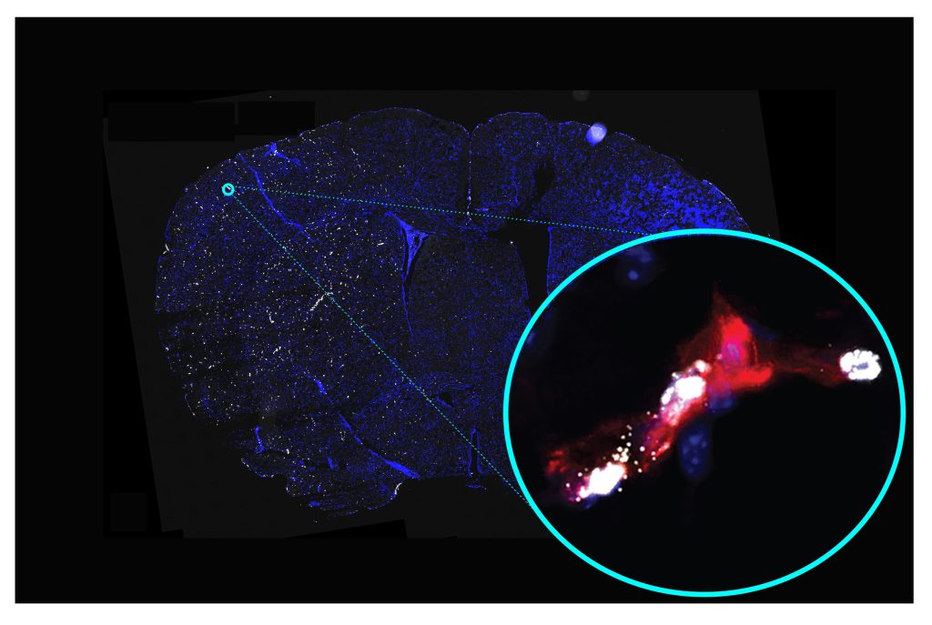
By intermittently giving mice the inflammation-causing bacterial toxin lipopolysaccharide, a recent animal study examined the effect of inflammation brought on by recurrent microbial infections on cognitive function.

The study discovered that early middle-aged mice with mild-to-moderate inflammation brought on by repeated lipopolysaccharide treatment had deficiencies in memory and learning.

These results in ageing mice imply that mild-to-moderate illness brought on by microbial infections may require more vigorous therapy than the existing standard of care, particularly in populations susceptible to cognitive impairment, such as the elderly.

The standard medical advise for those with mild to moderate infections is to obtain enough rest and drink more fluids. It’s interesting to note that a recent study in the journal Brain, Behaviour, and Immunity claims that repeated inflammation brought on by giving a bacterial toxin to middle-aged rats led to cognitive deficiencies. Along with these cognitive deficiencies, the hippocampus, a part of the brain important for learning and memory, experienced changes in the plasticity of its neurons.

Elderly people are more prone to microbial infections, which could exacerbate the decline in cognitive function in elderly people and result in dementia or moderate cognitive impairment.

The results of this study point to the possibility that older persons may require more intensive therapies in order to avoid these illnesses’ long-term consequences on cognitive function. It is crucial to remember that since this study used a mouse model, it is unknown whether the conclusions apply to people as well.

Brain aging contributed by inflammation

As a result of the biological changes brought on by brain ageing, a loss in several cognitive capacities is shown during the course of normal ageing. Similar to this, cognitive impairment seen in neurodegenerative diseases like Alzheimer’s disease is connected to pathological brain ageing.

According to studies, several variables, including inflammation brought on by microbial infections, may accelerate the aging of the brain. For instance, higher lifetime exposure to infectious agents is linked to worse cognitive function and a faster decline in cognitive function in older people.

Additionally, research using animal models has demonstrated that inflammation brought on by microbial exposure might affect cognitive function. Animals are injected with the toxin lipopolysaccharide (LPS), which is found in the outer membrane of gram-negative bacteria, to study the effects of inflammation brought on by microbial infections.

These investigations on animals have demonstrated that LPS injection can elevate cytokine levels in the brain, a family of inflammatory proteins, and result in abnormalities in cognitive function. Additionally, as people age, these negative effects of LPS become more obvious.

The majority of these studies have looked at how LPS affects brain and cognitive function when it is given continuously or in single doses. According to these research, even a single dose of LPS-induced inflammation can alter the brain permanently.

However, little study has been done on the effects of lifetime exposure to microbial illnesses on alterations in the brain and cognitive function. In genetically modified mouse models of Alzheimer’s disease, there is some indication that repeated injection of LPS can raise the risk of cognitive impairment.

EFfects of LPS-induced inflammation

In the current investigation, mice were given escalating dosages of LPS every 15 days for 2.5 months. Repeated injection of the same amount of LPS causes tolerance to develop, which includes the absence of an inflammatory reaction, according to prior studies.

The researchers worked around this problem by gradually increasing the LPS dose over the course of the five injections. The mice experienced mild illness after each LPS treatment, but they recovered within 15 days.

Two weeks after giving the last dosage of lipopolysaccharide, the researchers then tested the animals’ behaviour to see how well they could think. At 5–6 weeks after the last injection, the researchers also put the animals to death to look at how inflammation brought on by lipopolysaccharide affected the brain.

Saline-treated mice made comprised the control group. The mice were 10 months old when the study began, which corresponds to the transition from late adulthood to middle age.

The scientists discovered that mice given LPS injections displayed cognitive abnormalities in learning and memory retention of information acquired the day before.

Researchers observed alterations in the hippocampus in mice who received LPS injections on a sporadic basis. In Alzheimer’s disease, the hippocampus, which is important for memory and learning, exhibits the illness’s initial signs of degradation.

Interleukin-6 (IL-6) gene expression was upregulated in the hippocampus of LPS-treated mice as one of these modifications. This is in line with other research that found higher IL-6 levels following LPS treatment in cognition-related brain areas.

Additionally, the researchers discovered that giving LPS affected neuronal plasticity but not baseline signal transmission. Particularly, the LPS-treated animals displayed reduced long-term potentiation (LTP) between neurons in the hippocampus.

Linking flu frequency to dementia risk

Dr. Elizabeth Engler-Chiurazzi, a behavioral neuroscientist at Tulane University and co-author of the study, said the conclusions have significant ramifications for human brain health and disease.

“At the moment, staying in bed, getting lots of rest, drinking soup, and allowing your body to do its work of eradicating the infection is the standard of care for the common cold or the flu virus. According to my knowledge, this advice is offered to the entire population and is given regardless of the likelihood that dementia will develop in the future.

Dr. Elizabeth Engler-Chiurazzi stated, “Our results may be the first in a series of studies that could indicate that treatment for the common cold or other sources of intermittent infection among patients at high risk for cognitive decline/dementia may need to be more aggressive than the standard recommendations of rest and fluids.”

According to these findings, a history of more “flu-like” episodes may be a risk factor for cognitive problems in later life. According to Dr. Engler-Chiurazzi, some research conducted in people have started to investigate this connection. The results are consistent with our mouse observations.

It’s too soon to say for sure.

Dr. Engler-Chiurazzi issued a warning, noting that it’s possible that these findings won’t apply to people.

Although there are significant species differences in how these systems react, the immune system composition of mice and people is similar, according to the researcher. However, more research is required to determine how well these findings are replicated in human populations.

The mechanisms behind the cognitive losses following repeated LPS treatment will also be looked at by the authors.

Dr. Engler-Chiurazzi stated, “An immediate next step for our group is to repeat these studies and determine the extent to which common brain consequences seen in dementia, such as a leaky blood-brain barrier or activation of brain immune cells (microglia), are observed after repeated intermittent exposure to sickness-like inflammation in the body.”

Dr. Engler-Chiurazzi also mentioned that they have not looked at how viral infections affect cognitive function but are now doing so using an animal model.

REFERENES:

For Dementia medications that have been suggested by doctors worldwide are available here https://mygenericpharmacy.com/index.php?cPath=77_478

Colder temperatures may help clear linked Alzheimer’s.

Colder temperatures may help clear linked Alzheimer’s.

For many years, it has been known that lower temperatures encourage longevity in various animals.

The exact mechanism underlying this has remained a mystery, but recent study indicates low temperatures can trigger a biological process that enhances the removal of protein aggregations that are misfolded.

Alzheimer’s disease and other neurodegenerative disorders are among the aging-related diseases that have been linked to misfolded proteins.

The identification of this mechanism may improve our knowledge of how to treat human disorders brought on by protein misfolding.

Since more than 50 years ago, it has been recognised that lower temperatures make people live longer, but the exact mechanisms by which this is true have remained a mystery.

In recent years, researchers have uncovered the mechanism underlying the extended worm lifespan and demonstrated how it affects human cells.

Researchers from Cologne, Germany, published their findings in the journal Nature Aging and found that a molecule that breaks down protein clumps is more active at lower temperatures.

This activity may contribute to a decrease in the prevalence of dangerous misfolded proteins, which are thought to contribute to the development of a number of aging-related diseases like Alzheimer’s disease.

Transforming attitudes towards ageing

A few decades ago, it was thought that the buildup of poisons from oxidation was the cause of ageing. This changed in the 1990s once it was discovered that the genetic model organism C. elegans, a species of worm, could live longer at lower temperatures, according to Professor David Vilchez, the study’s primary author and director of the CECAD Research Center at the University of Cologne.

“We chose to concentrate on cold temperatures because it was recognized [more than 50 years ago] that cold temperatures can increase longevity Hence, it was discovered in flies, Drosophila proved in C. elegans, also proved in fish, and most recently proved in mice. Thus, it is actually one of the best methods for extending longevity for many people.

The potential for this discovery to offer insight on the mechanisms underlying ageing didn’t become obvious until the process’ genetic regulation was revealed in 2012 in a publication published in Cell, according to Prof. Vilchez.

There is a notion that claims that since 1860, human body temperatures have been declining by 0.03°C every decade, despite the fact that all of these research have been conducted on animal models. Others have linked this result to the fact that life expectancy has been rising since then, however this is just a correlation and the explanation has not been established.

Removing aggregations of improperly folded proteins

Researchers grew worms at 25oC and then relocated them to surroundings set at 15 degreeC, 20 degreeC, and 25 degreeC to study how cold temperatures affected the worms. They discovered that the activity of the molecule in charge of removing misfolded proteins from cells significantly increased at lower temperatures.

Subsequent investigation revealed that this was caused by the activation of a cell channel that functions at lower temperatures to promote the production of certain proteins implicated in the cellular process.

The quantity of misfolded proteins in worm cells was later shown to decrease at lower temperatures.

Researchers employed worms with changed genomes to mimic crucial aspects of two aging-related human disorders, Huntington’s disease and amyotrophic lateral sclerosis, in order to further examine this (ALS). These models were simple to create because, in contrast to multifactorial diseases like Alzheimer’s, each of these disorders are brought on by mutations in a single gene.

In worm models of Huntington’s disease and ALS, they discovered that the same mechanism was triggered at low temperatures and prevented the aggregation of improperly folded proteins.

Aid in the treatment of Huntington’s disease

An area of the Huntingtin gene, which codes for a particular amino acid, the building block of proteins, is repeated excessively in people with Huntington’s disease.

Dr. Natalia Pessoa Rocha, a researcher on Huntington’s disease at the University of Texas Health Science Center in Houston who was not involved in the study, described the polyglutamine tail that results from the extra amino acids on the protein. This might lead to proteins folding incorrectly.

Although the protein can fold, if it misfolds, it can collect into hazardous aggregates for cells. This is a very well-known mechanism in the aggregation of misfolded proteins that occurs in Alzheimer’s, Parkinson’s, and other neurodegenerative illnesses. Hence, this characterizes Huntington’s,” she stated.

Researchers discovered that lowering the temperature of human cells to 36 oC resulted in the same cell process being triggered, in addition to discovering that cold temperatures increase the clearance of misfolded proteins in worms.

At 35oC, however, the opposite was discovered, indicating that moderately low temperatures are the best for inducing this process in human cells. It was also discovered that overexpression of the genes in charge of this mechanism induced this pathway, which led to an increase in the molecules that help cells get rid of misfolded proteins.

In order to lessen the protein aggregation linked to sickness, etc., we were able to express the protein in human cells in vitro and truly simulate what happens at cold temperatures.

Future research on aging

Although it is a long way off, Prof. Vilchez stated that the results of this study may assist to direct future research towards treating neurodegenerative illnesses brought on by protein misfolding by identifying a molecule in the process that might be utilised as a medication target.

It is an extremely significant paper. They could pinpoint a crucial target for every disease caused by a protein problem. Not just Huntington’s, but particularly Alzheimer’s Parkinson’s, and other disorders, as I told you,” he said.

Nonetheless, there is still much to be done to convert this into human terms. People are almost always highly thrilled when a new target is there, so we need to be careful about how we convey this message, according to Dr. Pessoa Rocha. When human cells’ temperature was lowered to 36 °C, the same cell process was induced.

REFERENCES:

For Alzheimer’s disease medications that have been suggested by doctors worldwide are available here https://mygenericpharmacy.com/index.php?therapy=31

Perks of Psychotherapy for Individuals with Disabilities.

Perks of Psychotherapy for Individuals with Disabilities.

While some people with disabilities face major obstacles to accessing mental health care. Also, research demonstrates that this care can enhance their general quality of life.

More than 46% of American adults received counselling, medication, or self-directed treatment for mental health issues in just 2020. Millions of people receive therapy each year, making it one of the most successful types of mental health care.

But, therapy can do more than only treat mental health issues. It can also help people cope with life’s challenges, provide them a platform for self-expression. Also, generally improve their quality of life, especially if they have a disability.

In the sections that follow, we’ll look at how regular treatment can help persons with impairments as well as some of the obstacles and things to keep in mind for handicapped people seeking mental health care.

Barriers to treatment for disabled people

When seeking mental health treatments, disabled people frequently encounter more difficulties being treated. Some of these obstacles are caused by physical or financial constraints, while others are brought on by ableism and stigma.

The following are some of the obstacles that may keep people with disabilities from receiving the assistance they require:

Social stigma

People with disabilities can be negatively impacted by stigma in a variety of ways, especially because stigmas associated with mental health are equally prevalent.

People with disabilities who experience social stigma may experience poorer mental health and find it more challenging to obtain the assistance they require to seek mental health therapy.

Stigma against medical professionals

People with disabilities may find it challenging to receive the degree of treatment they require to get better due to social stigmas as well as stigma from healthcare professionals. The standard of care for people with disabilities suffers when medical practitioners let their own personal views about disabilities affect how they treat patients with impairments.

Financial limitations

It can be expensive to receive mental health care, especially if you have to pay out of pocket or don’t have insurance. Due to the fact that many disabled persons are unable to work and that those who do tend to earn less money, it may be difficult or even impossible for them to be able to afford therapeutic services.

Communication difficulties

There are more obstacles to getting access to mental health care for people with physical disabilities, and even for certain people with intellectual and mental impairments.

It might be difficult for some disabled persons to leave the house, especially if they don’t have access to transportation or accommodations. For disabled individuals who lack the equipment or expertise needed, even virtual care might be difficult.

People with disabilities may find it challenging to obtain the degree of care they require or to access mental health therapy due to all of these obstacles.

How can disabled people benefit from therapy?

The Centers for Disease Control and Prevention (CDC) report that persons with impairments experience mental anguish five times more frequently than adults without disabilities. Yet studies have repeatedly demonstrated that having a disability can significantly impact one’s mental health, particularly in terms of depression and anxiety.

In particular those that come along with the experience of living with a disability, therapy can help you better understand your own thoughts, emotions, and behaviours.

To improve your emotional well-being, therapy can show you how to identify and alter the connection between those thoughts and feelings. And treatment can provide you with the abilities you need to deal with and adapt to life with a disability, whether you have a physical impairment, a mental disability, or both.

Considerations for disabled people in therapy

When you have a disability, it’s crucial that your therapist is aware of the impact it might have on your life not just physically and mentally, but also socially, professionally, and in other ways.

Researchers in the profession identified a number of areas in 2014 that are crucial for therapists to take into account while working with clients who have disabilities. Many instances include:

  • respecting their knowledge of the impairment, how it affects them, and how they have experienced it
  • taking careful to speak about a person’s disability while using inclusive language
  • assisting in the identification and understanding of potential challenges faced by disabled people
  • incorporating the individual’s strengths into the therapy’s structure
  • adjusting goals and recommendations to accommodate their disabilities

The most crucial thing for you to do if you’re a therapist who deals with clients who have impairments is to keep learning about what it’s like to have that handicap and how it affects daily life.

Disability and intersectionality

In terms of oppression and discrimination, intersectionality refers to the way that a person’s different identities, such as gender, race, sexual orientation, and others, interact with one another to produce various, personal experiences.

Intersectionality is a major cause of many of the treatment-related obstacles that persons with disabilities face.

For instance, there will be more difficulties in obtaining mental health services for a disabled person who lacks health insurance due to a lack of resources. In addition, additional characteristics like gender and colour may exacerbate the prejudice and obstacles that disabled individuals must overcome in order to access services or get care.

Best psychological techniques

Many treatment modalities can be useful for people with all kinds of disabilities, including those that are physical, mental, intellectual, and others.

For instance, one of the most prevalent causes of disability in the United States is chronic pain, and numerous studies have looked into the advantages of psychotherapy for chronic pain.

According to a review, psychotherapy modalities like cognitive behavioural therapy (CBT) and cognitive functional therapy (CFT) can help persons with chronic pain live better overall.

These methods not only aid in a greater understanding of the disease that affects a person, but they also aid in coping mechanisms and lessen the emotional anguish that comes with having these conditions.

Moreover, therapy may even lower levels of handicap for those with mental health issues that are disabilities by easing generalised symptoms of the condition. Obsessive-compulsive disorder (OCD), for instance, is a serious mental health problem that can seriously impair a person’s capacity to function in day-to-day life.

Exposure and response prevention (ERP) and cognitive behavioural therapy (CBT) have both been demonstrated to be effective psychotherapy techniques for treating OCD. Both techniques also show promise in lowering impairment and improving quality of life for OCD sufferers.

Key message

In the United States, more than 60 million individuals struggle to fully participate in daily activities because they have a disability.

Also, a variety of variables may have an impact on a person with a disability’s mental health. These issues include lack of support and knowledge about living with a disability, social and professional stigma, increased care access hurdles, and increased barriers to care.

Therapy can help if you have a handicap since it can enhance your mental health, teach you crucial coping mechanisms, and provide you the care you require to feel supported.

REFERENCES:

For Alzheimer’s disease medications that have been suggested by doctors worldwide are available here https://mygenericpharmacy.com/index.php?cPath=77_478
New clues about how ketamine could lead to psychosis.

New clues about how ketamine could lead to psychosis.

Researchers looked into whether ketamine could cause mental changes like psychosis. They discovered that ketamine increases ambient noise, which may obstruct the brain’s ability to process sensory signals.

As rats were used in the study, more research is required to determine whether the results apply to people. Changes in reality perception, such as persistent delusions, hallucinations, and disorganized thought, are characteristics of schizophrenia. Almost 24 million people around the world are affected by the illness.

There is still no known cause for schizophrenia. Yet, research points to environmental, psychological, and genetic variables as potential causes of the illness.

By blocking NMDA receptors in the brain, the drug ketamine causes a mental state resembling psychosis in healthy humans. As a result, the central nervous system develops an imbalance of excitatory and inhibitory signals, which impacts sensory experience.

According to experts, schizophrenia-related perception abnormalities may be related to similar changes in NMDA receptors. Yet, it hasn’t been made clear how this might be the case.

Ketamine and psychosis

Recently, scientists investigated how ketamine alters sensory perception in rat brains.

They discovered that ketamine increased “background noise” in the brain, which reduced the clarity or intensity of sensory information. They remarked that this might help to explain why persons with schizophrenia or psychosis experience reality differently. The European Journal of Neuroscience published their findings.

These results, according to Dr. Sam Zand, a Las Vegas-based psychiatrist who was not involved in the study, “indicate that malfunction in NMDA receptors may play a role in the development of psychosis.”

“The work offers fresh understandings into the process by which ketamine may cause psychotic symptoms. The results might influence the creation of novel medications for psychosis that target NMDA receptors or brain noise, the researcher continued.

Study design

Seven male lab rats were used in the study to examine how ketamine affected their ability to perceive sensory information. To do this, they first implanted electrodes into rats’ brains to capture electrical activity.

They then recorded the brain’s reactions before and after administering ketamine while simulating their own whiskers. To be more precise, the scientists studied how ketamine affected beta and gamma oscillations in a neuronal network. They carries messages from sensory organs to the brain.

Gamma waves have a frequency range of 30-80 Hz, while beta oscillations have a frequency range of 17-29 Hertz (Hz). Processing sensory data requires the use of frequencies.

In the end, the scientists discovered that even before they stimulated the rats’ whiskers, ketamine enhanced power in both beta and gamma oscillations.

However, they also discovered that the amplitude of the rats’ beta and gamma oscillations dropped post-stimulus and after ketamine administration, which is associated with hampered perception.

They also observed that ketamine enhanced gamma frequency noise, which is related to a reduced capacity for sensory signal processing.

The researchers hypothesised that their findings suggest that increased background noise, which in turn may be brought on by damaged NMDA receptors leading to an imbalance of inhibition and excitation in the brain, may be a trigger for the distorted reality experienced in psychosis and schizophrenia.

According to Dr. Sofya Kulikova, senior research fellow at the HSE University in Perm, Russia, and one of the study’s authors, “The discovered alterations in thalamic and cortical electrical activity associated with ketamine-induced sensory information processing disorders could serve as biomarkers for testing antipsychotic drugs or predicting the course of disease in patients with psychotic spectrum disorders.”

Research limitations

The study was not conducted by Dr. Howard Pratt, a psychiatrist and mental health medical director at Community Health of South Florida. He made it clear that:

The main drawback of these results is that, although a strong association, causation has not yet been shown. There are many potential explanations for conditions like psychosis, including increases in dopamine, which is the focus of treatment for those with a diagnosis of schizophrenia. I’m interested to see what happens as the investigation expands past animal studies.

We also discussed the study’s shortcomings with Dr. James Giordano, the Pellegrino Institute professor of neurology and biochemistry at Georgetown University Medical Center who was not engaged in the study.

The fact that the study solely looked at ketamine-induced effects, he said, “is a key limitation. While valuable and practical for understanding ketamine’s activity in a rat model, it may not provide direct translation to comprehend non-drug-induced dissociative, and psychotic states in humans.”

Dr. Giordano went on to say that it is possible that the effects of ketamine on humans, while undoubtedly dissociative and exhibiting some psychotic traits, are not entirely representative of or identical to the neurological mechanisms underlying other forms of psychosis and schizophreniform disorders.

Possible clinical repercussions

The study’s ramifications, according to Dr. Giordano, are that “[t]hese findings are useful in that evidence of ketamine’s actions at defined brain networks may enable better understanding— and improved clinical applications—of its effects in humans.”

The researcher added, “In addition, by highlighting the functions of these brain nodes and networks involved in mediating dissociative experiences, we may create improved understanding — and possibly treatments for — specific types of drug-induced psychoses, and perhaps other psychotic illnesses, such as forms of schizophrenia, as well.

Larger Trials Needed

Dan Iosifescu, MD, MSc, associate professor of psychiatry at New York University School of Medicine in New York City, commented on the study and said that if the results “were based on a larger study” it would be very important because such patients are currently being denied access to a beneficial treatment due to a theoretical risk of psychosis.

A low risk of psychosis exacerbation following IV ketamine, according to Iosifescu, who is also the director of clinical research at the Kline Institute for Psychiatric Research in Orangeburg, New York, and was not involved in the study, is still possible given that the review is based on a small sample.

Veraart concurred, stating that “well-designed randomised controlled trials should be conducted to ascertain the efficacy, safety, and tolerability of ketamine in depressed individuals with a propensity to psychosis before administration on a large scale is pushed.”

The study received no particular funding. Outside of the submitted work, Veraart has received speaker honoraria from Janssen. Disclosures from the other authors are provided in the original publication. Iosifescu has advised clinics on the most effective ways to administer IV ketamine therapy as a consultant to the Centers of Psychiatric Excellence.

REFERENCES:

For Mental disease medications that have been suggested by doctors worldwide are available here https://mygenericpharmacy.com/index.php?cPath=77_478

Amino acid: A key in improving depression treatment.

Amino acid: A key in improving depression treatment.

The role of a receptor called GPR158 in depression was discovered by researchers at the University of Florida over the course of a 15-year investigation.

In a study, mice who had their GPR158 expression suppressed were less likely to experience stress-related sadness. The structure of GPR158 was discovered by the researchers, and this allowed them to connect it to the amino acid glycine.

Many drugs exist to treat depression, which affects millions of people, but it can be challenging to select the proper one.

Scientists at the Herbert Wertheim UF Scripps Center for Biomedical Innovation and Technology uncovered a finding that revealed how an amino acid is related to depression while studying neurotransmitters.

The revelation was the result of more than ten years of investigation into the functioning of brain cell signalling. Although though discovering a connection to depression was not the original aim of the study, the researchers are enthusiastic about their findings since they potentially influence how depression will be treated in the future.

Depression

In the United States, depression affects over 21 million adults each year, according to the National Institute of Mental Health (NIMH). The COVID-19 pandemic saw a sharp increase in depression rates, which have since persisted and are now a serious issue. Rates among children under 18 are also rising.

While some people suffer situational depression as a result of external factors (such losing a loved one), other people struggle with depression for extended periods of time and it can progress into Major Depressive Disorder.

The NIMH includes the following indications and symptoms of depression among others:

  • recurring sense of sadness
  • feeling a sense of emptiness
  • experiencing a decline in energy or weariness
  • having difficulties sleeping
  • feeling suicidal thoughts

Individuals who have persistent depressive symptoms may require medical attention. To treat the symptoms of depression, doctors may give drugs, offer treatment, or advise lifestyle modifications.

Tricyclic antidepressants (like imipramine or amitriptyline), selective serotonin reuptake inhibitors (like sertraline or escitalopram), and serotonin-norepinephrine reuptake inhibitors are a few examples of depression drugs (such as duloxetine or venlafaxine).

Those on antidepressants should consult with their healthcare providers frequently and inform them of any suicidal thoughts because these medications can have negative effects, including thoughts of suicide.

Brain cell receptors and depression

The goal of the writers was not to find a connection to depression at first. 15 years ago, they set out to study how brain cell receptors function, and they have achieved that goal.

Prof. Kirill Martemyanov, one of the study’s authors, stated: “Fifteen years ago we discovered a binding partner for proteins we were interested in, which led us to this new receptor.” “We’ve been unravelling this for a while now.”

Professor Martemyanov teaches at the University of Florida Health’s Department of Neurology.

The next several years saw the discovery of the GPR158 receptor by the researchers. In experiments with mice, they discovered that a mouse would be more resistant to stress-induced depression if that receptor was suppressed in the body.

Amino acid treating depression

A 2017 assessment of the literature concluded that glycine is “one of the most significant and straightforward non-essential amino acids in humans, animals, and many mammals.”

The scientists were shocked to learn that GPR158 was an inhibitor when they realised that glycine was the signal’s sender and that it binds to glycine; they changed its name to mGlyR as a result (metabotropic glycine receptor).

The identification of mGlyR should pave the way for fresh investigation into the management of depression, which Prof. Martemyanov intends to do.

Expert responses

Medical News Today spoke with Dr. Simon Faynboym, a physician who has experience working with the American Psychiatric Association.

Dr. Faynboym stated that the findings “essentially shows that glycine can interact with the GPR158 system.” The researchers demonstrate a biochemical mechanism, but the most significant lesson is that this process may represent the link between glycine and taurine’s potential antidepressant qualities.

Currently serving as a delegate to the California Medical Association is Dr. Faynboym. Dr. Faynboym acknowledged the significance of the study but also emphasized that depression is “very complex” and that other neurotransmitters are involved.

Dr. Faynboym remarked that there are numerous factors at play while dealing with depression. “Depression involves several neuronal networks, various neurotransmitters leaving and entering neurons at various rates, and impacts all areas of the brain. Because of the dynamics of the brain, one of the most complicated medical specialisations is mental health.

Dr. Faynboym emphasised the significance of this kind of research in light of this. Since they provide another glimpse behind the curtain of the big unknown that is the brain, research pieces like this one advance the science of psychiatry.

A psychiatrist from Palm Beach Gardens, Florida named Dr. Jessica Turner also discussed the study’s findings.

The medial prefrontal cortex is a well-known brain region associated with depression, and this work suggests targeting a specific receptor there, according to Dr. Turner. “We anticipate providing better, more efficient alleviation for people who are depressed in the future with more focused treatments.”

REFERENCES:

For more details, kindly visit below.

Common Dry-Cleaning Agent May lead to Parkinson’s Disease

Common Dry-Cleaning Agent May lead to Parkinson’s Disease

Trichloroethylene (TCE), a chemical, has been linked by some researchers to Parkinson’s disease. Dry cleaning, degreasing, and even decaffeinating coffee have all been common uses for TCE.

According to recent study, the chemical’s capacity to reach the brain and harm cell mitochondria may be the root of the problem.

The authors claim that the chemical’s influence might be “enormous” given how commonplace it is in the environment. TCE should be outlawed, and people should be shielded from more exposure, as suggested alternatives.

According to the Parkinson’s Foundation, the substania nigra, a region of the brain that includes cells that create the neurotransmitter dopamine, is affected by Parkinson’s disease, a neurodegenerative condition.

Parkinson’s disease patients endure tremors, slowness of movement, limb stiffness, and balance issues.

Muhammed Ali and Michael J. Fox are two well-known public celebrities who suffer with the illness.

According to the authors, up to a third of the groundwater in the United States has TCE pollution. The chemical is additionally present in Camp Lejeune, a Marine Corps base, and 15 Superfund sites in Silicon Valley.

What is trichloroethylene (TCE)?

TCE is a chemical that is a colourless liquid that does not exist in nature. It is well known to smell like chloroform.

This substance can be found in a wide range of goods and businesses, such as:

  • industry-wide dry cleaning
  • metal scrubbing
  • wiping cloths
  • carpet and garment stain removers
  • lubricants
  • aerosol adhesives

Using TCE-containing products or working in a TCE-containing plant are two ways that people can be exposed to the chemical.

TCE can also contaminate our air, water, and food and drink by leaking into the soil, water, and air near where it is used or disposed of. High levels of TCE exposure can cause the following symptoms:

  • dizziness
  • headaches
  • confusion
  • nausea
  • facial paralysis

Link between TCE and Parkinson’s disease

According to the study, which was published in the Journal of Parkinson’s Disease, there may be a “invisible” cause of Parkinson’s disease that is related to the widespread use of TCE.

Dr. James Beck, Chief Scientific Officer of the Parkinson’s Foundation, commented on the study, saying, “We have known for some years that TCE exposure and Parkinson’s disease are related. I believe that this opinion piece effectively highlights the risks associated with TCE exposure.

The researchers’ findings are discussed in the study together with the evidence tying TCE to Parkinson’s disease.

TCE is lipophilic, as the scientists explain in their research, which means that it has a propensity to dissolve in fatty tissues. This makes it simple for it to enter the brain and other bodily tissues where it can wreck havoc with cell mitochondrial function. This sort of toxin is extremely toxic to dopamine-producing cells, which may help to explain how exposure to TCE might cause Parkinson’s disease.

Seven other people, including the late Senator Johnny Isakson, are also profiled. Isakson employed TCE to degrease aircraft during his time in the military; as a result, he eventually developed Parkinson’s disease as well as renal cell carcinoma, a cancer associated to TCE exposure.

The matter was initially brought to his notice, according to co-author Dr. Ray Dorsey, when his colleague, Dr. Caroline Tanner, told him about the exposures at Camp Lejeune.

TCE poses a “enormous” risk to the general public’s health, he claimed. “At one time, it was used by 10 million Americans, including printers, embalmers, mechanics, dry cleaners, chip manufacturers, engineers, painters, metal workers, pilots and others. It has been absorbed into the environment by millions more people.

Avoiding exposure to TCE

The issue of TCE contamination, according to Dorsey, requires a few actions. It must be prohibited first, along with tetrachloroethylene (PCE), another industrial solvent.

Second, home remediation systems such to those used for radon must be deployed to alert and protect persons who are at danger of exposure.

The connection between TCE and Parkinson’s disease, he added, has to be explored further through research. People can be exposed to TCE through contaminated soil, food, water, air, or direct skin contact, according to the U.S. Centers for Disease Control and Prevention.

You are most likely to become exposed by drinking polluted water, but you can also become exposed through the air as it is released from contaminated water, as a third of all groundwater may be affected.

Working in a sector where TCE is manufactured or utilised, like the degreasing business, is a significant additional method that you could be exposed to it. It can enter the body by coming into direct touch with the skin or by inhaling the vapours.

You may also be exposed through contaminated soil, such as that found in landfills. Moreover, TCE can enter your body through the consumption of contaminated foods or contact with consumer products that contain it.

According to the CDC, TCE is a common solvent used in a variety of products, including cold metal cleaners, adhesives, lubricants, paints, varnishes, paint strippers, and paints. Labels for these goods should include instructions on how to reduce exposure.

The Occupational Safety and Health Administration (OSHA) also mandates that your company give you a material safety data sheet (MSDS) outlining the dangers and proper handling techniques for any chemicals you use at work.

When exposed to TCE

A doctor can check for TCE in your blood, breath, or urine if you’ve recently been exposed to the substance, according to the Agency for Toxic Substances and Disease Registry (ATSDR).

It can also be tested for in the environment you have visited. But, once you’ve been exposed, there is no cure to get it out of your system. Either your breath will expel it or your kidneys will excrete it into your urine.

They advise avoiding any known toxic regions, such as those with tainted water, soil, or air.

You should always use items containing TCE in well-ventilated areas with the appropriate personal protective equipment, such as chemical-protection gloves, safety goggles, and respirators, in addition to adhering to any safety advice on such products.

High TCE exposure can, in the short term, irritate people and even make them pass out or become fatally ill. If you have been exposed to the chemical extensively, it is crucial to seek immediate medical care.

The individual should also be transferred to clean air, and any contaminated clothing should be taken off, if it’s safe to do so. If the skin or eyes have been exposed, thoroughly rinse them with water.

Kidney cancer and Parkinson’s disease have both been associated with long-term TCE exposure. The ATSDR advises that the greatest form of protection is to shield yourself from exposure altogether.

REFERENCES:

For more details, kindly visit below.

Relation between native language and way of thinking.

Relation between native language and way of thinking.

Although though some key brain areas are involved in the processing of all languages, these regions exhibit varied activity patterns when processing other languages.

According to the various brain activation patterns seen when processing particular languages, people who speak different native languages would likely have diverse brain structures. According to a recent study, the wiring of the brain regions responsible for language processing differed between native Arabic and German speakers.

These findings imply that learning one’s native language during childhood changes the connections in the brain and may help to explain why people’s ability to think differently depending on their original language.

The native tongue of a person can influence how they think. For instance, Russian has two different terms that distinguish between bright and dark blue, whereas English only has one word for hues in the blue spectrum.

Intriguingly, native Russian speakers typically perform substantially better than native English speakers in examinations requiring the differentiation of light and dark blue. Similar findings have been made about how different native languages express directions and how well-rounded a person’s sense of orientation is.

There are changes in the wiring of language processing regions in the brains of native Arabic and German speakers, according to a recent study published in the journal NeuroImage.

This raises the possibility that acquiring one’s native language as a youngster could alter the brain’s structural makeup, potentially explaining the disparities in cognitive function between people who speak various first languages.

Language processing in the brain

Throughout the years, research has established that a complex network of interconnected brain regions, primarily in the left side or hemisphere of the brain, processes and produces language. Moreover, research has shown that specific brain regions are linked to processing certain components of language, such as semantics (word meaning), syntax (grammatical structure), and phonology (sounds associated with language).

Brain areas in the left hemisphere are principally where syntactic and semantic processing takes place. However, other language-related information, particularly that pertaining to sounds and intonation, is either processed in the right hemisphere or both of them.

In the world, there are close to 7,000 different languages spoken. Individuals with different native languages display varied patterns of brain activation during language processing, despite the fact that the same basic brain regions are engaged in the comprehension and production of all languages.

The brains of people who speak different native languages are structurally different, according to studies. This shows that learning one’s native language throughout childhood causes differences in the brain’s structure from individuals who speak a different first language.

There are two types of brain tissue: grey matter and white matter. The processing of information takes place in the grey matter, which is made up of the cell bodies of neurons or nerve cells. Axons and dendrites, which transmit information from one neuron to the next, make up white matter, on the other hand.

Gray and white matter density in the regions involved in language processing differs between English and Chinese speakers, according to a prior study. In a similar vein, research has revealed variations in the white matter tract patterns connecting several brain regions in native English, German, and Chinese speakers.

Differences in brain connectivity

Researchers compared the differences in the wiring of the brain areas responsible for language processing in speakers of Arabic and German in the current study. These languages were chosen by the researchers because of how differently semantics and syntax are used in each.

First of all, these languages are not related to one another; German is an Indo-German language, while Arabic is a Semitic language. Arabic is more pronounced semantically than German, which has a more complicated grammatical or syntactic framework.

Arabic words frequently lack short vowels, unlike German, and their pronunciation and meaning depend on their context. In addition, German script is written from left to right, whereas Arabic script is written and read from right to left.

Studies have revealed increased syntactic processing activity during German comprehension, which is consistent with this. Others, however, have demonstrated a larger engagement of semantic processing regions during Arabic comprehension. In these investigations, individuals analyse a specific language while magnetic resonance imaging (MRI) is used to look at brain patterns.

Left and right hemispheres connection

The purpose of the current investigation was to determine whether native Arabic and German speakers have structural differences in their brains at baseline as a result of the distinct demands that each language places on the brain.

The researchers specifically looked at whether baseline brain connectivity patterns differed between native German and Arabic speakers. Diffusion-weighted MRI was utilised to find the white matter tracts that link the various parts of the brain.

Both groups’ participants had increased connection in the left hemisphere, which is where language processing is predominately processed. Yet, compared to Arabic speakers, participants whose native language was German had better connections between brain areas involved in language processing in either hemisphere.

Native German speakers, on the other hand, displayed weaker connections between regions in the left and right hemispheres than native Arabic speakers. The right-to-left writing system and the semantic intricacy of Arabic may be to blame for the increased activation of both hemispheres.

Brain scans of German speakers also showed greater connectivity between left hemispheric brain areas involved in syntactic processing. In contrast, native Arabic speakers had stronger connections between the brain regions involved in semantic processing.

Can language change personality?

In conclusion, the current study demonstrates that lifetime exposure to one’s native language causes structural changes in the brain. Dr. Alfred Anwander, a neuropsychologist at the Max Planck Institute and the study’s author, stated:

“The study provides significant evidence for the impact of the environment and first language on brain development, particularly the shaping of the connections that directly affect how our brains process information during thought. This may indicate that unique personality is a product of life experiences rather than being “hard-wired” in our brains.

Dr. Anwander added that the study’s findings might be applied to develop strategies specifically for people with neurological disorders.

Childhood in especially, when the brain still exhibits a larger capacity for adaptation, may leave lasting imprints on our brains and lay the groundwork for the specialised nature of each unique brain. Dr. Alfred Anwander, study author, noted that this may have some implications for clinical situations where individualised treatment may be necessary, such as in neurological rehabilitation strategies, in addition to improving our understanding of how the brain processes various languages differently.

REFERENCES:

For more details, kindly visit below.

Dementia due to brain damage by high blood pressure?

Dementia due to brain damage by high blood pressure?

According to research, some aggressive blood pressure medicines can benefit brain health. As per them, the therapies function by opening up neural pathways in the perivascular regions of the brain.

According to experts, a balanced diet, regular exercise, enough sleep, and avoiding overstimulation are all important components of maintaining excellent brain health. Researchers may be able to detect people with severe cognitive impairment with the aid of additional research.

Dementia, a general term for a multitude of illnesses affecting memory loss and cognitive deterioration, affects more than 55 million individuals worldwide. Scientists are unsure of the exact aetiology of dementia, but they do know that certain factors can affect whether or not someone gets dementia. High blood pressure is one of these.

According to earlier studies, those who have high blood pressure are more likely to experience dementia, including Alzheimer’s disease.

Researchers from the University of Edinburgh in the United Kingdom and Jagiellonian University Medical College in Krakow, Poland, have now identified the precise regions of the brain that may be harmed by high blood pressure and are connected to the onset of dementia.

What is blood pressure? 

The amount of power the heart exerts to pump blood through the arteries is measured as a person’s blood pressure.

The doctor gets two distinct readings while taking your blood pressure. The highest figure reflects the systolic pressure experienced as the heart pumps blood into the arteries. The diastolic pressure, which the heart experiences between heartbeats, is represented by the bottom number. They are both expressed in millimetres of mercury (mmHg).

For instance, the systolic and diastolic pressures of normal blood pressure are both less than 120 and 80 mmHg, respectively (but more than 90mmHg systolic and 60mmHg diastolic).

High blood pressure is seen as beginning when the systolic and diastolic pressures both rise to 130 and 80, respectively. Stage 2 high blood pressure is defined as systolic and diastolic pressures of at least 90mmHg and 140mmHg, respectively.

High blood pressure

Hypertension, also referred to as high blood pressure, develops when the amount of effort required for blood to flow through the arteries is too great.

This may occur if plaque inside the artery walls, which contains cholesterol, causes the arteries to become damaged or narrower.

A person’s chance of acquiring high blood pressure may be increased by a number of variables, such as:

  • unsound diet
  • obesity
  • inactivity
  • Using tobacco or alcohol
  • genetics
  • certain illnesses, like diabetes

How does high blood pressure affect the brain? 

The lead author of this study and professor of cardiovascular medicine at Jagiellonian University Medical College in Krakow, Poland, and the University of Edinburgh in the United Kingdom, Prof. Tomasz Guzik, believes that having high blood pressure can negatively impact the structure and operation of the brain in a variety of ways.

“For example, hypertension can have a serious influence on your brain’s blood vessels, causing them to change in shape, stiffen, and form clogged arteries. “Small vessel disease” is a condition that results from the malfunction of smaller blood vessels in the brain caused by high blood pressure, which is passed from bigger to smaller blood vessels.

“High blood pressure not only impacts the flow of blood to the brain, but it also speeds up pathological processes including neurodegeneration and inflammatory activationTrusted Source. Dementia, memory loss, and other cognitive impairments are all caused by these conditions, said Tomasz Guzik, Ph.D.

Furthermore, according to Prof. Guzik, excessive blood pressure might harm the brain’s white matter.

He said that damage to this area can result in poor cognitive performance and raise the risk of stroke. “The white matter is formed of nerve fibres that transfer information across different brain regions,” he said.

According to him, this emphasises how crucial it is to keep blood pressure under control in order to avoid white matter damage and the accompanying cognitive problems.

Blood pressure study

The individuals were split into two groups by the scientists for their analysis. With a goal of 120 mm Hg systolic pressure, 243 patients in one group had highly intense treatment for high blood pressure.

The second group, consisting of 199 individuals, received routine care with a 140 mm Hg goal. Pre- and post-study MRIs were used to compare the number of perivascular spaces in each participant’s brain.

The innermost layer of the brain contains spaces filled with cerebrospinal fluid known as Virchow-Robin spaces, or perivascular spaces (PVS), according to Dr. Sandra Narayanan, a vascular neurologist and neuro-interventional surgeon at the Pacific Stroke & Neurovascular Center at Pacific Neuroscience Institute in California.

They may get bigger as a result of neurodegenerative processes, demyelinating diseases, inflammation, or ageing. They aid in clearing the brain of metabolic waste and water, she said.

The American Heart Association states that a normal blood pressure reading is less than 120/80 mm Hg. Elevated blood pressure is defined for adults as 120 to 129 mm Hg for the systolic pressure (the top number) and less than 80 mm Hg for the diastolic pressure.

High blood pressure is defined as a persistently high systolic pressure of 130 mm Hg or higher, or a diastolic pressure of 80 mm Hg or higher.

Hypertension-dementia link

Thousands of participants from the UK Biobank, COGENT, and the International Collaboration for Blood Pressure provided observational data for this study, which Prof. Guzik and his team conducted using a combination of brain magnetic resonance imaging (MRI), genetic analysis, and observational data.

After examination, scientists discovered alterations in nine regions of the brain connected to both deteriorating cognitive function and elevated blood pressure.

The putamen, which is in charge of learning and motor control, is one of these regions. Alzheimer’s disease and putamen dysfunction are related, according to earlier study.

White matter regions, the anterior thalamic radiation, the anterior corona radiata, and the anterior limb of the internal capsule were also affected by high blood pressure. 

Both basic and complicated behaviours are planned and carried out by the anterior thalamic radiation, whereas the anterior corona radiata supports decision-making and emotion regulation. Moreover, the internal capsule’s anterior limb supports motivation, decision-making, and cognitive processing.

What you can do to improve brain health?

According to Narayanan Sajd, “many risk factors for neurodegenerative illnesses, such senior age, tend to be progressive.” There are significant potential clinical consequences on quality and quantity of life for these debilitating and pervasive illnesses if some of the associated clinical aspects are changeable.

According to the study’s findings, maintaining a healthy blood pressure level is crucial for maintaining a healthy brain.

The Mediterranean diet, which is low in fat and abundant in fruits and vegetables, is one example of a nourishing diet that the Alzheimer’s Association recommends. It’s also crucial to challenge your intellect by learning something new.

Behavioral scientist, relationship expert, and developer of the Happiness Hypothesis Method Clarissa Silva advises incorporating the following into your life:

  • Exercise. Daily aerobic activity for at least 30 minutes encourages the growth of new neurons in the brain.
  • Sleep. Regular sleeping patterns prevent the onset of brain fog and can make it worse throughout the day.
  • Overstimulation. Work productivity may be hampered by multitasking, which also adds to our brain’s ongoing overstimulation. Neuroplasticity in the brain is enhanced by concentrating on one task at a time and taking a break before moving on to the next one.
  • Unattended trauma. Trauma affects cognitive performance, self-esteem, and decision-making. Working with a qualified specialist to address trauma can start the process of reversing current cognitive impairment and preventing further degenerative disorders.

According to Silva, limiting screen time can aid in neuron regeneration. “Setting up a routine. At first thing in the morning and an hour before going to bed, try to cut back on checking your gadget. The constant overstimulation that we experience throughout the day needs to be repaired in our brains.

REFERENCES:

For more details, kindly visit below.