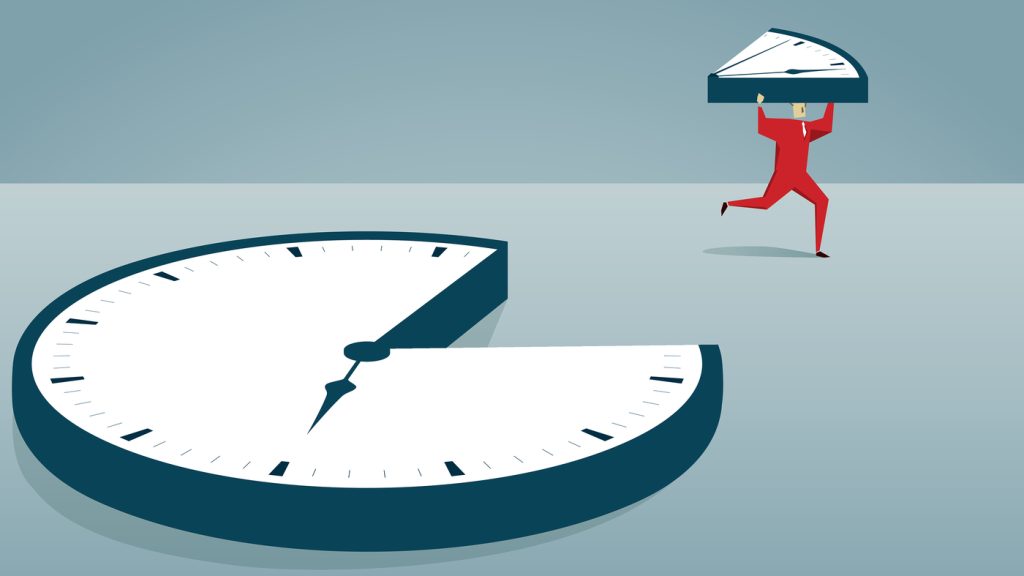
Browsed by
Author: blogadmin

Characteristics and prevention of emphysema with symptoms.

Characteristics and prevention of emphysema with symptoms.

A form of chronic obstructive pulmonary illness is emphysema (COPD). Lung air sacs deteriorate and stretch under these circumstances. This causes a persistent cough and respiratory problems.

Emphysema can be brought on by a variety of things, but smoking is by far the most common cause. Although there is no known cure, giving up smoking can better the prognosis.

Emphysema has been diagnosed in about 3.8 million Americans, or 1.5% of the total population. 7,085 persons (2.2 per 100,000) lost their lives to the illness in 2017.

What is emphysema?

A form of COPD is emphysema. Emphysema causes the air sacs and alveoli in the lungs to enlarge and the lung tissue to become less elastic.

The air sacs’ walls deteriorate, are damaged, shortened, collapsed, stretched, or overinflated. This implies that the surface area available for the lungs to get oxygen into the blood and expel carbon dioxide from the body is reduced. Although the damage is irreparable and permanent, there are techniques to treat it.

Causes and risk factors of emphysema

Emphysema affected 2 million adults in 2018, or 1.6% of those who are 18 years of age or older, according to the American Lung Association.

Males, non-Hispanic white persons, and people over 65 had greater rates than other groups. The disparity between the sexes has, however, been closing as female rates have been rising over the past few decades.

Emphysema is primarily caused by tobacco usage. The likelihood of getting emphysema increases with the amount of smoking you do. This includes cannabis smoking.

More over 480,000 Americans die as a result of smoking each year, and COPD, including emphysema, is to blame for 80% of those fatalities. Emphysema risk is also increased by exposure to secondhand smoke.

The following are other causes of and potential risk factors for emphysema development:

  • exposure to chemical vapours or lung irritants that are very polluting
  • Alpha-1 deficiency-related emphysema is an uncommon form of emphysema that is caused by the genetic disorder alpha-1 deficiency.
  • history of respiratory infections in children
  • a weakened immune system, particularly due to HIV
  • uncommon illnesses like Marfan syndrome.

Symptoms of emphysema

Emphysema’s main signs and symptoms include:

  • breathing difficulty or dyspnea
  • a persistent cough that is mucus-producing
  • breathing that makes a whistling or squeaky sound and wheezing
  • chest constriction

A person may initially experience these symptoms while engaging in physical activity. However, when the illness worsens, they may also begin to occur while you’re sleeping. COPD and emphysema both take time to develop.

Later on, an individual may have:

  • flare-ups and frequent lung infections
  • Wheezing, coughing, and shortness of breath are some of the symptoms that are getting worse.
  • reduction in weight and appetite
  • exhaustion and a decline in energy
  • Lack of oxygen can cause cyanosis, blue-tinged lips, or blue-tinged fingernail beds.
  • sleep issues with sadness and anxiety

How is emphysema diagnosed?

The first thing your doctor will do is ask you about your background and medical history, namely whether you smoke and whether you regularly come into contact with dangerous gases or pollutants at work or at home.

Emphysema can be found using a variety of tests, such as:

  • X-rays and CT scans are imaging procedures used to examine your lungs.
  • blood tests to assess the efficiency of your lungs’ oxygen transport
  • Using pulse oximetry, you can determine how much oxygen is in your blood.
  • lung function tests, which gauge how well your lungs carry oxygen to your bloodstream and how much air your lungs can take in and out,
  • tests to determine the amount of blood and carbon dioxide in your blood are known as arterial blood gas analyses.
  • ECGs are used to evaluate heart health and rule out heart illness.

Complications of emphysema.

Emphysema can develop into a serious condition if it is not adequately managed or treated. These may consist of:

  • either bacterial or viral pneumonia
  • many respiratory infections
  • failure of the right side of the heart is referred to as cor pulmonale.
  • When air gathers between the lungs and the chest cavity, it causes a pneumothorax, which can cause the lungs to collapse.
  • respiratory acidosis, or when the lungs are unable to get adequate oxygen, can result in coma
  • When the lungs can’t effectively oxygenate the blood, it’s called hypoxemia.

Emphysema treatment

Emphysema does not have a treatment. The goal of treatment is to lessen symptoms and stop the spread of the illness using drugs, therapies, or operations.

If you smoke, quitting is the first step in curing your emphysema. To help you quit smoking, you could require medicine. Think about going over a strategy with your doctor.

Medications

The disease can be treated with a number of drugs, including:

  • Breathing becomes easier and coughing and shortness of breath are reduced thanks to bronchodilators, which help open airways.
  • steroids, which reduce breathlessness
  • medicines, which combat infections that could worsen the situation

These drugs are all able to be breathed or given orally.

Therapies

By strengthening breathing muscles and reducing symptoms, pulmonary rehabilitation or light exercise like walking can improve breathing and make it easier to be physically active. Deep breathing techniques, yoga, and tai chi can all aid with symptom relief.

Breathing can be made simpler with oxygen therapy. People who have severe emphysema could require oxygen all the time.

Surgery

A lung transplant can replace the entire lung, and lung volume reduction surgery can be done to remove tiny portions of the diseased lung. Only those who have severe emphysema can undergo these uncommon operations.

Other therapies

You might lose weight if you have emphysema. It is advised to consume meals high in vitamins A, C, and E, such as fruits and vegetables, to boost your general health.

You can lessen your risk of contracting an infection that could aggravate emphysema by getting immunised against specific illnesses, such as pneumonia. These illnesses include COVID-19, influenza, and pneumonia.

If you don’t exercise as often as you used to, you can also feel anxious and depressed. You can connect with people who have the same ailment and go through similar experiences by joining a support group. This can make you more aware of the fact that you are not facing the sickness alone.

Perspective and prevention for Emphysema

Emphysema is primarily brought on by tobacco usage, thus quitting smoking is the best way to prevent it. Additionally, it’s critical to avoid exposure to toxic substances, gases, and areas with high pollution.

Depending on how severe their emphysema is, each person’s outlook is different. The condition has no known cure and just becomes worse with time, but you can stop it from getting worse.

Smoking cigarettes typically accelerates the condition, therefore stopping is crucial. Emphysema patients can experience life-threatening complications as their lungs and hearts deteriorate over time, making early disease detection crucial.

Maintaining good health requires a balanced diet and regular exercise. Emphysema can be managed with medications and treatments so that you can enjoy a long, healthy life.

REFERENCES:

  • https://www.healthline.com/health/emphysema
  • https://www.medicalnewstoday.com/articles/8934
  • https://www.mayoclinic.org/diseases-conditions/emphysema/symptoms-causes/syc-20355555
  • https://my.clevelandclinic.org/health/diseases/9370-emphysema

For more details, kindly visit below.

Causes and treatment for irritating Eye twitching.

Causes and treatment for irritating Eye twitching.

Myokymia, or twitching of the eyelids, can be brought on by dry eyes, eye irritation, eye strain, lack of sleep, or an excessive amount of caffeine. Eyelid spasms that are severe or persistent could be symptoms of various diseases.

Myokymia, often known as an eyelid twitch, is a recurrent, uncontrollable spasm of the eyelid muscles. Although it can happen in either the upper or lower lids, a twitch typically happens in the upper lid.

These spasms are typically minor and feel like a slight tugging on the eyelid. Some people may have a spasm that is severe enough to make both eyelids totally close. These spasms normally last for a minute or two and happen every few seconds.

Eyelid twitching episodes might happen at any time. It’s possible for the twitch to come and go for several days. After that, you might not twitch for several weeks or even months.

Although the twitches are usually mild and unharmful, you could find them annoying. Most spasms will go away on their own, rarely requiring medical intervention.

When they are accompanied by additional face twitches or uncontrollable movements, eyelid spasms can occasionally be a sign of a persistent movement problem.

Types of eyelid twitches

Three types of eyelid twitching can be distinguished:

  • General eyelid spasm
  • essential blepharospasm
  • hemifacial spasm

General eyelid spasm

Eyelid spasms occasionally are deemed normal and don’t always signify a major issue. These twitches can be caused by a number of environmental causes and typically go away when you rest. You might want to discuss your symptoms with your doctor if these twitches persist and interfere with your daily activities.

Benign essential blepharospasm

You can have benign essential blepharospasm, which is the terminology for persistent and involuntary winking or blinking, if the spasms become chronic (long-term).

Usually affecting both eyes, this illness is more prevalent in women than in men. Up to 50,000 Americans may be affected by it, and it often appears in middle to late adulthood. Over time, the condition is likely to get worse and may lead to:

  • hazy vision
  • increased light sensitivity
  • facial twitches

Hemifacial spasm

When only one eye is affected by the eyelid twitch, a hemifacial spasm may be present. This particular spasm is a neuromuscular problem that is typically brought on by a blood vessel placing too much pressure on a facial nerve.

In addition to being more prevalent in Asian people, this illness affects women more frequently than it does men. Untreated, it could result in:

  • eye twitching that occurs often and without control
  • not being able to open your eye
  • your entire facial muscles on one side start to twitch.

What causes eyelid twitches?

There are many different reasons why eyelids could twitch. Talking to your doctor about this symptom could be helpful if it is causing you any trouble.

Eyelid twitching or spasms could be brought on by or aggravated by:

  • corneal abrasion, eye strain, or irritation
  • irritations caused by the environment, such as wind, bright lights, the sun, or air pollution
  • weariness or little sleep
  • physical effort or tension
  • usage of coffee, cigarettes, or alcohol
  • wet eyes
  • adverse effects of medicine
  • sensitivity to light
  • Uveitis, or swelling of your eye’s middle layer
  • Eyelid inflammation is known as blepharitis.
  • Pinkeye, or conjunctivitis
  • migraine attacks

Complications of eyelid twitches

Rarely are spasms of the eyelids a sign of a more severe brain or nerve problem. These more serious illnesses nearly typically come with other symptoms in addition to eyelid twitching.

The following brain and nerve conditions might produce eyelid twitches:

  • Bell’s palsy, also known as facial palsy, is a disorder that makes one side of your face droop downward.
  • Dytonia, which results in sporadic muscle spasms and twisting or contorting of the bodily part in question
  • Due to cervical dystonia (spasmodic torticollis), your neck may occasionally spasm and your head may occasionally twist in an uncomfortable manner.
  • Multiple sclerosis (MS), a condition of the central nervous system that impairs movement and cognition and produces exhaustion and eye twitching
  • Parkinson’s disease, which can result in shaky limbs, rigid muscles, balance issues, and speech difficulties
  • Involuntary movements and verbal tics are hallmarks of the Tourette syndrome.

Eye Twitching Treatment

The majority of mild twitches vanish on their own. Getting enough sleep and limiting coffee, alcohol, and tobacco use may be beneficial. Try over-the-counter artificial tears if your eyes are dry or irritated.

Benign essential blepharospasm cannot be cured. However, your physician can provide symptom relief. Botulinum toxin is the most widely utilised therapy (Botox, Dysport, Xeomin). Hemifacial spasms are also treated by it.

To reduce the spasms, your doctor will inject little quantities into your eye muscles. The effect gradually fades away after a few months. You’ll require multiple treatments.

Your doctor might recommend drugs such as:

  • Clonazepam (Klonopin)
  • Lorazepam (Ativan)
  • Hydrochloride of trichexyphenidyl (Artane, Trihexane, Tritane)

These typically provide only transient comfort.

Alternative therapies consist of:

  • Biofeedback
  • Acupuncture
  • Hypnosis
  • Chiropractic
  • Nutritional treatment
  • coloured glasses

These therapies haven’t been shown to be effective in scientific trials. Your doctor might recommend surgery in some circumstances. Some of the muscles and nerves that surround your eyelid are removed during a treatment known as a myectomy.

A hemifacial spasm is brought on by pressure from an artery on your facial nerve, which can be relieved through surgery. The effects are long-lasting. However, there is always a potential of problems with surgery.

Eye Twitch prevention

Try keeping a notebook and documenting when your eyelid spasms occur if they are happening more regularly.

Take note of how much caffeine, alcohol, and tobacco you consume, as well as your level of stress and the amount of sleep you have been obtaining in the days before and during the eyelid twitching.

Try going to bed 30 minutes to an hour earlier if you notice that you experience more spasms when you don’t get enough sleep to help relieve the strain on your eyes and lessen your spasms.

REFERENCES:

  • https://www.healthline.com/health/eyelid-twitch
  • https://www.mayoclinic.org/symptoms/eye-twitching/basics/causes/sym-20050838
  • https://www.webmd.com/eye-health/why-your-eyes-twitch
  • https://my.clevelandclinic.org/health/symptoms/17663-eye-twitching

For more details, kindly visit below.

Important guide on causes and prevention of kidney stones.

Important guide on causes and prevention of kidney stones.

When dissolved minerals accumulate inside the kidneys, kidney stones begin to form. Low fluid intake, dietary elements, and a person’s medical background could all play a role in their development.

Some kidney stones can develop to the size of a golf ball, however most kidney stones are small and pass through the urinary canal undetected. Significant discomfort may be experienced as larger stones exit the body.

Kidney damage, infections, and urinary issues can result from kidney stones if they are not treated. In the United States, kidney stones are a prevalent issue, and the prevalence seems to be increasing. According to one study, dietary habits and climate change may be responsible for this increase.

Types of kidney stones

The crystals that make up kidney stones differ from stone to stone. Kidney stones come in several varieties, including:

Calcium Stones

The majority of stones are calcium stones. They can be manufactured of calcium phosphate or maleate, although they are frequently made of calcium oxalate.

Your risk of getting this kind of stone can be decreased by consuming less meals high in oxalate. Foods high in oxalate include:

  • fried potatoes
  • peanuts
  • chocolate
  • spinach

Getting adequate calcium in your diet can stop stones from forming, even though some kidney stones are comprised of calcium.

Uric acid stones

The second most typical kidney stone is this kind. People with gout, diabetes, obesity, and other forms of metabolic syndrome are susceptible to them.

When urine is too acidic, this type of stone can form. A diet high in purines can raise the acidity of urine. Animal proteins like fish, shellfish, and meats contain a colourless chemical called purine.

Struvite stone

UTI sufferers are more likely to get this sort of stone (UTIs). Large stones of this nature can clog the urinary tract.

Stones called struvite are brought on by kidney illness. Strucvite stones can be avoided by treating an underlying infection.

Cystine Stones

Cysteine kidney stones occur in about 1 in 7,000 individuals worldwide. The hereditary condition cystinuria causes them in both men and women.

An acid that exists naturally in the body, cystine, seeps from the kidneys into the urine when this sort of stone forms.

Symptoms of kidney stones

Kidney stones can be excruciatingly painful. It’s possible that kidney stones don’t show any symptoms until they start to pass through the ureters. Renal colic is the name for this excruciating discomfort. One side of your back or abdomen may be painful.

Pain may spread to a man’s groyne area. Renal colic can cause severe discomfort that comes and goes. Renal colic patients frequently exhibit restlessness.

Kidney stone symptoms frequently include:

  • either side of the abdomen, the groyne, or both
  • urine with blood in it
  • nausea and diarrhoea
  • an infection of the urinary tract (UTI)
  • If there is an infection, there will be fever and chills
  • a greater urge to urinate

A kidney infection might happen if kidney stones prevent urine from passing. The signs consist of:

  • a temperature and chills
  • tiredness and fragility
  • diarrhoea
  • hazy, repulsive urine

One should seek immediate medical attention if they experience any of these symptoms.

Causes of Kidney disease

People between the ages of 20 and 50 have the highest risk of developing kidney stones. Your likelihood of getting a stone might be increased by a variety of reasons. White Americans have a higher kidney stone prevalence than Black Americans in the US.

Sex also has an impact. According to the National Institute of Diabetes and Digestive and Kidney Diseases, more men than women experience kidney stones (NIDDK).

Kidney stones in the past could put you at danger. A history of kidney stones in the family also helps. Other danger signs consist of:

  • dehydration
  • obesity
  • consuming a lot of protein, salt, or sugar
  • state of hyperparathyroidism
  • having a gastric bypass
  • gastrointestinal conditions that enhance calcium absorption
  • taking prescription pharmaceuticals including calcium-based antacids, triamterene diuretics, and seizure meds

Risk factors for kidney stone

In addition to dehydration, the following factors raise the risk of kidney stones:

  • a personal or family history of kidney stones
  • being at least 40 years old, while they occasionally impact kids
  • sex, as they are more prevalent in men than in women.
  • a diet heavy in salt and protein
  • a sedentary way of life
  • obesity
  • diabetes
  • blood pressure is high.
  • pregnancy
  • recent digestive system surgery
  • Health disorders like persistent diarrhoea and inflammatory bowel illness that interfere with the body’s ability to absorb calcium

A number of drugs, including topiramate (Topamax) and allopurinol (Zyloprim), can also raise the risk. If patients have questions regarding any medications they are taking, they should consult their doctor.

Diagnosis of kidney Stone

A kidney stone’s presence can be determined by a number of tests.

  • Physical testing may reveal that the kidneys are the cause of the pain.
  • A urine test can detect infections or blood in the pee.
  • Complications can be found using blood tests.
  • Any structural alterations will be visible through imaging tests like a CT scan or ultrasound.

Imaging tests can assist physicians in determining:

  • whether there is a stone
  • any stones’ size and placement
  • if there are any obstructions
  • the state of the urinary system
  • whether or not the problems have impacted other organs

The absence of radiation makes an ultrasound during pregnancy preferable to a CT scan.

How are kidney stones treated?

Your healthcare professional will first decide if you even require therapy after a diagnosis. When you urinate, some tiny kidney stones might pass from your body. This may cause severe agony. Your treatment choices, if determined by your doctor to be necessary, include both medication and surgery.

  • Medications. Drugs could be recommended for:
    • lessen the pain. An over-the-counter drug like ibuprofen or, if you’re in the emergency room, an IV narcotic may be suggested by your healthcare professional.
    • Control nausea and vomiting.
    • To help the stones pass, relax your ureter. Tamsulosin (Flomax®) and nifedipine (Adamant® or Procardia®) are two drugs that are frequently recommended.

Ibuprofen should only be taken after consulting a healthcare professional. When used during an acute kidney stone attack, this medication can raise the risk of kidney failure, especially in people with a history of renal disease and related conditions such diabetes, hypertension, and obesity.

Surgery. To treat kidney stones, four different surgical procedures are available. The first three are minimally invasive, which means the doctor enters your body by a small incision or a natural entrance (like your urethra).

  • Ureteroscopy
  • Shockwave lithotripsy:
  • Percutaneous nephrolithotomy
  • Open stone surgery

How to prevent kidney stones?

A crucial preventive action is proper hydration. It is advised to consume enough liquid to produce at least 2.5 litres of pee every day. It helps to cleanse the kidneys when you pass more urine.

To assist you consume more fluids, you can substitute fruit juice, ginger ale, and lemon-lime soda for water. Citrate juices may aid in the prevention of stones if the cause of the stones is low citrate levels.

You can lessen your risk of kidney stones by limiting your intake of salt and animal proteins as well as oxalate-rich meals.

To aid in preventing the production of calcium and uric acid stones, your doctor may prescribe medicines. Consult your doctor to learn the best ways to prevent kidney stones if you’ve already had one or are at risk of getting one.

REFERENCES:

  • https://www.healthline.com/health/kidney-stones
  • https://www.medicalnewstoday.com/articles/154193
  • https://www.mayoclinic.org/diseases-conditions/kidney-stones/symptoms-causes/syc-20355755
  • https://my.clevelandclinic.org/health/diseases/15604-kidney-stones
  • https://www.kidneyfund.org/all-about-kidneys/other-kidney-problems/kidney-stones

For more details, kindly visit below.

General causes, symptoms, & remedies to treat hemorrhoids.

General causes, symptoms, & remedies to treat hemorrhoids.

Hemorrhoids develop when the veins around your anus swell, frequently as a result of constipation or straining during bowel movements. Hemorrhoids of a particular variety can be quite painful.

What is Hemorrhoids?

Hemorrhoids, often known as piles, are enlarged veins in the lower rectum and anus that resemble varicose veins. Internal haemorrhoids, which appear under the skin around the anus, are haemorrhoids that form inside the rectum (external hemorrhoids).

Nearly three out of every four adults will get haemorrhoids occasionally. There are numerous causes of haemorrhoids, however frequently the cause is not known. Fortunately, there are treatments for haemorrhoids that work. Home remedies and alterations in lifestyle help a lot of people.

Types of hemorrhoids

Your rectum may develop haemorrhoids inside or outside of it. Where the vein becomes enlarged determines the kind. Types consist of:

  • Externally, the skin around your anus develops swollen veins. The canal in your anus is where your faeces exit. The irritation and pain of external haemorrhoids might vary. They bleed once in awhile. They sometimes become filled with blood that could clot. Although this is not harmful, it may cause pain and swelling.
  • Internal: Your rectum develops swollen veins. The portion of your digestive system known as your rectum joins your colon (large intestine) to your anus. Although internal haemorrhoids might bleed, they often don’t hurt.
  • Hemorrhoids can prolapse either internally or externally, which means they can stretch and enlarge outside of your anus. They could bleed or result in pain.

What causes hemorrhoids?

Hemorrhoids are a result of straining, which puts pressure on the veins in your rectum or anus. They may come to mind as varicose veins on your bottom.

Anal and rectal veins might swell and inflame as a result of any strain that puts more pressure on your tummy or lower extremities. The following factors might cause haemorrhoids to form:

  • during a bowel movement, straining
  • long periods of sitting, particularly on the toilet
  • having persistent diarrhoea or constipation
  • having haemorrhoids in your family
  • carrying out regular heavy lifting or other physically demanding activities
  • being overweight
  • sexual activity that takes place anally, which might aggravate haemorrhoids
  • having a baby (an enlarged uterus presses on the vein in the colon, causing it to bulge)
  • being 50 years or older

Symptoms of hemorrhoids

The sort of haemorrhoids you have will determine your symptoms.

As a result of internal haemorrhoids,

  • the presence of blood on tissue following a bowel movement
  • skin that protrudes from the anus when a person urinates

Symptoms of an external haemorrhage include:

  • intense itchiness in the anus
  • unpleasant lump(s) or swelling(s) close to your anus
  • Anus aches or pains, especially when sitting

Often, haemorrhoids don’t hurt. However, external haemorrhoids can occasionally cause a blood clot to grow on the skin. A thrombosed haemorrhoid is what this condition is. Hemorrhoids inside the body can also prolapse. Therefore, they won’t close themselves off inside the anus. Hemorrhoids that have prolapsed or that have thrombosed can be quite painful.

Hemorrhoids are not life threatening and frequently go away on their own without treatment, despite the fact that they can be very unpleasant.

Whenever you experience bleeding or black stools, see your doctor. Bleeding must be examined because it can have causes other than haemorrhoids. If after a week of home treatment, haemorrhoids don’t improve, consult a doctor as well.

How are hemorrhoids diagnosed?

The diagnosis of haemorrhoids may be made simply by looking at your anus. Your doctor might perform a separate examination to look for any anomalies inside the anus in order to confirm the diagnosis.

Known as a digital rectal exam, this test is performed. Your doctor will probe your rectum with a finger that is gloved and lubricated during this examination. Your doctor may prescribe an extra test such an anoscopy, sigmoidoscopy, or colonoscopy depending on your risk factors for digestive disorders.

Each of these examinations entails your doctor using a tiny camera to look for any irregularities in your colon, rectum, or anus. The interior of your anus is inspected by an anoscopy, the final two feet (50 centimetres) of your colon are investigated by a sigmoidoscopy, and the entire colon is investigated by a colonoscopy.

In these exams, a tiny fiber-optic camera is introduced into a tiny tube that is placed in your rectum. Your doctor may inspect the haemorrhoid up close with the help of this test since it provides a clear picture of the interior of your rectum.

Treatment options for hemorrhoids

Hemorrhoids can be treated at a doctor’s office or at home.

Pain reduction

Spend at least 10 minutes each day in a warm tub of water to reduce pain. To ease the discomfort of external haemorrhoids, you can also sit on a warm water bottle.

Use an over-the-counter (OTC) medication suppository, ointment, or cream to ease the burning and itching if the pain is intolerable. Hemorrhoid suppositories are available both online and offline.

Fiber-rich foods and supplements

You can also use an over-the-counter fibre supplement if you have constipation to assist soften your stools. The supplements psyllium and methylcellulose are two examples of this kind.

A home remedy

Your discomfort from haemorrhoids can be reduced by over-the-counter topical medications like hydrocortisone or haemorrhoid cream. Witch hazel pads can also provide relief from haemorrhoids.

  • Both hydrocortisone and haemorrhoid cream are available online.
  • It may also be beneficial to soak your anus for 10 to 15 minutes each day in a sitz bath.
  • Every day, take a shower or bath and wash your anus with warm water to maintain proper cleanliness. However, avoid using soap because it can make haemorrhoids worse. Additionally, when wiping after a bowel movement, avoid using toilet paper that is dry or scratchy.
  • Your anus can benefit from applying a cold compress to lessen haemorrhoid swelling. Acetaminophen, ibuprofen, or aspirin are examples of painkillers that can help with discomfort or pain.

Medical procedures

Your doctor might advise obtaining a rubber band ligation if self-care measures are unsuccessful in treating your haemorrhoids. During this operation, the doctor ties a rubber band around the haemorrhoid to stop blood flow to it.

As a result, the haemorrhoid loses circulation and must contract. A medical practitioner should be the only one to carry out this surgery. Don’t attempt this alone.

Injection treatment, also known as sclerotherapy, may be used by your doctor if rubber band ligation is not a possibility for you. Your doctor directly injects a drug into the blood vessel during this treatment. The haemorrhoid becomes smaller as a result.

Prevention of hemorrhoids

Steer clear of straining when having a bowel movement to stop or stop haemorrhoids from getting worse. Try to consume more water as well. Getting adequate water can prevent the hardness of your faeces.

To stop haemorrhoids from forming, use the bathroom as soon as you begin to feel a bowel movement coming on. Avoid sitting for extended periods of time, especially on hard surfaces like concrete or tile, to prevent constipation. Exercise frequently.

The likelihood of having haemorrhoids later on can be reduced by eating meals high in dietary fibre. Good sources of dietary fibre include:

  • whole grain
  • brow risotto
  • oatmeal
  • pears
  • carrots
  • buckwheat
  • bran

Dietary fibre aids in the development of intestinal bulk, which softens the stool and makes it simpler to pass.

REFERENCES:

  • https://www.healthline.com/health/hemorrhoids
  • https://www.medicalnewstoday.com/articles/73938
  • https://www.mayoclinic.org/diseases-conditions/hemorrhoids/symptoms-causes/syc-20360268
  • https://my.clevelandclinic.org/health/diseases/15120-hemorrhoids
  • https://www.webmd.com/digestive-disorders/understanding-hemorrhoids-basics

For more details, kindly visit below.

How common is chikungunya virus and its symptoms?

How common is chikungunya virus and its symptoms?

Humans become infected with the chikungunya virus through mosquito bites. Fever and joint ache are two symptoms. Although it is rarely fatal, the symptoms can be very bad, very persistent, and very crippling.

More than a fifth of the world’s countries now have cases of what was formerly thought to be a tropical disease. This page will describe the chikungunya virus, including its causes, symptoms, treatments, diagnosis, and prevention measures.

The bite of a female mosquito carrying the chikungunya virus is the main method of transmission. It is generally not thought to be communicable, but in a few rare instances, the virus can be spread by coming into contact with someone who has the infection through their blood.

How is it spread and found?

Chikungunya, which was formerly restricted to Africa and Asia, has rapidly spread since 2004. Today, more than one-third of the world’s population resides in areas at risk for infection. The Americas, Africa, Asia, Europe, and the Caribbean islands, as well as those in the Indian and Pacific oceans, are among these locations.

When a mosquito bites a person, the mosquito bites carry the chikungunya virus. Chikungunya cannot be contracted from another person. However, when a mosquito bites a person who is infected, the virus is spread. To prevent the virus from infecting others, refrain from acquiring further mosquito bites if you have the infection. Avoid travelling as well.

Symptoms of chickungunya

A fever is usually the initial symptom of chikungunya, followed by a rash. Typically, 4 to 8 days after a mosquito bite, an infected person becomes unwell (but the range can be 2 to 12 days).

These signs include:

  • abrupt onset of a fever (typically above 102 degrees F)
  • Joint aches
  • Headache
  • Myalgia
  • Arthritis
  • Conjunctivitis
  • Nausea
  • Vomiting
  • rash with maculopapules (characterized by a flat red area on the skin covered with elevated bumps)

According to the Centers for Disease Control and Prevention, 3 to 28% of chikungunya infections are asymptomatic even though the majority of those who contract the virus exhibit symptoms.

Causes and Risk Factors of Chikungunya

A virus called chikungunya is spread to people by infected mosquitoes. The virus that causes chikungunya (CHIKV) is to blame.

The virus cannot be passed on directly from one sick individual to another. When a mosquito feeds on a person who has the virus circulating in their blood, the sickness is transmitted. Through its bite, the mosquito can acquire the virus and pass it on to another person.

No infants have been discovered to have acquired the chikungunya virus through breastfeeding, according to the CDC. Mosquitoes carrying the Aedes aegypti and Aedes albopictus species are the primary vectors of the chikungunya virus. The mosquitoes that spread the dengue virus are the same ones.

Urban mosquito A. aegypti has been predominantly linked to CHIKV epidemics in Africa and Asia. However, A. albopictus has been introduced as a second important CHIKV spreader since an incident on Réunion in 2005.

A. albopictus has a larger geographic range and can endure in moderate areas. Contrary to A. aegypti, which primarily inhabits tropical and subtropical regions, A key risk factor for chikungunya is the presence of mosquito breeding grounds close to places where people live.

Complications of chikungunya

Possible complications include:

  • Uveitis is an inflammation of the part of the eye’s ocular structure that is between the inner retina and the outer fibrous layer, which is made up of the sclera and cornea.
  • Retinal inflammation is known as retinal retinitis.
  • Heart muscle inflammation is known as myocarditis.
  • Hepatitis is liver inflammation.
  • Kidney inflammation is known as nephritis.
  • Hemorrhage means to bleed.
  • Meningoencephalitis is an inflammation of the brain and surrounding cerebral tissue membranes.
  • Spinal cord inflammation is known as myelitis.
  • Muscle weakness is a hallmark of the rare peripheral nervous system disorder Guillain-Barré syndrome.
  • Loss of function in the cranial nerves is known as cranial nerve palsies.

Diagnosis of chikungunya

Since symptoms of chikungunya are frequently difficult to distinguish from those of other illnesses, only a blood test can provide a conclusive diagnosis.

Dengue fever has a greater fatality rate than chikungunya, which is 0.1 percent, and must be ruled out as soon as possible because to its higher mortality rate of up to 50% if left untreated.

A person with the aforementioned symptoms should see a doctor as soon as feasible if they have recently been to a region where either of these diseases is prevalent.

Treatment of chikungunya

Although the virus is rarely lethal, its symptoms can be very bad and incapacitating. The majority of people get over their fever in a week, but joint pain might last for months. 20% of patients still experience persistent joint discomfort one year afterwards.

Chikungunya is not specifically treatable with medication; instead, doctors advise rest and drinking lots of fluids.

Fever and joint pain can be reduced with over-the-counter drugs. These consist of:

  • naproxen
  • ibuprofen
  • acetaminophen
  • Physiotherapy may be beneficial for aches that remain longer.

Vaccination for chikungunya

Although there is currently no vaccination or antiviral medication available, the illness is often transient and seldom lethal. The goal of medication is to treat the symptoms rather than the underlying problem. Currently, a phase 2 clinical trial of a chikungunya vaccine is being funded by the National Institutes of Health (NIH). As opposed to inactivated or weakened viruses, the vaccine uses so-called virus-like particles (VLPs).

The immunological responses induced by VLP-based vaccinations can resemble those produced by naturally acquired immunity after viral infection. VLPs can’t reproduce themselves and are not contagious. VLP vaccines do not require the use of entire viruses, hence high-level biocontainment facilities are not required for their preparation.

Prevention of Chikungunya

Since mosquito bites are the primary way of chikungunya transmission, avoiding mosquito contact is one of the greatest preventative measures. The following actions can be followed to stave off chikungunya:

  • using DEET (N, N-Diethyl-meta-toluamide) or picaridin-containing insect repellent to skin and clothing.
  • covering one’s entire body with garments.
  • attempting to spend as much time indoors as you can, particularly in the morning and evening.
  • avoiding trips to places where epidemics are happening.
  • It may be useful to use products that contain PMD (p-Menthane-3,8-diol) or oil of lemon eucalyptus.
  • using air conditioning to keep rooms cool prevents mosquitoes from entering.
  • sleeping with a mosquito net over you.
  • utilising pesticide vaporizers and mosquito coils.

Although chikungunya seldom results in death, the symptoms can be debilitating and last for a long time. The secret is avoiding mosquitoes.

REFERENCES:

  • https://www.medicalnewstoday.com/articles/306828
  • https://www.webmd.com/a-to-z-guides/what-is-chikungunya
  • https://www.mayoclinic.org/diseases-conditions/infectious-diseases/expert-answers/what-is-chikungunya-fever/faq-20109686
  • https://www.everydayhealth.com/chikungunya/guide/

For more details, kindly visit below.

Latest note on hairloss types and its remedy.

Latest note on hairloss types and its remedy.

Biologists have recently unravelled many of the mysteries surrounding hair growth and loss in disorders like pattern baldness and alopecia areata. In this Special Feature, we examine the most recent advancements and therapies while also debating the viability of hair loss prevention.

Alopecia, or hair loss, is a fairly common condition. Although it’s more common in older people, anyone can get it, even kids.

The American Academy of Dermatology estimates that between 50 and 100 hairs fall out on average per day (AAD). Your head has roughly 100,000 hairs, so that slight loss is not visible. In most cases, new hair grows in to replace the lost hair, although this isn’t always the case.

Hair loss can occur suddenly or gradually over many years. It could be either transient or permanent, depending on the underlying cause.

Hair loss symptoms

Although excessive hair loss is the primary sign of alopecia, it might be trickier to spot than you might expect.

The symptoms listed below can give some indications:

  • Expanding portion. If you separate your hair, you can begin to notice that your part is expanding, which may indicate hair thinning.
  • Hairline is receding. Likely an indication of thinning hair is if you find your hairline is higher than usual.
  • Untied hair. After using a brush or comb, inspect it. Are there more hair clumps than usual? If so, hair loss might be indicated by this.
  • Bald spots. These might come in different sizes and develop over time.
  • Drain blockages. Your shower or sink drains can be obstructed with fallen hair.
  • Itching or pain. You might also feel pain or itching on your scalp if you have a skin issue that is the root of your hair loss.

Types and causes of hair loss

There are various types of hair loss, some of which are more frequent than others and have unique underlying reasons.

Depending on the type of hair loss, heredity, internal causes, or external sources may be to blame. Here are a few examples of hair loss in different contexts:

Androgenic alopecia

Androgenic alopecia, often known as “pattern alopecia,” is inherited hair loss such as male pattern baldness or female pattern baldness. It can affect both sexes. Up to 50% of people have hair loss due to it, making it the most prevalent reason.

Androgenic alopecia-related hair loss typically occurs gradually. While some people may start losing their hair before adolescence, others may not start noticing symptoms until their middle ages.

Female pattern baldness frequently manifests as scalp thinning all throughout and may seem as widening or thinning around the part. Although it usually starts after the age of 65, it can start earlier in life for some ladies.

Male pattern baldness often causes a “M” shape to the head due to gradual hair loss above the temples and thinning towards the crown.

Alopecia areata

Due to the autoimmune disease alopecia areata, your immune system attacks your hair follicles, causing little to big bald patches to appear. It might result in total hair loss in some circumstances.

Some persons with alopecia areata also have hair loss from their brows, eyelashes, or other body regions in addition to scalp hair loss.

Anagen effluvium

In anagen effluvium, hair is lost quickly. Typically, radiation or chemotherapy treatments are to blame for this.

Following the end of the treatment, hair normally grows back.

Telogen effluvium

An emotional or physical shock, such as a traumatic event, a prolonged period of high stress, or a serious disease, can cause a type of sudden hair loss known as telogen effluvium.

It may also occur as a result of hormonal changes, such as those that take place in:

The following are additional telogen effluvium causes:

  • malnutrition, which may include vitamin or mineral deficiencies
  • some endocrine conditions
  • switching between hormonal birth control methods
  • due to the anaesthetic after surgery
  • serious infections like COVID-19 or acute illnesses

It may also result from a number of drugs, including:

  • anticoagulants
  • anticonvulsants
  • retinoids taken orally
  • beta-blockers
  • thyroid-related drugs

Once the underlying reason has been treated, this kind of hair loss frequently returns on its own.

Capitis tinea

Tinea capitis, often known as scalp ringworm, is a fungal infection that can harm the hair shaft and scalp. It results in tiny, scaly, irritating bald spots. If left untreated, the patch or patches will enlarge and fill with pus over time.

Additionally, scarring from trusted sources might result from these patches, also known as kerion.

Additional signs include:

With antifungal medicine, it is curable.

Pull-out alopecia

Too much strain and tension on the hair, frequently from wearing it in tight fashions like braids, ponytails, or buns, causes traction alopecia.

How is hair loss diagnosed?

It’s recommended to make an appointment with a medical practitioner if you detect any changes in your hair because so many reasons can result in hair loss. To help identify the causes, they’ll probably utilise a combination of your health history, including any recent illnesses, surgeries, life stressors, and family history, as well as a physical exam.

They could take a biopsy of the skin on your scalp if they suspect an autoimmune or skin disorder. For laboratory testing, numerous little skin samples must be carefully removed.

It’s crucial to bear in mind that hair development is a complicated process, and you might need a number of tests to figure out the root of your hair loss. If it is initially difficult to determine what the potential underlying causes might be, a biopsy might also be performed.

Additionally, they could request blood testing to look for any nutrient shortages or indications of an underlying illness.

How To Stop & Control Hair Fall?

Medications

If you have been experimenting with hair-regrowth drugs, you must be familiar with minoxidil. This over-the-counter medication is thought to promote hair growth. Don’t use high doses because they can cause acne and scalp irritation.

Another drug that aids in the contraction of hair follicles to stop hair breakage when pulled is phenylephrine.

Another drug that promotes hair growth and is accessible only with a prescription is finasteride.

Occupational Therapy Options

Transplanting hair

Hair follicles from a donor spot on your body are surgically removed and then transplanted to balding areas as part of the hair transplant procedure. This treatment, which is minimally invasive, is frequently used to treat male pattern baldness.

Scalp Shortening

As the name suggests, a scalp reduction surgery is surgically removing balding patches from the scalp. It is used to both men and women and resembles scalp lifting.

Laser Treatment

Low-level lasers can help those who are undergoing chemotherapy or have genetic hair loss. Red light therapy activates epidermal stem cells, which is how it works.

Plasminogen-rich plasma

Your blood is centrifuged to separate the platelets from the rest of the blood. Your scalp is subsequently stimulated with this platelet-rich plasma to promote hair growth.

Substitute medical care

Despite the lack of scientific proof, people have been adopting natural remedies to help prevent hair loss for centuries. Common all-natural remedies for hair growth include onion juice, hibiscus leaves, coconut oil, green tea, and aloe vera massages.

Diet

Eat a diet high in protein. Amla can help you avoid hair loss and hair ageing, so include it in your diet. Our resident physician, Dr. Zeel, advises you to consume less salt and spice.

She asserts that “moong beans and coconut are particularly effective at halting hair loss. Early greying and hair loss are definitely caused by excessive salt consumption. Regular consumption of raw aloe leaf juice can help regulate hormones and stop hormone-related hair loss.

Hair Fall Prevention Tips

  • Change to a healthy diet.
  • Get enough rest.
  • Avoid having tight haircuts.
  • Take a tablet of multivitamins.
  • When you are outside in the smog, cover your hair with a scarf.
  • Avoid using hair styling methods as much as you can.
  • Regular oil massages will strengthen the roots, reduce stress, and soothe the mind.
  • Hot water hair washing might result in hair loss. It is always advised to wash your hair with cool water.

REFERENCES:

  • https://www.medicalnewstoday.com/articles/hair-loss-the-latest-science-on-causes-treatment-and-prevention
  • https://www.healthline.com/health/hair-loss
  • https://www.mayoclinic.org/diseases-conditions/hair-loss/symptoms-causes/syc-20372926
  • https://skinkraft.com/blogs/articles/hair-loss

For more details, kindly visit below.

How Contagious is Adenovirus and its symptoms?

How Contagious is Adenovirus and its symptoms?

Human adenoviruses are viruses that can lead to a number of conditions, including pneumonia, acute bronchitis, pink eye, the common cold, and other ailments. Mild or severe disease can be brought on by adenoviruses.

However, the Centers for Disease Control and Prevention (CDC) assert that adenovirus-related serious disease is less frequent. Human adenoviruses are viruses that typically affect the upper or lower respiratory tract or the eyes, according to a 2018 review paper.

According to the CDC, adenoviruses can cause a number of diseases, including:

  • the typical cold
  • flu-like signs
  • fever
  • an upset stomach
  • chronic bronchitis
  • pneumonia
  • Pink eye, or conjunctivitis
  • acute gastroenteritis and gastrointestinal tract inflammation

There are 49 different forms of adenovirus that can harm people. However, according to a 2020 article, the majority of adenovirus infections are asymptomatic. In other words, those who contract these viruses might not exhibit any symptoms.

There are numerous adenoviruses that can lead to major outbreaks, according to the CDC. These consist of:

  • three, four, and seven strains of adenovirus, which cause severe respiratory diseases
  • Adenovirus type 14, which also causes severe respiratory infections, is more common in US military personnel.
  • Types 8, 19, 37, 53, and 54 of the adenovirus, which can cause extremely contagious conjunctivitis
  • Adenovirus types 40 and 41, which primarily affect children and cause gastroenteritis

How the adenovirus spreads?

Adenoviruses are common viruses that cause traditional cold-like symptoms that many people get every year.

Adenovirus symptoms typically appear between 2 and 14 days after the initial exposure.

Symptoms of adenovirus infection can include cough, sore throat, bronchitis, diarrhea, pneumonia, and pinkeye. With a healthy immune system, these symptoms are usually mild and pass quickly.

“The adenovirus is typically associated with respiratory infections, as well as gastrointestinal infections,” said Dr. Helena Gutierrez, medical director of the University of Alabama at Birmingham and Children’s Pediatric Liver Transplant Program.

Dr. William Schaffner, an infectious disease expert at Vanderbilt University Medical Center, agrees that adenovirus is quite common and is mostly responsible for outbreaks of pinkeye.

“However, there are also a number of papers that show adenovirus, once it gets into a unit that takes care of immunocompromised patients, can cause more serious outbreaks,” Schaffner said. “They frequently result in pneumonia, and in this case, a number of deaths.”

Despite this recent increase in cases of hepatitis, Schaffner doesn’t believe the general public (or those with a healthy immune system) should be overly concerned. Many cases are mild.

Adenovirus, once it enters a facility that cares for immunocompromised patients, however, can also produce more serious outbreaks, according to a number of articles, Schaffner said. They frequently cause pneumonia and, in this instance, several fatalities.

Schaffner thinks the general public (or those with a healthy immune system) shouldn’t be overly concerned despite the recent rise in hepatitis cases. Many cases are minor ones.

Symptoms of Adenovirus

Various adenovirus types can have different effects on you:

  • Cough, runny nose, fever, chills, and bronchitis
  • Colds and other respiratory infections: Swollen glands, runny and stuffy nose, cough, sore throat, and
  • Coughing up bark, having trouble breathing, and making a high-pitched noise when you breathe in
  • Ear infection: fever, irritability, and ear pain
  • Red eyes, discharge from the eyes, crying, and the sensation that something is in your eye are all symptoms of pink eye (conjunctivitis).
  • Pneumonia: fever, cough, and breathing difficulties
  • Infections of the stomach and intestines: nausea, vomiting, headaches, fever, and cramping in the stomach
  • Meningitis and encephalitis can cause swelling of the brain and spinal cord.
  • nausea, vomiting, stiff neck, fever, and headache (this is rare)
  • Urinary tract infections: frequent urination, burning and pain, and blood in the urine

Consult your child’s paediatrician if you suspect one of these viruses may be present in them. If a baby under three months old exhibits signs of an adenovirus infection, always call the doctor.

If your child exhibits any of these more severe signs, call the doctor right away:

  • difficulty breathing
  • eyes are swollen around them
  • a fever that persists for several days
  • Dehydration symptoms, such as fewer tears or fewer wet diapers

Are adenoviruses contagious?

Adenoviruses spread easily. They can quickly spread by:

  • Close contact: Hand shaking, kissing, and hugging are all ways that the virus can pass from one person to another.
  • Sneezing and coughing can spread the infection through the air. Sneezes, coughs, and other respiratory discharges can spread to other people through the air.
  • Objects and surfaces: If you don’t immediately wash your hands after touching a contaminated surface, the virus can enter your body through contact with your eyes, nose, or mouth.
  • The virus can spread through an infected person’s poop (stool). As an illustration, changing your baby’s diaper can expose you to an infection.
  • Water: Water that has not been chlorine-treated can help transmit the infection. For instance, if an adenovirus-infected individual swims in a pool without enough chlorine, they can spread the infection. Though uncommon, this kind of spread does exist.

Adenoviruses are immune to a variety of widely used disinfectants. As a result, they can spread disease for a very long time on surfaces.

Furthermore, even after you have recovered from an infection, these viruses can continue to release from your body (shed) for days or even weeks. So even if you are symptom-free, the virus can still spread.

Treatment of Adenovirus

Since antibiotics primarily treat bacterial infections, they cannot treat viral infections. Within a few days, children frequently recover on their own from illnesses. Some diseases, such as pneumonia or pink eye, can persist for a week or longer.

Children with a weakened immune system could require hospital care to aid in their recovery.

To assist your child in feeling better, you can:

  • Drink a lot of liquids. Children lose fluids due to diarrhoea, vomiting, and fever. Dehydration is a possibility. For kids to stay hydrated, water or 100% fruit juice are the best options. You might also give an electrolyte-containing children’s solution a shot.
  • Eliminate congestion Help your child to frequently blow their nose. Put a few drops of saline spray or drops into the nostril of an infant. Mucus should then be removed using a bulb syringe.
  • Turn on a humidifier with cool mist. Your child’s breathing will be made easier thanks to the moisture.
  • lower the fever. To reduce pain and a fever, ask your doctor if you can give your kid acetaminophen (Tylenol) or ibuprofen (Motrin).
  • Avoid giving children aspirin-containing goods because they may cause Reye syndrome, an uncommon but deadly illness.

Prevention of adenovirus

Making sure that both adults and kids frequently wash their hands is a simple preventative strategy. The best practise is to wash your hands:

  • during and after the cooking process
  • prior to eating
  • touching someone who is vomiting or having diarrhoea both before and after
  • cutting and wound care before and after
  • following a bathroom visit
  • after a diaper change or after assisting someone to the restroom
  • after sneezing, coughing, or blowing one’s nose
  • Upon contact with any animal, animal products, or animal waste (such as soiled cat litter),
  • after handling any trash

If someone has conjunctivitis, they should also refrain from sharing towels or pillows. This is done to stop individuals of the same home from contracting the adenovirus.

REFERENCES:

  • https://www.healthline.com/health-news/what-is-the-adenovirus
  • https://www.medicalnewstoday.com/articles/adenovirus-infection
  • https://www.webmd.com/children/adenovirus-infections
  • https://my.clevelandclinic.org/health/diseases/23022-adenovirus

For more details, kindly visit below.

Bottom line on bronchitis and its various treatment.

Bottom line on bronchitis and its various treatment.

In people with bronchitis, the airways that connect the mouth, nose, and lungs experience swelling and irritation. A cough, wheezing, and trouble breathing are bronchitis symptoms. Additionally, individuals may struggle to clean their airways of thick mucus or phlegm.

You can have acute or chronic bronchitis. Acute bronchitis typically goes away on its own, but chronic bronchitis persists and never fully heals. Bronchitis can be avoided by not smoking or by quitting altogether.

Symptoms and Types of Bronchitis

You can have acute or chronic bronchitis. If the condition is acute, it only occurs once before a person recovers. Even if it may occasionally get better and worse, if it is chronic, a person must always deal with it.

Both acute and chronic bronchitis have the following symptoms:

  • a chronic cough that occasionally produces mucous
  • wheezing
  • chills and a low fever
  • a sensation of chest constriction
  • an upset stomach
  • bodily pains
  • breathlessness
  • headaches
  • clogged sinuses and nose

If the bronchial tubes take a long time to heal completely, a person with bronchitis may have a cough that lasts for several weeks or even a few months. Chronic bronchitis symptoms can recur frequently. This occurs frequently for many people during the winter.

Nevertheless, bronchitis is not the only illness that results in a cough. A persistent cough could indicate asthma, pneumonia, or a number of other illnesses. For a diagnosis, anyone with a recurrent cough should visit a doctor.

Acute bronchitis

A certain amount of time is required to treat acute bronchitis. It frequently has a similar pattern to a viral infection, such the flu or a cold, and it might be caused by the same virus.

The individual could:

  • a cough, whether or not it is mucous
  • chest pain or discomfort
  • fever
  • bodily aches and a little headache
  • breathing difficulty

In most cases, symptoms disappear after a few days or weeks.

Chronic bronchitis

Although chronic bronchitis is a continuing condition, it exhibits symptoms similar to acute bronchitis. According to one definition, someone has chronic bronchitis if they cough productively every day for two or more years in a succession and for at least three months out of the year.

It is classified as a kind of chronic obstructive pulmonary disease (COPD) in which the bronchial tubes create a lot of mucus, according to the National Library of Medicine. Either it stays put or it leaves but keeps returning.

According to the Centers for Disease Control and Prevention (CDC), someone who develops emphysema combined with chronic bronchitis will be given the diagnosis of COPD. A dangerous and maybe fatal condition, this one.

Who does bronchitis affect?

Everyone can get bronchitis, however several factors put you at greater risk:

Diagnosis of bronchitis

A stethoscope will be used by the doctor during the physical examination to listen for any strange sounds coming from the lungs.

They might also inquire of someone about:

  • their signs, particularly the cough
  • their medical background
  • any recent cold or flu symptoms
  • or not they smoke
  • exposure to air pollution, dust, fumes, or secondhand smoke

The physician could also:

  • to check for bacteria or viruses in the lab, obtain a sputum sample.
  • assess the person’s blood oxygen levels.
  • suggest a pulmonary lung function test, a chest X-ray, or blood testing

Treatment of bronchitis

A bronchitis patient may be advised by a doctor to:

  • rest
  • ingest liquids
  • ingest over-the-counter (OTC) drugs, like ibuprofen

Taking over-the-counter medication will help you get rid of a cough and any related pain. Acute bronchitis will eventually go away, frequently untreated.

For a period, the signs of chronic bronchitis may go away or get better. But if there is exposure to smoking or other triggers, they will return or get worse again.

Options that could be useful include:

  • Cough medicine: While medication can offer comfort, especially at night, coughing is still helpful in clearing mucus from the bronchial tubes.
  • Online retailers sell cough medications.
  • Honey consumption: Two spoonfuls of honey may help with cough symptoms.
  • Using a humidifier: It can help to reduce wheezing, increase airflow, and release mucus.
  • Bronchodilators: These help to remove mucus by widening the bronchial passages.
  • Mucolytics: These facilitate sputum production by thinning or loosening mucus in the airways.
  • Steroid and anti-inflammatory medications: These can aid in reducing inflammation, which can lead to tissue damage.
  • Oxygen therapy: In serious conditions, a person may require additional oxygen to help them breathe easier.

Behavioural cures

The following are additional bronchitis treatment options:

  • removing, say, a lung irritant by not smoking
  • strengthening the chest muscles through exercise will aid with breathing
  • pulmonary rehabilitation improves breathing technique

Breathing exercises like pursed-lip breathing can assist slow breathing down and improve its efficiency.

Antibiotics

A doctor may advise antibiotics if a bacterial infection causes acute bronchitis. In some circumstances, taking antibiotics may also help avoid a secondary illness. However, a person with a virus should not use these medications.

Most medical professionals won’t recommend antibiotics until they know that bacteria are to blame for the patient’s ailment. Antibiotic resistance is one of the causes of this, as excessive antibiotic use makes it more difficult to treat an infection over the long term.

How to prevent pneumonia?

Even though you may not be able to prevent bronchitis, there are certain things you can do to reduce your risk. These consist of:

  • avoiding irritants such as smoke
  • obtaining a seasonal influenza vaccine
  • Using handwashing to stop the transmission of germs
  • obtaining lots of sleep
  • eating a balanced diet

REFERENCES:

  • https://www.healthline.com/health/bronchitis-symptoms
  • https://www.medicalnewstoday.com/articles/8888
  • https://www.mayoclinic.org/diseases-conditions/bronchitis/symptoms-causes/syc-20355566
  • https://my.clevelandclinic.org/health/diseases/3993-bronchitis
  • https://www.webmd.com/lung/understanding-bronchitis-basics

For more details, kindly visit mbelow.

Explore each types of Colitis and its numerous symptoms.

Explore each types of Colitis and its numerous symptoms.

When your big intestine becomes inflamed, colitis develops. It can result in discomfort as well as other symptoms like ulcers, bloating, and diarrhoea. Different therapies are needed for various forms of colitis.

Your colon, sometimes referred to as your large intestine, is inflamed when you have colitis. You will have abdominal discomfort and agony if you have colitis. This discomfort could be modest and recurrent over time, or severe and striking out of nowhere.

On the colon’s lining, ulcers can develop in serious situations. These ulcers may bleed, resulting in the production of pus and mucous. There are many different drug options, and doctors can customise the course of treatment to match specific needs.

Types and causes of colitis

Ulcerative colitis

One of two disorders categorised as inflammatory bowel disease is ulcerative colitis (UC). The second is Crohn’s illness. The inner lining of your large intestine becomes inflamed and develops bleeding ulcers as a result of the chronic condition known as UC. Typically, it starts in the rectum and progresses to the colon.

The most typical colitis diagnosis is UC. Experts don’t know why the immune system overreacts to bacteria and other chemicals in the digestive tract, which causes it to happen.

Typical forms of UC include:

  • Proctosigmoiditis, a condition that affects the rectum and lower colon,
  • Left-sided ulcerative colitis, which starts from the rectum and affects the left side of the colon.
  • Having an effect on the entire large intestine, pancolitis

Pseudomembranous colitis

The expansion of the bacterium Clostridium difficile causes pseudomembranous colitis (PC) (C. diff). In the intestine, this type of bacteria is typically present, but because “good” bacteria exist to balance it out, it doesn’t create any issues.

Antibiotics in particular are known to kill beneficial bacteria. This enables C. diff to take control and release inflammatory toxin-producing toxins.

Ischemic colitis

When blood supply to the colon is abruptly cut off or reduced, ischemic colitis (IC) develops. A abrupt blockage may be brought on by blood clots. The most common cause of recurrent IC is atherosclerosis, or the accumulation of fatty deposits in the blood arteries supplying the colon.

This kind of colitis frequently results from underlying diseases. These may consist of:

  • An inflammatory condition of the blood vessels is called vasculitis.
  • diabetes
  • stomach cancer
  • dehydration
  • losing blood
  • heart disease
  • Blockage or hindrance
  • injury or trauma

Although it’s uncommon, taking certain medications, including fibrates and nonsteroidal anti-inflammatory drugs, can cause IC (NSAIDs). To fully comprehend all drugs that might be a contributing component, more research is required.

Microscopic colitis

A physician can only diagnose microscopic colitis by using a microscope to examine a sample of colonic tissue. A doctor will search for inflammation-related indicators, such as white blood cells called lymphocytes.

Occasionally, doctors would divide microscopic colitis into lymphocytic and collagenous colitis. When a large number of lymphocytes are found, a doctor says the patient has lymphhocytic colitis. The tissues and lining of the colon, however, are not abnormally thickened.

Collagenous colitis is a condition in which the outermost layer of tissue of the colon’s lining thickens abnormally as a result of an accumulation of collagen. Doctors are unsure of the specific cause of microscopic colitis. They are aware that certain persons are more susceptible to the disease than others.

People who are more vulnerable are:

  • habitual smokers
  • individuals who were born as females
  • persons who have had an autoimmune disease in the past
  • those over the age of 50
  • those who take specific medications, such as some varieties of:
  • proton pump blockers (PPIs)
  • inhibitors of selective serotonin reuptake (SSRIs)
  • Aspirin and other NSAIDs (nonsteroidal anti-inflammatory medications)

The following are the signs and symptoms of microscopic colitis:

  • persistent diarrhoea
  • stomach bloating
  • abdominal pain

Babies with allergic colitis

Infants are susceptible to the disorder known as allergic colitis, which typically appears in the first few months following birth. Infants with the syndrome may have the following symptoms:

  • reflux
  • uncontrollable spitting up
  • fussiness
  • potential blood specks in a baby’s poop

Doctors are unsure of the specific cause of allergic colitis. One of the most widely accepted explanations is that some components in breast milk cause allergic or hypersensitive reactions in newborns with allergic colitis. A review of papers published in 2020 suggested that formula, cow’s milk, or breast milk protein allergies may be at blame.

Infants with similar symptoms may also develop an allergic colitis called eosinophilic colitis. Similar to that, its causes are unknown, but they are probably connected to a protein allergy.

Symptoms of Colitis

Typically, diarrhoea is ulcerative colitis’s initial sign. Stools becoming gradually looser, and some people may get cramping in their stomachs and have a strong urge to use the restroom.

The onset of diarrhoea may be gradual or abrupt. The degree and spread of inflammation affects the symptoms.

The following list of ulcerative colitis symptoms is common:

  • abdomen ache
  • crimson and mucus-filled diarrhoea

Some folks might also go through:

  • drowsiness or tiredness
  • slim down
  • reduced appetite
  • anaemia
  • an increased temperature
  • dehydration
  • a persistent urge to urinate

Early in the morning is usually when symptoms are worse. Mild or nonexistent symptoms may last for months or years at a time. However, depending on the area of the colon that is afflicted, they frequently come back without therapy.

Risk factors of colitis

Following are a few well-known risk factors for ulcerative colitis:

  • Age: Although ulcerative colitis can affect anybody, it is more prevalent in people between the ages of 15 and 30.
  • Ethnicity: White people and persons of Ashkenazi Jewish origin are more likely to develop the illness than other ethnic groups.
  • Genetics: Although new research has discovered particular genes that may contribute to ulcerative colitis, the relationship is unclear because environmental variables also play a part.

Diagnosis of Colitis

Your symptoms may be frequently asked about by your doctor, along with when they first appeared. The doctor will do a complete physical examination and use diagnostic procedures like:

  • A flexible tube with a camera is inserted through the anus to see the rectum and colon during a colonoscopy.
  • Similar to a colonoscopy but only displaying the rectum and lower colon, sigmoidoscopy
  • stool specimens
  • scans of the abdomen using MRI or CT technology
  • ultrasonography, which, depending on the area being examined, can be useful
  • An X-ray of the colon taken after it has been injected with barium, which improves image clarity, is known as a barium enema.

Treatment of colitis

Treatments, which attempt to lessen symptoms, can differ depending on things like:

  • a kind of colitis
  • age
  • overall state of health

Bowel rest

Limiting your oral intake can be beneficial, especially if you have IC. It could be required to administer fluids and other nutrients intravenously at this time.

Medication

To help you control the symptoms of colitis, your doctor may recommend a number of drugs. These medicines may consist of:

  • Corticosteroids or 5-aminosalicylates are examples of anti-inflammatory drugs that can be used to alleviate pain and swelling.
  • immune system suppressants such cyclosporine, azathioprine, or tofacitinib (Xeljanz) (Gengraf, Neoral, Sandimmune)
  • Biologics like ustekinumab, adalimumab, and infliximab (Remicade) (Stelara)
  • antibiotics for infection treatment
  • taking painkillers
  • antibiotics for diarrhoea
  • spasmolytic medications
  • nutritional deficiency supplements

Surgery

Your colon or rectum may be completely or partially removed during surgery for colitis. In the event that other therapies fail, this might be required.

Prevention of colitis

Surgery is the only surefire method of avoiding a flare-up of colitis. You can lessen the likelihood of flare-ups by doing the following if you want to avoid surgery:

  • To keep note of the things that can make symptoms worse, keep a food journal.
  • Find out from your doctor whether you should alter how much fibre you consume.
  • Ask your doctor if eating more frequent, smaller meals will benefit you.
    If you can, up your level of activity.
  • Learn stress-reduction techniques like yoga, meditation, and mindfulness practises.
  • Always follow the directions on your prescriptions, and let your doctor know if you haven’t.
  • Make sure your doctor is aware of all of your other prescription and over-the-counter medications, as well as vitamins.

REFERENCES:

  • https://www.healthline.com/health/colitis
  • https://www.medicalnewstoday.com/articles/163772
  • https://my.clevelandclinic.org/health/diseases/10351-ulcerative-colitis
  • https://www.mayoclinic.org/diseases-conditions/ulcerative-colitis/symptoms-causes/syc-20353326
  • https://www.webmd.com/ibd-crohns-disease/ulcerative-colitis/what-is-ulcerative-colitis

For more details, kindly visit below

Characteristics of peptic ulcers and its complications.

Characteristics of peptic ulcers and its complications.

What are peptic ulcers?

If you have open sores in your stomach or upper small intestine, you may have a peptic ulcer. That occurs when the mucus lining your digestive tract’s protective layer is removed by stomach acids. There can be no symptoms, or there might be discomfort or a sharp ache. Internal bleeding brought on by peptic ulcers can occasionally need receiving blood transfusions in a medical facility.

Researchers predict that 8.09% of people worldwide experienced at least one peptic ulcer in 2019; which is a 25% increase since 1990. According to the same study, fewer occurrences have been documented in the US since the first half of the 20th century. Between 1990 and 2019, they started to rise once more.

Peptic ulcers come in three different varieties:

  • Internal stomach ulcers are referred to as gastric ulcers.
  • Esophageal ulcers are so named because they appear inside the oesophagus.
  • Duodenal ulcers are ulcers that form in the duodenum, the upper portion of the small intestine.

Symptoms of peptic ulcers

Peptic ulcers may not show any symptoms at all or they may just produce minor discomfort. Others can feel a stomach ache that is both dull and searing.

Additional signs can include:

  • meal is tough to swallow
  • nausea
  • vomiting
  • feeling full or bloated
  • sticky, dark stool (if bleeding)

Ulcers can result in severe symptoms, bleeding, and indications. These are symptoms of an ulcer that is bleeding:

  • throwing up blood
  • faeces that are dark crimson in colour or that are black and tarry
  • nausea and vomiting that is particularly intense and continuous

A medical emergency is indicated by these symptoms. The patient has to see a doctor right away or dial 911.

Causes of peptic ulcers

There are numerous potential causes of peptic ulcers, however the two most frequent ones are as follows:

H. pylori cause ulcers

It is unclear why H. pylori bacteria only cause ulcers in some people, despite the fact that many people naturally carry them. H. pylori spread via water and food. They reside in the mucus that covers the lining of the stomach and duodenum and generate the enzyme urease, which reduces the acidity of stomach acid, neutralising it.

The stomach creates more acid to make up for this, which irritates the stomach lining. Additionally, the germs irritate the stomach and impair its immune system.

Nonsteroidal anti-inflammatory drugs cause ulcers

NSAIDS are a class of drugs frequently used for headaches, menstrual cramps, and other symptoms. They can also aid in lowering inflammation and fever. Ibuprofen and aspirin are two examples. Many NSAIDs are accessible over-the-counter.

They lessen the stomach’s capacity to produce a thick layer of mucus for protection. As a result, stomach acid has a greater potential to harm Trusted Source. NSAIDs may also hinder blood flow to the stomach, which would hinder the body’s capacity to repair cells.

Other causes of peptic ulcers

Among the additional probable causes of peptic ulcers are:

  • Genetics: Many people who have peptic ulcers have relatives who also have the condition, indicating that hereditary factors could be at play.
  • Smoking: It’s possible that people who smoke tobacco frequently have a higher risk of developing peptic ulcers than people who don’t smoke, though it’s unknown how smoking affects peptic ulcers specifically.
  • Alcohol consumption: Alcohol consumption can aggravate the lining of the stomach and raise acidity.
  • Use of corticosteroids: Studies have shown that those who use high or ongoing dosages of corticosteroids are also at higher risk.

Studies on the link between stress and an increased risk of ulcers are contradictory, but one study revealed that psychological stress may make peptic ulcers more common in a way that cannot be explained by other factors like NSAID use.

Complications of a peptic ulcer

Ulcers that are left untreated may worsen over time. They may trigger additional, more severe health issues like:

  • Perforation: An infection results from a hole that forms in the stomach or small intestine lining. A perforated ulcer can cause abrupt, excruciating stomach pain.
  • Internal bleeding: Because bleeding ulcers can cause severe blood loss, hospitalisation is often necessary. Lightheadedness, wooziness, and dark stools are symptoms of a bleeding ulcer.
  • Scar tissue: After an accident, thick tissue known as scar tissue forms. Food has a tough time moving through your digestive tract because of this tissue. Vomiting and weight loss are indications that there is scar tissue.

All three issues are significant and might necessitate surgery. If you suffer any of the following symptoms, get emergency medical help right away:

  • unexpectedly acute abdominal ache
  • extreme perspiration, fainting, or confusion, as these could be symptoms of shock
  • blood in the stool or vomit
  • a difficult-to-touch abdomen
  • stomach ache that gets better when lying perfectly motionless but gets worse when you move around

Diagnosis of peptic ulcers

During a regular exam, a doctor will frequently become suspicious of a peptic ulcer due to a patient’s description of their symptoms.

The following tests can support a diagnosis:

  • an H. pylori blood test, while a positive result does not usually indicate a current infection
  • a breath test that detects H. pylori using a radioactive carbon atom.
  • a stool antigen examination to find H. pylori in the stools
  • using a GI (upper gastrointestinal) X-ray) to look for ulcers

The most popular device used by doctors to make diagnoses is Upper endoscopy or esophagogastroduodenoscopy. In order to check for the presence of an ulcer in the stomach and upper intestine, a tube must be inserted through the mouth.

The doctor may take a biopsy (a small sample of tissue) to be examined under a microscope if the test reveals an ulcer. A biopsy can check for cancerous tissue and test for H. pylori. A second endoscopy may be required a few months later to check on the ulcer’s progress.

Treatment of peptic ulcers

The goal of treatment will be to either completely eradicate the H. pylori infection or reduce stomach acid levels to allow the ulcer to heal. The following are typical treatments that a doctor might suggest:

  • Antisecretory substances. To help reduce the production of stomach acid, these include proton pump inhibitors and H2 receptor antagonists.
  • Triple treatment. To get rid of H. pylori, two antibiotics plus a proton pump inhibitor are used.
  • Staying away from NSAIDs. If someone regularly uses NSAIDs, doctors advise against using them.
  • Surgery. A doctor may advise surgery in cases of bleeding, with an average recovery time of 8 to 12 weeks.

REFERENCES:

For more details, kindly visit below.