Browsed by
Category: Brain disease

Epilepsy risk is 2.5 times higher in hypertensive people.

Epilepsy risk is 2.5 times higher in hypertensive people.

Researchers looked into the connection between epilepsy and high blood pressure. They discovered that taking antihypertensive drugs lowers the risk of epilepsy. This is increased by high blood pressure by about 2.5 times.

They come to the conclusion that while high blood pressure is a risk factor for epilepsy, further research is required to fully understand the mechanisms that underlie the association.

Ablestock

After stroke and dementia, epilepsy is the third most prevalent neurological illness that affects elderly individuals.

According to research, late-onset epilepsy has increased in prevalence over the past 20 years. As the population ages, the prevalence of the disorder will undoubtedly continue to climb. This makes epilepsy a substantial public health concern.

Despite this, 32-48% of cases of epilepsy have unknown underlying causes. According to several studies, vascular risk factors may raise the chance of developing late-onset epilepsy. According to another study, epilepsy may be caused by vascular risk factors starting in a person’s 30s.

Policymakers may be better able to develop public health initiatives and preventive actions to lower and manage rates of the condition if they have a better understanding of the involvement of vascular risk factors in late-onset epilepsy.

A recent investigation of the relationship between vascular risk factors and the start of epilepsy was conducted by researchers in the United States under the direction of the Boston University School of Medicine.

They discovered a connection between hypertension and a nearly 2-fold increased incidence of late-onset epilepsy. For those who did not use medication to control their blood pressure, this risk was significantly larger.

Data evaluation

Data from the Framingham Heart Study (FHS), an ongoing, community-based study that started in 1948, was used by the researchers. Through health exams every four years, the Offspring Cohort follows the health outcomes of 5,124 of the children of the original participants.

The researchers combined data from 2,986 individuals who underwent their fifth checkup between 1991 and 1995, were at least 45 years old at the time, and whose health records contained information on vascular risk factors.

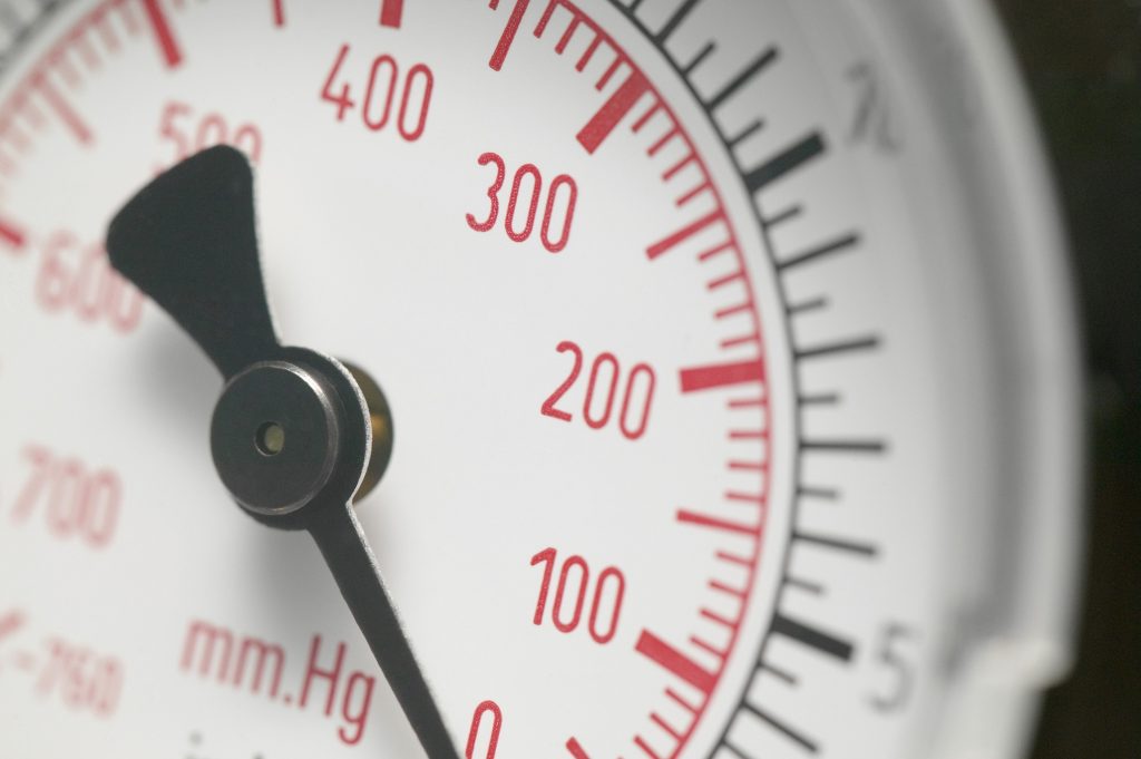
Systolic and diastolic blood pressure were measured as vascular risk factors. The researchers defined high blood pressure as having a systolic pressure of at least 140 millimeters of mercury (mm Hg) and a diastolic pressure of at least 90 mm Hg, as well as taking antihypertensive drugs.

Additionally, the researchers looked for:

  • diabetes
  • cholesterin amounts
  • smoking history
  • a cardiovascular condition
  • stroke
  • BMI, or body mass index

The ICD-9 codes associated with epilepsy or seizures, self-reported seizures, routine chart reviews for neurological diseases, and antiepileptic medication use were utilised by the researchers to screen participants for epilepsy or seizures.

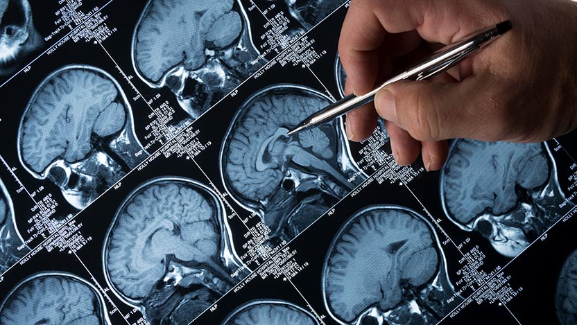
To identify cases of epilepsy, the researchers also analysed electroencephalography (EEG), cardiac, and other pertinent data, as well as brain imaging.

The study found 55 cases of epilepsy in the group, of which 26 were confirmed, 15 were likely, and 14 were suspected. The average age of the subjects at the time of a possible diagnosis was 73.8 years old.

The study’s findings revealed a nearly 2-fold increased risk of epilepsy in people with hypertension. The other risk variables, however, were unrelated to epilepsy.

They also discovered that the probability of having epilepsy increased by 17% for every 10 mm Hg change in systolic blood pressure.

Underlying processes

The connection between epilepsy and hypertension is explained by several theories. The renin-angiotensin system (RAS), which controls blood pressure, may be one mechanism.

According to research, rats who have had repeated seizures had 2.6–8.2 times the RAS components of seizure-free mice. Antihypertensive medications that decreased RAS component levels postponed the start of seizures and decreased their frequency.

More investigation is required, though, as other studies indicate the system may only have a modest impact on the relationship between epilepsy and hypertension.

Small vascular disease (SVD), a disorder in which the walls of small arteries and capillaries are damaged and do not adequately convey oxygen-rich blood to numerous organs, is another potential underlying mechanism, claim the researchers. According to a recent study, the duration of high blood pressure is a reliable indicator of developing SVD in later life.

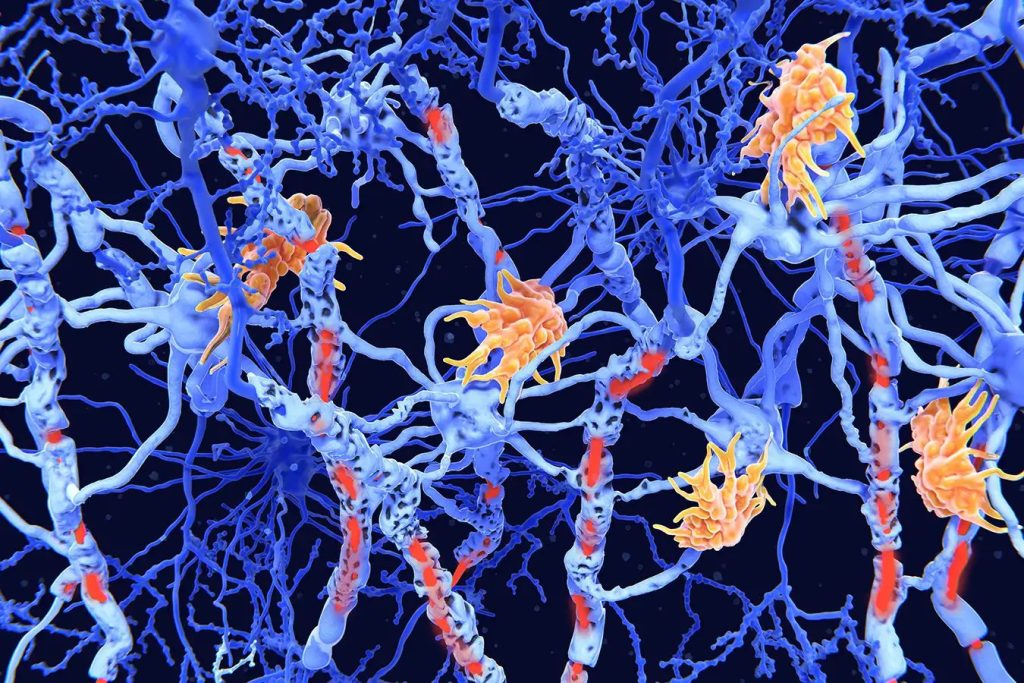
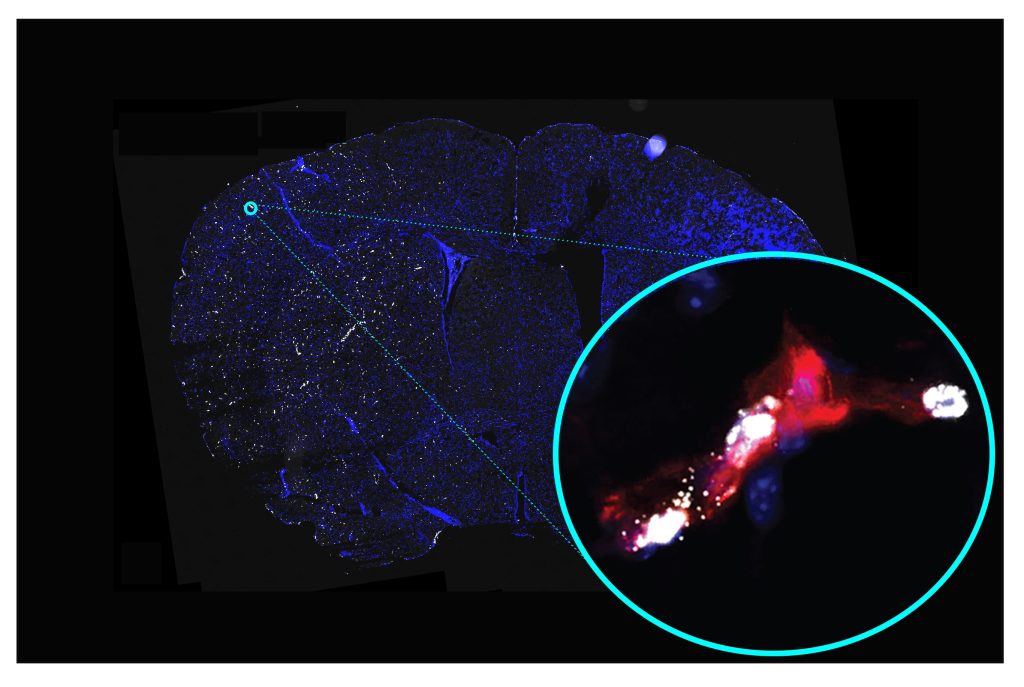
The researchers also mention that temporal lobe epilepsy and SVD are related. Cortical microinfarcts, tiny lesions in the cortical tissue, and the disruption of U fibers are a few potential explanations. These conditions may cause excessive excitability and hence seizures. U fibers link adjacent regions of the cerebral cortex.

The researchers draw the conclusion that hypertension is a standalone predictor of late-onset epilepsy and carries a 2-fold chance of getting seizures after 45 years.

They do acknowledge that their study has some limitations. It might not accurately represent all races and ethnicities because it was mostly made up of white people. The study’s observational design, according to the researchers, precludes the ability to establish causality.

Dr. Jason Hauptman, a neurosurgeon at Seattle Children’s Hospital, told us that these findings were particularly intriguing because there has been debate over whether elevated blood pressure (hypertension) is a standalone risk factor for stroke.

living with high blood pressure and epilepsy

A healthy blood pressure reading is less than 120/80 mmHg; if you are unsure of your current blood pressure, ask your doctor. High blood pressure is closely linked to a variety of health risks, including cardiac arrest, so it is important to try and maintain this level. The good news is that there are lots of easy lifestyle adjustments you can make to lower your blood pressure, like:

  • healthy eating
  • not a smoker
  • controlling stress
  • lowering the consumption of alcohol
  • Exercising
  • consuming less salt

Speak to your doctor if you have concerns about your epilepsy and high blood pressure. They can assist you in creating a management strategy to lower your blood pressure through dietary and activity modifications. while also making sure that you are controlling your seizures.

REFERENCES:

For Epilepsy disease treatments that have been suggested by doctors worldwide are available here https://mygenericpharmacy.com/index.php?therapy=49

New brain imaging techniques for the treatment of epilepsy.

New brain imaging techniques for the treatment of epilepsy.

In a recent study, researchers identified a brain circuit that can be targeted with brain stimulation by mapping abnormalities in the brain linked to epilepsy.

They stated that they hoped their discoveries could lessen the symptoms that come along with seizures.

They stated that the brain mapping method might also aid in predicting whether stroke survivors will experience seizures.

According to a recent study published in the journal JAMA Neurology, deep brain circuit stimulation may be able to identify whether people who have had a stroke may eventually acquire the disease and assist treat epilepsy.

Scientists from Brigham and Women’s Hospital in Massachusetts examined five datasets that had more than 1,500 individuals with brain injuries for their study.

The lesions have several diverse causes, such as tumors, trauma, and stroke.

The ability to explore across many brain regions and forms of brain injury for common network connections associated with epilepsy as a result allowed researchers to do so.

Brain mapping: What is it?

There are specific functions for each region of the brain. The surgeon wants to comprehend how the brain regions close to the seizure onset operate before doing any type of brain surgery, including epilepsy surgery. This enables your team to determine how much of the seizure focus can be safely removed.

The process of brain mapping can be used to pinpoint the functions of various brain areas.

Different people have different locations for different bodily processes (such as movement, voice, vision, and more). Tumours, seizures, or other brain abnormalities may alter which regions of the brain are in charge of a certain function. Sometimes general laws don’t apply.

By activating particular brain regions, one can create a “map” of each person’s brain. The map reveals to medical professionals which regions of the brain are in charge of vital processes like speech, sensation, or movement.

Brain mapping for epilepsy

The sites of brain damage in epilepsy patients and those without the condition were compared by the researchers.

According to the researchers, the brain was filled with lesions connected to epilepsy. They did, however, have a common network.

The researchers pointed out that epilepsy may be brought on by disruption of brain connections rather than the site of the damage. The basal ganglia and cerebellum, two deep-brain regions, were the locations of the linkages.

According to the researchers, identifying lesions in a brain network may aid in determining whether or not a person may experience epilepsy following a stroke. They claimed that common brain pathways could connect various damages and result in epilepsy.

The researchers point out that earlier studies have linked deep brain regions to modifying and regulating seizures in epilepsy-prone animals. They might have a braking effect on the brain.

How have scientists used deep brain stimulation?

The researchers examined the deep brain stimulation results in 30 patients with drug-resistant epilepsy.

If the stimulation was linked to the same brain network they discovered when mapping brain lesions, they claimed that the benefit would be greater.

Dr. Frederic Schaper, an assistant scientist at the Centre for Brain Circuit Therapeutics at Brigham and Women’s Hospital and an instructor of neurology at Harvard Medical School in Massachusetts, said, “In our study, we analysed existing data from patients that received deep brain stimulation for drug-resistant focal epilepsy.”

Although all patients had electrodes for deep brain stimulation implanted in the anterior thalamus, Schaper informed us that each patient’s precise electrode placement and stimulation sites varied slightly. “We found that patients with deeper brain stimulation sites that were more connected to deep brain regions in the cerebellum and basal ganglia had better seizure control than patients who were less connected to these regions.”

“This finding suggests an important role for brain networks distant from the anterior thalamic deep brain stimulation site in the mechanism of action of deep brain stimulation for epilepsy and seizure control,” he continued.

Deep brain stimulation principles

The American Association of Neurological Surgeons defines deep brain stimulation as a surgical procedure in which electrodes are placed in particular parts of the brain. Then, in order to assist manage aberrant brain activity, these electrodes transmit electrical impulses.

The amount of stimulation is managed via an implanted programmable device that resembles a pacemaker. The device is connected to the brain’s electrodes by a wire.

The full mechanism through which deep brain stimulation reduces seizure frequency is unknown, according to Schaper. “Previous research in people and animal models indicates that deep brain stimulation disturbs the brain networks responsible for seizures. It is uncertain, nevertheless, whose brain networks are in charge of [deep brain stimulation]-induced seizure control.”

Schaper mentioned that deep brain stimulation is a safe and efficient treatment for drug-resistant focal epilepsy and has received approval from federal regulators.

Improving epilepsy symptoms

In this investigation, brain networks were sought after. They claimed that deep brain stimulation can lessen epilepsy symptoms if it activates just one node in the network.

“This study is quite exciting,” said Dr. Jean-Philippe Langevin, a neurosurgeon and the director of the Restorative Neurosurgery and Deep Brain Stimulation Programme at the Pacific Neuroscience Institute at Providence Saint John’s Health Centre in California. He was not involved in the research.

“The scientists discovered that networks were more associated with epilepsy than brain lesions. “They could influence epilepsy symptoms if they could concentrate stimulation within the networks,” Langevin told us.

The roadways in the brain are called brain networks. The roadside stops are called lesions. The researchers discovered that the entire network was influenced when electrical currents were applied anywhere along a network of streets.

According to Langevin, “Deep brain stimulation works for other diseases.” These include essential tremors, Parkinson’s condition, dystonia, obsessive-compulsive disorder, and dystonia. For certain conditions, “working within a single network would also hold true.”

“This is exciting because, in the future, when patients come to us with seizures, a scan can look at how the network is connected, making it easier to use [deep brain stimulation],” continued Langevin. “The scans do exist, but we don’t typically use them in the study.”

Symptoms of a seizure

Different people experience different pre-seizure warning symptoms.

But there are a few widespread indications:

  • a sense of impending disaster
  • For every seizures, the same tone or sound is produced.
  • trouble generating ideas
  • having trouble finding the right words
  • Having underwater-like audio perception
  • experiencing déjà vu or believing that nothing is familiar
  • feeling queasy in the stomach
  • having the impression that everything is deformed, either larger or smaller than it should be.

It is suggested that you lay on your side if you are experiencing a seizure. Someone else should roll a seizure victim over if they are unable to move.

Additionally, a person experiencing a seizure ought to be relocated to a location where they won’t damage themselves. For instance, a space free of any furnishings.

Ensure that they are not wearing anything tight around their neck, such as a necktie, scarf, or button-up shirt. If so, you ought to take these things off.

Never abandon a person experiencing a seizure. Until the seizure is finished, be at their side.

REFERENCES:

For Epilepsy medications that have been suggested by doctors worldwide are available here https://mygenericpharmacy.com/index.php?therapy=49

High levels of lean muscle may help prevent Alzheimer.

High levels of lean muscle may help prevent Alzheimer.

High quantities of lean muscle may help prevent Alzheimer’s disease, according to recent studies. To determine whether this connection is causal, more study is required.

Resistance training and a sufficient intake of protein in the diet are advised by experts as ways to build lean muscle mass. Previous studies have shown a link between obesity and an elevated risk of Alzheimer’s.

A recent study that was published in BMJ Medicine suggests that having a lot of lean muscle may prevent Alzheimer’s disease. The authors of the study pointed out that additional study is required to comprehend the biological mechanisms underlying it.

Researchers gathered data on 450,243 participants’ genetics, lean muscle mass, cognition, and health for this study from the U.K. Biobank. They next used a method known as Mendelian randomization to search for genetic relationships between lean muscle mass and genetic variants.

The quantity of lean muscle and fat tissue in the arms and legs was measured using bioimpedance, an electric current that moves through the body at varying speeds. The researchers next discovered 584 genetic variations related to lean muscle mass, but none of these were located in an area of the genome known to include genes connected to an elevated risk of Alzheimer’s disease.

The chance of developing Alzheimer’s disease did, however, appear to be reduced in individuals who had high amounts of lean muscle mass and associated genetic variations.

These results were confirmed by measuring the quantity of lean muscle mass and body fat tissue in a second cohort of 252,879 individuals without Alzheimer’s disease and 7,329 individuals with the condition.

The findings indicated that lean mass was associated with enhanced cognitive performance, but this association did not account for lean mass’s protective effect against the onset of Alzheimer’s disease.

The study’s objectives

The majority of the data utilized by the researchers came from the UK Biobank, a continuous database that collects health and genetic data on 500,000 people. The researchers used data from hundreds of thousands of people.

Mendelian randomization, which employs gene variations as a proxy for specific risk variables, was used to analyze the data.

Additionally, bioimpedance measurements which determine the speed at which an electrical current travels through the body based on its composition in terms of muscle and fat were used to calculate each person’s lean mass.

A total of 584 genetic variations were connected to lean muscle mass, but none of them were discovered in the region of the brain linked to Alzheimer’s susceptibility.

Participants’ chance of developing the disease was statistically significantly (12%) lower on average for those with higher amounts of (genetically proxied) lean muscle.

The analysis was performed using data from an additional 260,208 individuals, of whom 7,329 were identified as having Alzheimer’s disease, in order to confirm these findings. They measured lean muscle mass over the entire body, not only in the arms and legs.

Again, they discovered that having more lean muscle was linked to a lower risk of Alzheimer’s.

Unexpectedly, the analysis did not reveal a correlation between body fat and the probability of developing Alzheimer’s disease once lean mass was taken into account. Body fat was associated with inferior performance in cognitive activities.

The significant contrast between the protective effect of lean mass on dementia risk and the lack of an effect of fat mass on dementia risk, according to Daghlas, startled him.

How Alzheimer’s disease and lean muscle are related?

The fact that the processes underlying the association between lean muscle and Alzheimer’s disease are still unknown is another drawback of the study.

The researchers made brief speculations about possible connections. For instance, Alzheimer’s has long been linked to cardiovascular illness, though Daghlas cautioned that the connection is “complicated.”

According to Daghlas, heart disease problems like stroke and hypertension are what fuel vascular dementia. Though this is a contentious idea, he added, “the most recent causal evidence suggests weaker evidence for an effect of vascular risk factors on Alzheimer’s disease risk.”

Lean mass may very possibly lower the incidence of vascular dementia through lowering the risk of cardiovascular disease, according to Daghlas, however more research is needed in this area. “This can be looked into in upcoming studies.”

In the publication, the researchers also made the speculative claim that “new” processes, such as centrally acting myokines, may be at work.

Myokines are proteins that muscles produce that have an impact on other tissues, according to Daghlas. “Experimental studies have demonstrated that they are induced by exercise and have a positive impact on brain function.”

Other muscle-related issues, in addition to these, “may explain the larger picture,” according to Dr. Anna Nordvig, a neurologist at NewYork-Presbyterian and Weill Cornell Medicine who was not involved in the study. Examples include “bone hormones, cardio vs. strength training differences, sex hormones, glymphatic drainage depending on exercise, and sleep, to name a few.”

In the end, further clinical intervention studies are required to demonstrate the link between lean muscle and Alzheimer’s disease and the mechanisms underlying it.

The best way to build lean muscle mass

Having lean muscle mass has several health benefits in addition to possible advantages for the brain.

Resistance training using weights, bands, and pleiomorphic activities are advised by Dr. Joseph C. Maroon, clinical professor, vice chairman, and Heindl scholar in neuroscience at the Department of Neurosurgery at the University of Pittsburgh.

Additionally, he recommends supplementing with B-hydroxy B-methylbutyrate (myHMB), a good source of dietary protein.

This naturally occurring compound aids in the maintenance of a healthy weight and the development of lean muscle mass in humans. B-hydroxy helps muscles recover from hard activity, improves athletic performance, and enhances muscle and strength, according to him.

According to Dr. Sullivan, the best food, the best kind and frequency of exercise, the optimum amount of rest, and stress management are the primary factors that influence muscle growth.

These are the recommendations she makes:

Exercise: four to five quick strength-training sessions each week. In comparison to two or three longer aerobic exercises each week, this will produce greater lean muscle mass.

Diet: Put your attention on lowering insulin resistance by eating fewer carbohydrates and more protein, the building block of muscle.

Sleep: 8 to 9 hours of sleep per night are recommended if you want to recuperate from this kind of workout completely.

Stress management: With the rise in inflammation and blood sugar that stress hormones like cortisol induce, chronically high stress can sabotage any self-improvement effort. Long-term high cortisol levels can contribute to persistent muscle tension and lactic acid buildup, which can inhibit muscle growth. Moving your body more, going outside, eating more good foods, being an aggressive communicator, and finding your purpose are the simplest ways to relieve chronic stress.

How to lower your risk of developing Alzheimer’s?

Although there is no cure for Alzheimer’s, doctors think there are a number of steps you may take to lower your risk.

“Protect” and “stimulate” are the two categories that these fall under, according to Nordvig, and “physical activity falls into both of these.”

“Protect” includes monitoring factors like blood pressure, sugar levels, weight, nutrition, and sleep, she said. These are topics covered at a yearly checkup.

We should also work to safeguard ourselves from environmental risk factors associated to Alzheimer’s, according to Dr. Rena Sukhdeo Singh, a vascular neurologist at the University of Maryland Shore Regional Health.

The incidence of dementia has also been connected to fine particulate matter in air pollution.

According to Sukhdeo Singh, systemic inflammation also contributes to the development of Alzheimer’s disease. Numerous factors, including medications, a diet high in sugar and processed foods, smoking, and binge drinking, can contribute to chronic inflammation.

Optimising additional daily inputs that influence cognition is what “stimulate” entails. For instance, Sukhdeo Singh proposed, “learning a new skill, hobby, language, or instrument,” or taking part in shorter activities like “sudokus, puzzles, and number games.”

Unfortunately, there are some factors that we have no control over. Age, genetics, and sex are non-modifiable risk variables, she added.

Restrictions of this research

Researchers solely considered lean muscle mass for this investigation. However, there are other things to think about.

The protein amyloid, which is damaging to the functioning of the brain, is found in higher amounts in adipose tissue, but the researchers neglected to test these signs of inflammation and insulin resistance, according to Maroon. “This probably diminishes the significance of their findings.”

Furthermore, “while their positive finding was statistically significant, the effect size was modest in lean muscle mass reducing the risk of dementia and only explained 10% of the variance,” added Dr. Sullivan.

The link between more lean muscle mass and a lower incidence of Alzheimer’s disease has to be further investigated.

According to Nancy Mitchell, a registered nurse, “For now, people with lower muscle mass tend to be obese, which is a risk factor for type 2 diabetes.”

We refer to Alzheimer’s disease as “diabetes of the brain” because it has been hypothesized that high blood sugar harms the nerve endings in the regions of the brain that are most impacted by cognitive decline. Therefore, the link may actually be between a reduced risk of obesity and diabetes. This can be a study drawback in and of itself because greater specificity is still needed. Not all correlations indicate causation. Nancy Mitchell is a nurse practitioner.

REFERENCES:

For Alzheimer’s disease medications that have been suggested by doctors worldwide are available here https://mygenericpharmacy.com/index.php?therapy=31

Can Hormone therapy for menopause increase Dementia risk?

Can Hormone therapy for menopause increase Dementia risk?

Menopausal hormone therapy is used by about 45% of all women to lessen menopause symptoms.

According to prior studies, some types of hormone replacement treatment may make women more susceptible to developing serious illnesses.

Menopausal hormone therapy is linked to an increased risk of dementia and Alzheimer’s disease, according to researchers from Copenhagen University Hospital, says Rigshospitalet.

These results go against earlier research that suggested HRT might reduce a woman’s risk of dementia.

Menopausal hormone therapy, often known as hormone replacement therapy (HRT), is used by about 45% of all women worldwide to cope with menopause symptoms.

HRT can cause adverse effects like nausea and migraines. According to earlier studies, women who use specific forms of HRT may be more susceptible to strokes, gallbladder problems, and malignancies including breast and endometrial.

Menopausal hormone therapy is now linked to a higher risk of dementia and Alzheimer’s disease, according to study from Copenhagen University Hospital, as per Rigshospitalet.

These results go against earlier research that suggested HRT might reduce a woman’s risk of dementia. The BMJ recently published an article based on this study.

What occurs throughout menopause?

Every woman experiences menopause, which is the end of the monthly cycle and the last time the ovaries release eggs.

Menopause usually begins in a person between the ages of 45 and 55. Perimenopause, often known as the menopausal transition, can persist between seven to fourteen years.

A woman who is beginning menopause may experience symptoms like:

  • a hot flash
  • morning sweats
  • irregular or absent
  • vulvar aridity
  • difficulty sleeping
  • mood swings like anxiousness and depression

Menopause is a natural part of ageing, but it comes with some changes that some people may desire to minimise. Menopause-related symptoms may be treated with the following methods:

  • HRT
  • hormonal birth control at a low dose
  • low-dose mood stabilisers
  • prescription or over-the-counter drugs for vaginal dryness

Additionally, several lifestyle modifications can assist in relieving some symptoms:

  • routine exercise
  • wholesome diet
  • meditation techniques
  • restricting alcohol
  • giving up smoking
  • counselling for mood changes
  • maintaining a healthy sleep routine

What is hormone therapy for menopause?

The purpose of HRT is to enhance and balance the levels of the female hormones progesterone and oestrogen in the body.

Although the body’s ovaries naturally produce both of these hormones, their production declines after menopause, leading to menopausal symptoms.

There are two primary types of menopausal hormone treatment that a doctor could recommend, depending on a woman’s situation and requirements:

  • treatment with just oestrogen
  • combined treatment utilising progesterone and oestrogen

HRT can be applied topically or vaginally, and comes in tablet, nasal spray, skin patch, and vaginal cream or suppositories forms.

The following are possible HRT adverse effects:

  • bloating
  • headaches
  • breast discomfort
  • nausea
  • acne
  • mood changes
  • uterine bleeding

How Does HRT Affect the Risk of Dementia?

Dr. Nelsan Pourhadi, the study’s lead author and a researcher at the Danish Cancer Society and the Danish Dementia Research Centre in the Department of Neurology at Copenhagen University Hospital – Rigshospitalet in Copenhagen, Denmark, claims that the study’s objectives were dual and based on understudied facets of the subject matter.

“First, we sought to look into whether menopausal hormone therapy use, as advised by guidelines, increased the incidence of dementia.” Second, he told us, “we were looking into continuous versus cyclic therapy regimes.”

Dr. Pourhadi and his team used data from a national registry database for this investigation. The study’s controls were about 56,000 age-matched women without a dementia diagnosis and approximately 5,600 women with dementia. Danish women between the ages of 50 and 60 in 2000 who had no history of dementia or any conditions that would exclude the use of HRT were included in the data, which covered the years 2000 to 2018.

The subjects’ average age at the time of dementia diagnosis was 70. In comparison to controls, 32% of women with dementia and 29% of controls had used estrogen-progestin therapy starting at an average age of 53 before receiving a diagnosis. For dementia-stricken women, therapy lasted an average of 3.8 years, compared to 3.6 years for males.

Analysis revealed that women who got estrogen-progestin therapy had a 24% higher incidence of Alzheimer’s disease and all-cause dementia. Even ladies who started the treatment at age 55 or younger experienced this.

The Women’s Health Initiative Memory Study (WHIMS), the largest clinical experiment in the field, found similar results, according to Dr. Pourhadi.

Does HRT alter the risk of dementia?

Researchers have previously searched for a link between HRT and the risk of dementia.

Menopausal hormone therapy may aid in lowering a woman’s risk of dementia, according to earlier studies. Menopausal hormone therapy use was associated with a lower chance of developing all neurological disorders, including Alzheimer’s disease and dementia, according to a study released in May 2021.

Additionally, a study published in June 2022 discovered that women with depression who used HRT after menopause had a lower risk of developing Alzheimer’s disease and vascular dementia.

Several research have shown a connection between HRT use and an elevated risk of dementia. HRT was linked to a higher incidence of dementia, according to research released in September 2022.

And according to a study that was just released in April 2023, women who had HRT more than five years after the onset of menopause or who started menopause early had greater levels of tau protein in their brains, which is thought to be one of the main causes of Alzheimer’s disease.

When questioned why prior and current studies may have conflicting results, Dr. Pourhadi responded, “It is crucial to emphasise that our findings are in line with those of the largest clinical trial on the topic, WHIMS. The majority of prior clinical trials were hindered by issues like poor selection, limited sample sizes, brief follow-up periods, and results that were purely dependent on cognitive testing rather than a clinical evaluation of dementia.

Furthermore, earlier observational studies, particularly short-term ones, were unable to evaluate the use of menopausal hormone therapy close to menopause, the author continued. The discrepancies between the findings of those studies and those of our study “may be explained by these differences.”

Can HRT lead to dementia?

Dr. Pourhadi explained that because this study is an observational one and not a causal one, it is impossible to establish a link between menopause hormone therapy and dementia.

Therefore, more investigation is required to determine whether or not the observed link may be assumed to be causal. Additionally, it is important to distinguish between the various menopausal hormone therapy delivery methods such as tablets, patches, and gels,” he continued.

Dr. Mindy Pelz, a specialist in holistic medicine who specialises in women’s and hormonal health but was not part in this study, concurred.

It’s vital not to overestimate the results of this new observational study. Correlation does not imply causality, and prior research has shown that menopausal hormone therapy lowers the incidence of dementia, so it’s conceivable there’s a variable missing that we haven’t thought of yet, the researcher added.

She told us that many women go for hormone replacement medication to deal with their symptoms when they have cognitive deficiencies after menopause, which could be a sign of dementia in the future.

Dr. Jewel Kling, assistant director of women’s health internal medicine at the Mayo Clinic in Arizona and a non-participant in this study, informed us after reviewing the findings that because this was an observational study using data from a national registry, we could not draw any conclusions about the cause-and-effect relationship between menopausal hormone therapy and dementia risk.

The only way to conclude causation is through a randomised control design, which this wasn’t. “(We) can only claim that there was a relationship identified between the two in their data. According to the study’s design, there are numerous additional factors that could potentially explain this association, the researcher said.

REFERENCES:

For Mental disease medications that have been suggested by doctors worldwide are available here https://mygenericpharmacy.com/index.php?cPath=77_478

MS severity: Genetic markers may lead to better treatment.

MS severity: Genetic markers may lead to better treatment.

Globally, 2.8 million people will have Multiple Sclerosis (MS) in 2020. The symptoms of MS can worsen over time and result in chronic problems, and the condition presently has no known cure.

The first genetic indicator of MS severity and progression has been identified by researchers at the University of California, San Francisco.

This discovery, according to scientists, may help in the creation of new medications that can delay the advancement of the illness.

Multiple sclerosis (MS), a condition of the central nervous system that affects mobility and vision, will impact roughly 2.8 million individuals worldwide by the year 2020.

MS presently has no known cure. Each person is uniquely affected by the illness, both in terms of when symptoms initially appear and how severe they are.

The severity of the illness can worsen as the body experiences a cycle of symptom flare-ups and remissions, resulting in persistent mobility problems, visual loss, and even partial or complete paralysis.

The first genetic signature connected to MS severity and progression has now been identified by researchers from the University of California, San Francisco.

This discovery, according to scientists, may help in the creation of new medications that can delay the advancement of the illness.

Research targets MS progression

The University of California, San Francisco’s Dr. Sergio Baranzini, professor of neurology and co-senior author of the study, explained that they chose to look for a genetic variation associated with faster MS progression because the disease develops differently in each patient after diagnosis.

“Neurological progression is a common feature in persons with MS, which is inexorable and independent of whether relapses are controlled or not,” he said.

“Some people have a very aggressive disease that can impact their mobility and neurological function in a few years, while others experience a much more benign course,” he said. We already knew that genetics has a significant influence on risk, but the wide range of outcomes revealed that genetics may also affect severity.

Data from The MultipleMS Consortium and The International Multiple Sclerosis Genetics Consortium (IMSGC), two sizable MS research consortiums, were used by Dr. Baranzini and his team.

For a genome-wide association study (GWAS), data from both groups were pooled to represent more than 12,500 MS patients.

From there, researchers combed through more than 7.5 million genetic variants before discovering one linked to accelerated disease progression in MS patients.

This particular mutation is situated between two genes named DYSF and ZNF638 that had no known association with MS. ZNF638 aids in the control of viral infections whereas DYSF aids in the restoration of damaged cells.

Possibility of new treatments for MS

Since there is currently no treatment for MS, doctors employ a variety of drugs to treat the symptoms, delay the disease’s course, and help avoid relapses.

The results of this study, according to Dr. Baranzini, will open the door for a new class of medicines that will treat progression and probably target the central nervous system.

Dr. Baranzini made the point that genetic information considerably reduces the risks associated with drug development because developing medicines represents a considerable risk for the pharmaceutical business, where only a tiny percentage of drugs reach the market.

“This discovery will set up several development programs that will target the unmet need of disease progression in MS,” he said.

“All relapse-controlling medications are immunomodulatory, which is consistent with the genetics of the more than 200 MS risk variations. The central nervous system should be the target of this new class of therapies, according to the genetics of illness severity.”

Dr. Baranzini explained that since it has been proven that genetics contribute to the severity of an illness, the IMSGC is currently preparing for a new genetic study including even more participants.

The researcher continued, “Our prior experience with disease susceptibility suggests that a larger study translates into more findings, and we are pleased to uncover new genetic variants that could aid in the development of more efficient treatments for MS.”

How is MS being treated right now?

The capacity to move, think, talk, and see clearly can all be significantly impacted by MS since it affects the brain and nerve system.

The majority of scientists concur that MS is caused by the body’s immune system wrongly attacking the central nervous system, even though the exact origin of MS is still unknown.

The myelin that wraps the nerve fibres in the brain and spinal cord is damaged by this onslaught. When myelin is destroyed, it is unable to protect the exposed nerve fibre, which prevents messages from travelling from the nerves to the brain.

Additional MS risk factors include:

  • age – between the ages of 20 and 50, most persons acquire a diagnosis
  • MS is more prevalent in women than in men.
  • an MS family history
  • smoking
  • a lack of vitamin B12 or vitamin D
  • exposure to specific viral illnesses, such as mononucleosis or the Epstein-Barr virus

Why the recent study is beneficial?

We discussed the significance of the study with Dr. Krupa Pandey, director of clinical research at the Neurosciences Institute in New Jersey, director of the Hackensack University Medical Centre MS Centre, and associate professor of neurology at the Hackensack Meridian School of Medicine who was not involved in the current investigation.

She said, “There are a few ways in which this study is helpful. Finding a connection between genes and the potential severity of a disease is a positive step.”

“It is also beneficial since it offers more proof that environmental variables, like smoking, truly do assist people with genetically susceptible diseases to get sicker. This is a fantastic illustration of how a disease may be affected by both nature and upbringing, said Dr. Pandey.

The expert went on to say that similar discoveries “may lead to future findings that can help us counsel patients on how to tailor not just medication regimens but modify lifestyle-related factors.”

“It is also helpful for companies looking at MS therapies [to] enroll patients with higher risks for progression to see if the drug is effective,” she said.

REFERENCES:

For Nerve damage medications that have been suggested by doctors worldwide are available here https://mygenericpharmacy.com/index.php?cPath=30

Are brain fog, sleep, and pain improved by Cannabis?

Are brain fog, sleep, and pain improved by Cannabis?

Cannabis, according to researchers, may be able to lessen the negative effects of chemotherapy as well as cancer discomfort. In a recent study, cancer patients said that using cannabis helped them feel less pain, sleep better, and think more clearly.

According to experts, federal rules need to be altered so that more studies may be done on cannabis’ advantages and impacts on medical ailments.

According to a study conducted at the University of Colorado at Boulder and published in the journal Exploration in Medicine, people with cancer who use cannabis to alleviate symptoms experience less pain, sleep better, and have clearer thinking.

This is one of the first observational studies to examine the potential effects of cannabis products obtained from a dispensary on chemotherapy side effects and cancer symptoms.

University researchers are only permitted to possess and distribute cannabis products that are authorized by the government or that meet pharmaceutical standards in the United States, which makes it challenging to conduct studies on dispensary goods.

The researchers at the University of Colorado, however, developed a novel strategy. When 25 cancer patients bought their products, they watched how they responded.

During a baseline visit, the researchers evaluated the patient’s pain tolerance, sleep quality, and cognitive function. After that, the participants went to a dispensary and bought a cannabis edible product of their choice. They selected several different products, such as:

  • Chocolates
  • Gummies
  • Tinctures
  • Pills
  • a baked good

Additionally, the THC and CBD potencies varied widely.

Information from the cannabis and cancer pain study

The researchers traveled to each person’s residence in a mobile lab.

Before being asked to consume the cannabis product they had chosen at home, each person had their physical and mental capabilities evaluated in the van. After consuming medical marijuana, they completed another test.

Within an hour of using the items, the patients claimed that their pain levels had greatly decreased. Additionally, it made them feel “high” and affected their cognitive function. They claimed to feel higher the more THC was present.

After two weeks of consistent use, the individuals underwent a follow-up examination. At that time, the patients indicated that their pain, sleep, and cognitive abilities had all decreased. Improvements were evident in various cognitive domains, including reaction times, according to objective measurements.

The researchers found that lower levels of pain led to an improvement in cognitive performance. The improvement in cognition increased as the discomfort decreased.

Patients who consumed more CBD reported significant reductions in pain and better sleep. CBD reduces inflammation. The authors of the study point out that while cognition was temporarily reduced, it can be enhanced by treating pain.

How cannabis might benefit cancer patients

Some dosages and cannabis types, according to experts, may be beneficial for persons receiving cancer therapy.

Dr. Wael Harb, a hematologist and medical oncologist at Memorial Care Cancer Institute at Orange Coast Medical Centre in California who was not involved in the study, said, “This study adds to the growing body of research that examines the potential benefits of cannabis use in cancer patients.”

According to Harb, “the results highlight the potential for cannabis to reduce pain, enhance cognition, and improve the overall quality of life for patients.” These findings have important therapeutic ramifications since they imply that cannabis may be used as an auxiliary or alternative therapy for cancer patients, particularly those who are in pain or have cognitive deficits.

However, “it is important to note that the study has limitations, such as a relatively small sample size, which may not represent the larger population of cancer patients,” the author continued.

The study also uses self-reported information, which is prone to bias. To validate these results and investigate any potential hazards or negative consequences related to cannabis usage in cancer patients, additional research with bigger, more diverse samples and more objective assessment technologies is required.

What do you need to understand about medical marijuana?

The director of the UCI Centre for the Study of Cannabis, Daniele Piomelli, Ph.D., stated that “this small study supports what oncologists have known for a long time: many cancer patients (half of them, according to some surveys) use cannabis to cope with nausea, pain, and sleeplessness caused by cancer drugs“.

He informed us that the National Cancer Institute, a division of the National Institutes of Health, is aware of this and that urgently required larger investigations are about to begin.

According to the National Institutes of Health, the cannabis plant has two major compounds (cannabinoids) that are employed in medical procedures. THC and CBD are these.

These have not been given use approval by the Food and Drug Administration. The group has, however, approved a small number of drugs that do:

  • Cannabidiol (Epidolex)
  • Dronabinol (Marinol, Syndros)
  • Nabilone

Currently, marijuana use is either decriminalized or legal in 46 states. Anyone considering using medical items should first examine the regulations in their locality as each state has different legislation.

Dr. Olivia Seecof, clinical assistant professor of medicine and attending physician in supportive oncology at NYU Langone Perlmutter Cancer Centre in New York, said she was “very excited” about the study because it was “one of the first studies to address some of the issues surrounding recommending medical cannabis products in an evidence-based way.”

During outpatient supportive oncology appointments, I do certify patients for medical cannabis. I had to enroll in the New York State Medical Cannabis Programme and complete further training/certification to be able to do that,” she explained to us.

The market for medical marijuana is expanding.

Its US market value in 2021 was close to $27 billion. Market Research Future estimates that by 2030, it would reach $248 billion.

According to a Reuters article, some proposed measures in Congress would decriminalize or legalize marijuana.

It would be decriminalized under one statute, leaving state regulation to their discretion. More research will be possible thanks to the regulation reform, giving doctors and other healthcare providers the data they need to treat patients.

Because of cannabis’ murky legal status and negative connotations, many doctors are still uneasy about marijuana, according to Piomelli. “But the profession can no longer bury its head in the sand with so many patients using it. To better meet the requirements of their patients, doctors, nurses, and other medical professionals need to have more knowledge about the advantages and drawbacks of cannabis.

REFERENCES:

For Mental disease medications that have been suggested by doctors worldwide are available here https://mygenericpharmacy.com/index.php?cPath=77_478

Detect the Cognitive ability by a test before its arrival.

Detect the Cognitive ability by a test before its arrival.

According to researchers, a quick card memory test can identify cognitive decline years before symptoms appear. According to experts, the test would make it possible for those who are more likely to develop dementia to receive treatment and preventative measures sooner.

One specialist advocates administering the test to everyone over 45. Researchers claim to have created an easy test that can forecast a person’s future risk of developing cognitive impairment.

The test only applies to persons without pre-existing cognitive and memory impairments, the researchers write in their study, which was just published in the journal Neurology.

The study’s lead author and clinical professor at the Albert Einstein College of Medicine in New York City, Ellen Grober, Ph.D., said in a statement that there is “increasing evidence” that some people who appear to be healthy and have no cognitive or memory issues may fact be displaying very mild indicators of early cognitive impairment. In our study, a quick and accurate memory test indicated the likelihood that individualsnormally thought to have normal cognition will experience cognitive impairment.

Cognitive impairment study

969 participants in the study, whose average age was 69, took a basic memory test and then underwent follow-up exams over ten years.

There were two phases of the test.

The participants were first instructed to examine four cards, each of which had four drawings of various objects.

Then, the respondents were required to identify each item as a member of a specific category. For instance, when asked to name a fruit, participants might respond “grape.”

In the following stage, participants were required to recall the things to gauge their memory capacity. They were given category cues, which calculated the amount of memory storage, for the items they couldn’t recall.

Result of the tests for cognitive impairment

Using the Stages of Objective Memory Impairment (SOMI) approach, researchers categorized the participants into five groups with stages zero through four based on their test results.

Stage 0 was for patients with no memory issues (47% of cases).

Memory retrieval problems, which researchers noted can occur five to eight years before dementia, were more difficult to recall in stages one (35%) and two (13%) of the disease. When given indications, these participants could recall information.

The individuals in the third and fourth stages (5% overall) had trouble remembering all the objects, even with hints. These stages, according to the researchers, occur 1 to 3 years before dementia.

234 of the 969 subjects experienced cognitive impairment in the end.

Subjects at stages one and two were twice as likely to experience cognitive impairment compared to those at SOMI stage zero, even after accounting for factors such as age, gender, education level, and the APOE4 gene, which affects a person’s chance of developing Alzheimer’s disease.

Cognitive impairment was three times as likely to develop in those in stages three and four.

The significance of testing for cognitive impairment

The SOMI method continues to forecast an elevated risk of cognitive impairment even after accounting for indicators of Alzheimer’s disease including amyloid plaques and tau protein tangles.

According to research, 72% of people in the third and fourth stages will have cognitive impairment after ten years, compared to 57% of people in the second stage, 35% of people in the first stage, and 21% of people in stage zero.

Our findings confirm the SOMI system’s application in locating those most at risk for cognitive decline, according to Grober. “Researchers looking for remedies can benefit from spotting cognitive impairment early on. By working with their doctor and implementing strategies to support healthy brain aging, those persons who are discovered to be at elevated risk may also benefit.

Neura Health’s virtual headache and migraine clinic’s medical director, Dr. Thomas Berk, a neurologist, pointed out that present testing only reflect the brain’s current condition.

“Predicting neurological change years later is very difficult,” said Berk. When someone has neurological testing, “we are getting a snapshot of their current brain function, not what their brain will look like years later,”

“This does give some evidence for being able to assess the future risks of developing memory issues,” he continued.

There is unquestionably a need for “a simple and fairly rapid test,” particularly in light of the growing body of evidence that early intervention can have a positive impact, according to Dr. Dale Bredesen, head of the University of California Los Angeles’ Easton Centre for Alzheimer’s Disease Research.

Standard neurocognitive tests can take hours, making them impractical for screening, and common quick tests like the MMSE aren’t sensitive enough to catch these early alterations, according to Bredesen.

Using fresh memory tests

According to Bredesen, the subjects of present testing are those who already have cognitive problems.

“Simple tests like the one described in this report should be included for everyone over the age of 45, to identify those who should be evaluated further, and potentially treated,” he advised.

One physician claimed that the SOMI system made him think of a well-known kid’s game.

According to Dr. Clifford Segil, a neurologist at Providence Saint John’s Health Centre in California, “I advise my patients to make a mental image of a scene with all three words to help in their recall when they have to recall the three words.” “I would suggest the same for this proposed card cognition exam. Currently used cognitive exams include additional memory tests.

“Go Fish,” which is utilised as a learning tool for children rather than a cognitive exam for senior people, is similar to the suggested test to be employed in my elderly population, which interests me as a parent and a practising adult neurologist, Segil added.

“When you start to worry that you might be losing your memory, you should be evaluated by a neurologist to see if your complaints are generally age-appropriate normal or something else,” he said.

REFERENCES:

For Cognitive disease medications that have been suggested by doctors worldwide are available here https://mygenericpharmacy.com/index.php?cPath=77_478

Atrial fibrillation catheter therapy and dementia risk.

Atrial fibrillation catheter therapy and dementia risk.

Researchers looked into how medications and catheter ablation for atrial fibrillation affected people’s risk of developing dementia.

They discovered that catheter ablation therapy lowered the risk of dementia more effectively than medicine alone.

For the correlation to be verified and the cause of it to be understood, more research is required.

Atrial fibrillation (Afib)

A cardiac disorder called atrial fibrillation (Afib) is characterised by erratic beating in the upper chambers of the heart, which restricts blood flow to the lower chambers. Patients who have afib may experience discomfort and have a five-fold increased risk of stroke.

183,321 death certificates in the US in 2019 listed AFib as a cause of death. By 2030, 12.1 million persons in the United States are predicted to be affected by the illness.

AFib has been linked in studies to dementia and cognitive decline. Additionally, studies have shown that patients with dementia and cognitive impairment who also have AFib suffer from a greater rate of cognitive decline than those who do not.

Understanding whether treating Afib patients lowers their likelihood of developing dementia may improve patient outcomes.

Recent studies evaluated the effects of Afib medication versus catheter-based therapy on the risk of dementia. They discovered that compared to medicine alone, catheter-based Afib therapy was associated with improved cognitive function.

“Despite having negative brain imaging results, I have encountered multiple patients with long-term persistent atrial fibrillation who had cognitive impairment, decreased swallowing capacity, and slower motor performance.

Based on this, I do not find [the results] surprising,” Dr. Vicken Zeitjian, a San Antonio, Texas-based cardiologist board-certified in nuclear cardiology and echocardiography who was not involved in the study, told.

He went on to say that “these results further demonstrate that catheter ablation is a superior method of atrial fibrillation management than medical management alone.” The research will be presented at the 75th Annual Meeting of the American Academy of Neurology.

Medicine versus catheter ablation

887 Afib patients with an average age of 75 were included in the study by the researcher. Before enrollment, 193, or 21.8%, of the participants got catheter ablation, whereas the remaining patients just received Afib medication.

Catheter ablation is the process of utilising radiofrequency to remove tiny patches of cardiac tissue that may be the source of an irregular heartbeat.

The individuals’ cognitive function was evaluated at baseline, one year later, and two years later. Out of a possible 30, a 23 or below score indicated cognitive impairment.

The study’s findings showed that those who underwent catheter ablation had an average cognitive score of 25, compared to those who did not, who had a score of 23.

The researchers discovered that those who received catheter ablation were 36% less likely to experience cognitive impairment than those who were just given medication after controlling for conditions like heart disease, kidney disease, and sleep apnea.

However, they pointed out that there were no appreciable variations in the incidence of heart attacks between patients who underwent catheter ablation or medication alone.

They also discovered that using warfarin and other anticoagulants had no appreciable impact on cognitive deterioration.

The risk of dementia and catheter ablation

We enquired about how catheter ablation might lower the risk of dementia from Dr. Aaron Ritter, director of the Memory & Cognitive Disorders Programme at Hoag Hospital in Newport Beach, California, who was not involved in the study. Although the study doesn’t say how, he pointed out that there could be a number of causes.

“For me, ablation may be a more conclusive or long-lasting therapy than pharmaceutical management, which necessitates a commitment to a daily prescription schedule, perhaps twice daily. We have to wonder if the compliance issue is important to the outcome in those with memory issues, he said.

“Furthermore, we may also hypothesise that ablation may be more a successful treatment for atrial fibrillation, and as a result, individuals may have fewer blood clots or better consistent delivery of blood and oxygen to the brain,” he added.

Future research should, he hoped, incorporate measurements of blood flow, which would help researchers understand why ablation performed better in this trial.

less cognitive impairment overall

In the recent study, data for 887 older persons with AFib were analysed. Participants were 75 years old on average, about half were women, and more than 87% were white.

Approximately 22% of patients underwent catheter ablation. Compared to those who just received medicine for their AFib, these individuals were more likely to have both a persistent AFib and an implantable cardiac device.

Before the study began, participants performed cognitive function tests that evaluated their short-term memory, attention, concentration, and language skills. One and two years later, they conducted similar tests once more.

These tests merely evaluated whether a person had problems completing particular mental activities; they did not determine whether a person had dementia.

In contrast to individuals who only received pharmacological treatment during the two-year research, those who underwent catheter ablation had a 36% lower chance of experiencing cognitive impairment.

Dementia is caused by many things.

The results of the current study should not be taken too seriously because they have not yet undergone peer review, said Dr. Keith Vossel, a neurologist and the director of UCLA’s Mary S. Easton Centre for Alzheimer’s Research and Care in Los Angeles.

Additionally, the study must be published before its influence on clinical practise can be evaluated, the researcher added.

However, “it does this add to other research supporting the possible use of certain treatments for reducing dementia risk,” he said to Healthline.

The fact that blood flow to the brain was not measured in the present study’s limitations to determine if patients treated with catheter ablation or medication had different blood flow patterns to the brain.

Limitations

Additionally not engaged in the study, Dr. Fanny Elahi, a physician-scientist who is an assistant professor of neurology, neuroscience, pathology, molecular, and cellular-based medicine at Mount Sinai, stated:

“Although these findings are intriguing, more investigation is required to determine the connection between catheter ablation (CA) and dementia. With cognition as a co-primary endpoint and the study appearing to be observational rather than randomised, I wonder if the study is tainted by the baseline health of individuals who receive CA.

The main drawback is that cerebral blood flow measurements were not provided to go along with the cognitive testing. The authors’ use of a 30-point cognitive test, a somewhat constrained measure of cognition, is another drawback, says Dr. Ritter added.

Implications

cardiologist at Staten Island University Hospital, Dr. Rina Shah, who was not involved in the study, stated:

“It is significant to note that dementia and the prevalence of AFib both rise with ageing. AFib, however, can result in a variety of other medical issues, including hemodynamic deterioration and cardiomyopathies if rates are difficult to manage. We can assist prevent or delay the harmful effects of AFib by administering catheter ablation to patients earlier.

The study, according to Dr. Elahi, emphasises the significance of connections between the brain and the body.

“We must care for the whole person if we want to increase brain health. Implementing molecular and imaging biomarkers may help to further define the impact if there is one, she said.

“I am a strong believer in knowing the biological impacts of such therapies since we probably need several shots on goal to battle brain ageing and cognitive loss. Finding out why something works allows us to use synergistic methods to enhance the intervention, she added.

REFERENCES:

For Cardiovascular medications that have been suggested by doctors worldwide are available here https://mygenericpharmacy.com/index.php?therapy=11

Researchers identify potential new stroke treatment targets

Researchers identify potential new stroke treatment targets

When blood flow to a portion of the brain is stopped or reduced by a hemorrhage or obstruction, a stroke results. Although some stroke survivors recover completely, many still struggle with long-term repercussions and are at increased risk of having another stroke.

Brain damage following a stroke is thought to be a result of changes in small blood arteries that exist in addition to the blockage.

In the injured small blood arteries in the brain, a recent study identified multiple changes in gene activity that may serve as targets for pharmacological therapy to enhance stroke recovery.

An artery in the brain becomes clogged or bursts, resulting in a stroke. Beyond the blockage or bleeding, the brain cells are starved of oxygen and nutrients and suffer damage or degeneration. Researchers have been looking for strategies to lessen damage after a stroke and hasten recovery.

Researchers from Weill Cornell Medicine have now discovered alterations in gene activity in small blood arteries after a stroke. The results imply that these alterations could be targeted with current or upcoming medications to lessen brain damage or enhance stroke recovery.

The research was released in PNAS. Weill Cornell Medicine assistant professor of pathology and laboratory medicine and the lead author, Dr. Teresa Sanchez, told:

“By providing a knowledge platform of the molecular alterations in the cerebral microvasculature, our study has improved our understanding of the pathophysiology of stroke. This is critical to developing novel therapeutic strategies for this devastating condition.”

Stroke symptoms

The majority of strokes are ischemic strokes, in which a blood clot obstructs a blood vessel leading to the brain. This prevents nutrition and oxygen from reaching brain cells.

Immediate signs could be:

  • bewilderment and difficulty speaking
  • Headache, maybe accompanied by dizziness or nausea
  • numbness or a lack of movement in certain body areas, especially on one side
  • vision issues
  • Walking difficulties, a loss of coordination, and vertigo.

It’s crucial to get therapy and a diagnosis right away to reduce long-term damage. Many stroke victims, however, continue to experience physical and psychological after effects.

Over 795,000 people experience a stroke each year in the United States. Also, the condition is one of the main causes of long-term disability. This is according to the Centres for Disease Control and Prevention (CDC).

Long-term damage from a stroke

Despite the fact that only 10% of stroke survivors experience a near-complete recovery, survivors frequently experience a variety of symptoms, such as:

  • weakness or paralysis on one side of the body.
  • thinking, memory, and speaking issues.
  • difficulty swallowing and chewing.
  • issues controlling one’s bowels and bladder.
  • Depression.

Inflammation and long-term alterations in the brain’s small blood arteries, result in constrained blood flow to brain cells and leaking over the blood-brain barrier. They are the root of many of these symptoms.

In a recent study, gene activity in mouse cerebral microvasculature after a stroke was observed to vary. Similar modifications were found in stroke patients from people as well.

Gene activity changes following a stroke

The researchers discovered 541 genes whose activity was altered similarly in mice and people after stroke, and they also discovered many clusters of genes with various roles.

According to Dr. Teresa Sanchez, “Our work has also clarified the shared transcript alterations between human and mouse stroke and identified common changes in pathways associated with vascular/endothelial dysfunction, sphingolipid metabolism, and signalling.

They discovered genes linked to vascular disease, general inflammation, brain inflammation, and the specific form of vascular malfunction. This results in leaky cerebral microvessels. The blood-brain barrier, which controls the flow of chemicals between the blood and brain cells, is weakened as a result of these leaky arteries.

The activity of molecules that regulate the blood-brain barrier was shown to vary after stroke, according to the researchers.

Dr. Sanchez stated that a stroke causes “robust changes in the genes regulating the blood-brain barrier and endothelial activation, i.e. upregulation of genes causing blood-brain barrier leakage and downregulation of genes protecting the blood-brain barrier.

Additionally, they found that after a stroke, there was a disruption in the activity of genes that regulate sphingolipid levels, which are fat molecules involved in a broad variety of biological activities, including inflammation.

Potential therapeutic routes

Some of these molecular alterations, according to the researchers, might make for fresh medication therapy targets. They draw attention to the elevated sphingolipid concentrations in the cerebral microvasculature and hypothesize that sphingolipid targeting may be therapeutic after stroke.

We questioned Dr. Sanchez on the possibility of using medicines to either prevent these changes or undo the harm already done.

Since endothelial dysfunction is a major contributor to stroke and, concurrently, stroke-induced cerebral ischemia causes additional injury to the endothelium, which further compromises cerebral blood flow and exacerbates brain injury,” she said.

According to Dr. Allder, the discoveries could influence research into other neurological conditions:

I can see how it might make post-stroke treatments more accessible, but I also see how it might open up new treatment options for dementia and post-brain injuries, particularly repetitive brain injuries.”

More research is required

Therefore, the results may possibly suggest novel therapeutic avenues, but Dr. Iyer emphasised the need for more study:

The key drawback of this research is that human genome and transcriptome models aren’t usually transferable from mouse models. However, this study reveals a hitherto unrecognised cellular signalling route that is unquestionably ready for further study.

Preclinical research is currently being conducted by Dr. Sanchez and her team to see whether it would be advantageous for stroke patients to reverse some of the specific microvascular abnormalities that were observed in their study. They are doing this by employing candidate medications or genetic techniques.

REFERENCES:

For Stroke medications that have been suggested by doctors worldwide are available here https://mygenericpharmacy.com/index.php?cPath=77_99

Possible link between migraine and carpal tunnel syndrome.

Possible link between migraine and carpal tunnel syndrome.

Researchers looked into the prevalence of migraine headaches in patients who have nerve decompression surgery.

They discovered that people who have surgery to decompress a nerve at particular points on their bodies may be up to 70% more likely to get migraines than others who have the procedure elsewhere.

To determine whether nerve decompression can treat migraines, more research is required.

There may be pain and a loss of function when the nerves in the hands and arms contract around muscles and soft tissues. Between 5% and roughly 9% of the population are affected by various types of nerve compression in these places.

Surgery is frequently used to treat the illness and might result in full or partial symptom relief.

The muscles, blood arteries, and bone in the vicinity of the head’s surrounding nerves can also compress those nerves. Improvement or alleviation from migraine and headaches may result by decompressing these nerves.

What is Carpal tunnel syndrome?

Carpal tunnel syndrome is caused by compression of the median nerve. On the hand’s palm side, the carpal tunnel is a small opening encircled by bones and ligaments. Numbness, tingling, and weakness in the hand and arm are signs of median nerve compression.

Carpal tunnel syndrome can be caused by repetitive hand motions, health issues, and wrist morphology.

The tingling and numbness are typically reduced with appropriate care, and wrist and hand function is recovered.

Symptoms

The following list of signs and symptoms of carpal tunnel syndrome includes:

Feeling tingly or numb. Numbness and tingling in the fingers or hand may be apparent. Normal afflicted fingers include the thumb, index, middle, and ring fingers, but not the little finger. In certain fingers, you might experience something like to an electric jolt.

The wrist may feel the sensation before it moves up the arm. These symptoms may awaken you from sleep and frequently happen while you are holding the phone, newspaper, or steering wheel.

To try to alleviate their symptoms, many people “shake out” their hands. Over time, the numb sensation could persist continuously.

Weakness. You can feel weak in your hands and drop things. This can be because the thumb’s pinching muscles, which are similarly regulated by the median nerve, are weak or because the hand is numb.

Migraine and carpal tunnel syndrome

When the nerve that travels from the forearm to the palm of the hand is squeezed at the wrist, carpal tunnel syndrome develops.

In comparison to 16% of those without carpal tunnel syndrome, 34% of those with the condition get migraines, according to a cross-sectional study with 25,880 participants. It might be more effective to screen patients for the disorders if it is known whether nerve compression around the head is related to nerve compression in the hands and arms.

Researchers looked at how frequently people who had nerve decompression surgery for the hands and arms were diagnosed with migraines.

According to their findings, people who have particular types of nerve compression are more prone to suffer from migraine headaches. Not a part of the study, Dr. Chantel Strachan is an internist at ColumbiaDoctors and an assistant professor of medicine at Columbia University Irving Medical Centre in New York. She said.

“I wouldn’t jump to advise carpal tunnel release in every migraine patient. The choice to proceed with surgical treatment for nerve compression is specific to the patient and should be carefully considered with the patient’s medical care team.

Journal of Plastic and Reconstructive Surgery published the findings.

Most likely to experience migraine

Data from 9,558 patients who underwent nerve decompression surgery of the hands and arms between 2009 and 2019 were analysed for the study.

Participants were also evaluated by the researchers for the presence of migraine.

Of the subjects, the median nerve was decompressed in about 71% of cases. Surgery is done on the wrist to release pressure on the nerve, which lessens carpal tunnel syndrome symptoms.

A decompression of the ulnar nerve was done on about 14% of subjects. That is an elbow nerve decompression. 6.5% of patients underwent decompression procedures at various body locations.

In the end, the researchers discovered that people with multiple nerve decompression and median nerve decompression were respectively 30% and 70% more likely to experience migraines than people with ulnar nerve decompression.

Nerve compression and migraine

Dr. Sean Ormond, a specialist in anesthesiology and interventional pain management, did a study to learn more about the potential connection between nerve decompression and migraine.

He mentioned that there are a number of possibilities, but that the causes of nerve compression in the arms and hands and migraine are not entirely known.

“Both upper extremity nerve compression syndromes and migraine may share common risk factors, such as obesity, sedentary lifestyle, poor posture, or repetitive stress injuries,” stated Dr. Ormond.

The affected area may experience inflammation as a result of nerve compression. It is also recognised that inflammation contributes to the pathophysiology of migraines. The presence of inflammation in one place of the body may cause inflammation to spread throughout the body, potentially aggravating migraines, the doctor added.

Ormond observed that although further research is need to establish this, some people may be more prone to higher nerve compression and migraine due to a hereditary tendency.

According to Dr. Strachan, migraine sufferers may become more sensitive to pain due to nerve damage.

Limitations of the migraine study

Dr. Strachan pointed out that because the study was retrospective in nature, the results suggest association rather than causality.

She stated that different providers and their subspecialties, such as primary care, neurology, and pain, may have utilised different criteria to diagnose migraine.

The association between migraine and pain from nerve compression may be the consequence of other variables, as the researchers stated in their report that there is a general overlap across chronic pain disorders.

REFERENCES:

For Migraine disease medications that have been suggested by doctors worldwide are available here https://mygenericpharmacy.com/index.php?therapy=17