Browsed by
Category: Mental health

The importance of the stomach in Parkinson’s research.

The importance of the stomach in Parkinson’s research.

There is presently no cure for Parkinson’s disease, which affects millions of people worldwide. The specific etiology of this disorder is still unknown. Some academics are now focusing on the gut to comprehend the underlying mechanics. Why, and what might this study show? In this episode of our podcast In Conversation, we talk about how Parkinson’s disease may be influenced by gut health.

Parkinson’s disease is a neurological condition that affects mobility, balance, and muscle control in millions of people worldwide. However, it can also cause mood changes, digestive problems, a decline in memory and other cognitive abilities, and other symptoms.

The World Health Organisation (WHO) reports that the prevalence of Parkinson’s disease has doubled globally over the past 25 years and that the condition has caused “5.8 million disability-adjusted life years” globally.

Some of the current treatments for Parkinson’s disease include dopaminergic medications, deep brain stimulation, speech, and occupational therapy, but researchers are always looking for new and improved therapies.

Researchers are working to gain a better understanding of the mechanisms underlying Parkinson’s disease to pave the path for more effective treatments.

In the last 12 months, several research have concentrated on one specific element of Parkinson’s disease, particularly gut health. But why, and what insights may it provide into Parkinson’s, can gut health provide?

In the most recent episode of our In Conversation podcast, we welcomed two guests: Dr. Ayse Demirkan and Gary Shaughnessy, to learn more about the most recent research and how the disease can affect particular people.

Why trust your gut?

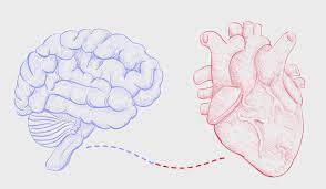
There has been a growing body of research over the past few years suggesting that the brain and the gut are capable of two-way communication. This is known as the gut-brain axis by researchers.

The gut-brain axis has been linked to a variety of brain-related illnesses, including depression and dementia. And while the relationship between the gut and the brain may be less obvious in other disorders, it is actually more evident in Parkinson’s disease, which is also sometimes accompanied by gastrointestinal symptoms like constipation.

The Braak hypothesis is one view on Parkinson’s disease. According to a reliable source, there are typically two ways for an unknown infection to enter the brain, one of which involves the gut.

The vagus nerve, the longest cranial nerve that connects the brain to, among other organs, the intestines, may be one route by which pathogens enter the body, travel through the gut, and then advance to the brain. Parkinson’s disease may then start to manifest as a result of this.

In our podcast, Dr. Demirkan recognized that it may initially seem strange to think about using your gut to learn more about Parkinson’s disease, but that the Braak hypothesis offers an intriguing lens through which to examine potential underlying mechanisms.

“Through the Braak hypothesis, there comes the idea that the disease actually starts in the intestines, and then through the vagus nerve, it spreads to the other tissues, and towards the brain,” she said.

She claims that for one straightforward reason alone, Parkinson’s disease is the neurological disorder that is most intriguing to examine regarding gut health because its gut microbiome stands out the most.

Parkinson’s disease has a distinct gut microbiota.

Dr. Demirkan and her colleagues recently discovered that people with Parkinson’s disease have unique gut microbiomes that were characterised by dysbiosis, the phenomenon of imbalance between so-called good and bad bacteria.

According to their research, those with Parkinson’s disease have gut flora that is different from those who do not have it by about 30%.

Dr. Demirkan stated in the podcast that “we found one-third of these microbes in the gut of people with Parkinson’s disease to be different.”

As a result, this strongly suggests dysbiosis. Also different were the bacteria’s modes of operation and the types of genes they possessed. We observed a decrease in the number of short-chain fatty acid manufacturers, such as gut-friendly bacteria. Escherichia coli and other harmful bacteria were identified in greater numbers, and numerous bacterial pathways were disrupted, which may have an impact on the health of the neuronal tissues,” according to Dr. Ayse Demirkan.

In the guts of Parkinson’s disease patients, Dr. Demirkan and her colleagues discovered that levels of bacteria like Bifidobacterium dentium, which can result in infections like brain abscesses, were noticeably raised.

Desulfovibrio bacteria may be related to Parkinson’s disease, according to research from the University of Helsinki that was published in the May 2023 issue of Frontiers. These microorganisms release hydrogen sulfide, which can cause different types of inflammation.

Desulfovibrio was mentioned in another study from The Chinese University of Hong Kong that was published in Nature Communications in May 2023. This study found an “overabundance” of these bacteria in persons with REM sleep behavior disorder and early Parkinson’s disease indicators. The goal of the study was to find a way to diagnose Parkinson’s disease earlier.

What potential mechanisms exist?

The question that arises is: What mechanisms might mediate gut bacteria’s impact on neurological health, assuming that they do in fact contribute to Parkinson’s disease?

Given that some of the bacteria that are overabundant in this condition are pro-inflammatory, which means they can cause inflammation, one theory raised in the studies on the connection between the gut and the brain in Parkinson’s is that systemic inflammation may be one of the processes involved.

Research reveals that immunosuppressant medicine may reduce the chance of Parkinson’s disease, which raises the possibility that a medication of a similar kind may potentially assist manage the disease.

Parkinson’s disease is characterised by chronic brain inflammation, and some studies appear to suggest that systemic inflammation may exacerbate chronic brain inflammation and speed up the course of the disease.

In fact, some inflammatory disorders have been associated with an increased risk of Parkinson’s. For instance, a 2018 Danish study found that those with inflammatory bowel disease (IBD) had a 22% higher chance of developing Parkinson’s disease than their non-inflammatory counterparts.

In the podcast, Dr. Demirkan concurred that “bad” bacteria in the stomach may be the source of inflammation associated with Parkinson’s disease. She emphasised that more investigation is required before drawing clear conclusions because this proposed mechanism is not yet established.

Could nutrition help Parkinson’s patients with dysbiosis?

It could be logical to assume that nutrition could aid in the fight against gut dysbiosis and perhaps offer a simple option for symptom treatment if gut bacteria may play a role in Parkinson’s disease.

While there are certain dietary suggestions and nutritional supplements that may help some people with symptom alleviation, it’s still not clear how much food can actually do to change how this condition develops.

According to one study from 2022, diets rich in flavonoids, which are natural pigments present in many fruits, may be associated with a lower risk of mortality from Parkinson’s disease.

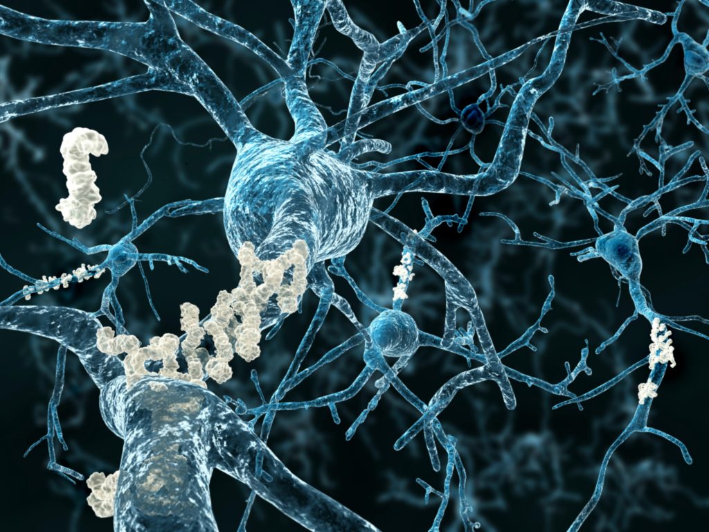
Additionally, an earlier study from 2018 suggested that a protein called parvalbumin, which is present in many types of fish, may help prevent Parkinson’s disease by preventing alpha-synuclein from clumping together in the brain, which occurs in the brains of people with Parkinson’s and disrupts signals between brain cells.

Dr. Demirkan did, however, show some scepticism when asked about the ability of food and vitamins to control gut flora in Parkinson’s patients.

She emphasised that it is challenging to provide generic advice that would truly be useful because different persons have various risk factors for Parkinson’s as well as varied forms of the disease.

“I find it very challenging to offer advice to anyone because each of us has a unique gut microbiota. Therefore, I believe that preventing the condition is one thing and that long-term maintenance, along with the various consequences of the disease, is another. I can’t really offer any advice because of this, although research indicates that consuming more sugar is problematic.

Can exercising treat Parkinson’s disease?

Nevertheless, some evidence suggests that exercising can help people with Parkinson’s disease manage their symptoms.

According to a study from 2022 that was published in Neurology, those with early-stage Parkinson’s disease may benefit from regular, moderate-to-vigorous exercise since it can delay the disease’s progression.

According to research published in 2017, doing at least 2.5 hours of exercise each week can assist Parkinson’s patients become more mobile while also delaying the onset of the disease.

Dr. Demirkan concurred that using exercise as a management tool for Parkinson’s disease can be beneficial. Exercise by itself is a fantastic technique to mould our brain and body, she claimed.

“There are some significant physiological consequences that we can consider in terms of reversing [Parkinson’s] disease. Your body must endure a lot of stress when you run a marathon, for instance. For instance, you may notice that your body temperature rises steadily and in a feverish manner. One thing is that there is a long-term rise in core heat, and it should unquestionably have a significant impact on the stomach,” she said.

In fact, some study indicates that the heat stress experienced during exercise may decrease intestinal blood flow, which may ultimately have an impact on the gut microbiota by allowing for the expansion of some bacteria while suppressing others.

In terms of the optimum type of exercise for persons with Parkinson’s disease, a Cochrane study that was released in January 2023 came to the conclusion that pretty much all types of exercise can assist those with this illness to live better lives. The authors of the review state that the available evidence “probably has a large beneficial effect” on the quality of life of aqua-based training.

REFERENCES;

For Parkinson’s disease medications that have been suggested by doctors worldwide are available here https://mygenericpharmacy.com/index.php?therapy=64

High levels of lean muscle may help prevent Alzheimer.

High levels of lean muscle may help prevent Alzheimer.

High quantities of lean muscle may help prevent Alzheimer’s disease, according to recent studies. To determine whether this connection is causal, more study is required.

Resistance training and a sufficient intake of protein in the diet are advised by experts as ways to build lean muscle mass. Previous studies have shown a link between obesity and an elevated risk of Alzheimer’s.

A recent study that was published in BMJ Medicine suggests that having a lot of lean muscle may prevent Alzheimer’s disease. The authors of the study pointed out that additional study is required to comprehend the biological mechanisms underlying it.

Researchers gathered data on 450,243 participants’ genetics, lean muscle mass, cognition, and health for this study from the U.K. Biobank. They next used a method known as Mendelian randomization to search for genetic relationships between lean muscle mass and genetic variants.

The quantity of lean muscle and fat tissue in the arms and legs was measured using bioimpedance, an electric current that moves through the body at varying speeds. The researchers next discovered 584 genetic variations related to lean muscle mass, but none of these were located in an area of the genome known to include genes connected to an elevated risk of Alzheimer’s disease.

The chance of developing Alzheimer’s disease did, however, appear to be reduced in individuals who had high amounts of lean muscle mass and associated genetic variations.

These results were confirmed by measuring the quantity of lean muscle mass and body fat tissue in a second cohort of 252,879 individuals without Alzheimer’s disease and 7,329 individuals with the condition.

The findings indicated that lean mass was associated with enhanced cognitive performance, but this association did not account for lean mass’s protective effect against the onset of Alzheimer’s disease.

The study’s objectives

The majority of the data utilized by the researchers came from the UK Biobank, a continuous database that collects health and genetic data on 500,000 people. The researchers used data from hundreds of thousands of people.

Mendelian randomization, which employs gene variations as a proxy for specific risk variables, was used to analyze the data.

Additionally, bioimpedance measurements which determine the speed at which an electrical current travels through the body based on its composition in terms of muscle and fat were used to calculate each person’s lean mass.

A total of 584 genetic variations were connected to lean muscle mass, but none of them were discovered in the region of the brain linked to Alzheimer’s susceptibility.

Participants’ chance of developing the disease was statistically significantly (12%) lower on average for those with higher amounts of (genetically proxied) lean muscle.

The analysis was performed using data from an additional 260,208 individuals, of whom 7,329 were identified as having Alzheimer’s disease, in order to confirm these findings. They measured lean muscle mass over the entire body, not only in the arms and legs.

Again, they discovered that having more lean muscle was linked to a lower risk of Alzheimer’s.

Unexpectedly, the analysis did not reveal a correlation between body fat and the probability of developing Alzheimer’s disease once lean mass was taken into account. Body fat was associated with inferior performance in cognitive activities.

The significant contrast between the protective effect of lean mass on dementia risk and the lack of an effect of fat mass on dementia risk, according to Daghlas, startled him.

How Alzheimer’s disease and lean muscle are related?

The fact that the processes underlying the association between lean muscle and Alzheimer’s disease are still unknown is another drawback of the study.

The researchers made brief speculations about possible connections. For instance, Alzheimer’s has long been linked to cardiovascular illness, though Daghlas cautioned that the connection is “complicated.”

According to Daghlas, heart disease problems like stroke and hypertension are what fuel vascular dementia. Though this is a contentious idea, he added, “the most recent causal evidence suggests weaker evidence for an effect of vascular risk factors on Alzheimer’s disease risk.”

Lean mass may very possibly lower the incidence of vascular dementia through lowering the risk of cardiovascular disease, according to Daghlas, however more research is needed in this area. “This can be looked into in upcoming studies.”

In the publication, the researchers also made the speculative claim that “new” processes, such as centrally acting myokines, may be at work.

Myokines are proteins that muscles produce that have an impact on other tissues, according to Daghlas. “Experimental studies have demonstrated that they are induced by exercise and have a positive impact on brain function.”

Other muscle-related issues, in addition to these, “may explain the larger picture,” according to Dr. Anna Nordvig, a neurologist at NewYork-Presbyterian and Weill Cornell Medicine who was not involved in the study. Examples include “bone hormones, cardio vs. strength training differences, sex hormones, glymphatic drainage depending on exercise, and sleep, to name a few.”

In the end, further clinical intervention studies are required to demonstrate the link between lean muscle and Alzheimer’s disease and the mechanisms underlying it.

The best way to build lean muscle mass

Having lean muscle mass has several health benefits in addition to possible advantages for the brain.

Resistance training using weights, bands, and pleiomorphic activities are advised by Dr. Joseph C. Maroon, clinical professor, vice chairman, and Heindl scholar in neuroscience at the Department of Neurosurgery at the University of Pittsburgh.

Additionally, he recommends supplementing with B-hydroxy B-methylbutyrate (myHMB), a good source of dietary protein.

This naturally occurring compound aids in the maintenance of a healthy weight and the development of lean muscle mass in humans. B-hydroxy helps muscles recover from hard activity, improves athletic performance, and enhances muscle and strength, according to him.

According to Dr. Sullivan, the best food, the best kind and frequency of exercise, the optimum amount of rest, and stress management are the primary factors that influence muscle growth.

These are the recommendations she makes:

Exercise: four to five quick strength-training sessions each week. In comparison to two or three longer aerobic exercises each week, this will produce greater lean muscle mass.

Diet: Put your attention on lowering insulin resistance by eating fewer carbohydrates and more protein, the building block of muscle.

Sleep: 8 to 9 hours of sleep per night are recommended if you want to recuperate from this kind of workout completely.

Stress management: With the rise in inflammation and blood sugar that stress hormones like cortisol induce, chronically high stress can sabotage any self-improvement effort. Long-term high cortisol levels can contribute to persistent muscle tension and lactic acid buildup, which can inhibit muscle growth. Moving your body more, going outside, eating more good foods, being an aggressive communicator, and finding your purpose are the simplest ways to relieve chronic stress.

How to lower your risk of developing Alzheimer’s?

Although there is no cure for Alzheimer’s, doctors think there are a number of steps you may take to lower your risk.

“Protect” and “stimulate” are the two categories that these fall under, according to Nordvig, and “physical activity falls into both of these.”

“Protect” includes monitoring factors like blood pressure, sugar levels, weight, nutrition, and sleep, she said. These are topics covered at a yearly checkup.

We should also work to safeguard ourselves from environmental risk factors associated to Alzheimer’s, according to Dr. Rena Sukhdeo Singh, a vascular neurologist at the University of Maryland Shore Regional Health.

The incidence of dementia has also been connected to fine particulate matter in air pollution.

According to Sukhdeo Singh, systemic inflammation also contributes to the development of Alzheimer’s disease. Numerous factors, including medications, a diet high in sugar and processed foods, smoking, and binge drinking, can contribute to chronic inflammation.

Optimising additional daily inputs that influence cognition is what “stimulate” entails. For instance, Sukhdeo Singh proposed, “learning a new skill, hobby, language, or instrument,” or taking part in shorter activities like “sudokus, puzzles, and number games.”

Unfortunately, there are some factors that we have no control over. Age, genetics, and sex are non-modifiable risk variables, she added.

Restrictions of this research

Researchers solely considered lean muscle mass for this investigation. However, there are other things to think about.

The protein amyloid, which is damaging to the functioning of the brain, is found in higher amounts in adipose tissue, but the researchers neglected to test these signs of inflammation and insulin resistance, according to Maroon. “This probably diminishes the significance of their findings.”

Furthermore, “while their positive finding was statistically significant, the effect size was modest in lean muscle mass reducing the risk of dementia and only explained 10% of the variance,” added Dr. Sullivan.

The link between more lean muscle mass and a lower incidence of Alzheimer’s disease has to be further investigated.

According to Nancy Mitchell, a registered nurse, “For now, people with lower muscle mass tend to be obese, which is a risk factor for type 2 diabetes.”

We refer to Alzheimer’s disease as “diabetes of the brain” because it has been hypothesized that high blood sugar harms the nerve endings in the regions of the brain that are most impacted by cognitive decline. Therefore, the link may actually be between a reduced risk of obesity and diabetes. This can be a study drawback in and of itself because greater specificity is still needed. Not all correlations indicate causation. Nancy Mitchell is a nurse practitioner.

REFERENCES:

For Alzheimer’s disease medications that have been suggested by doctors worldwide are available here https://mygenericpharmacy.com/index.php?therapy=31

Can Hormone therapy for menopause increase Dementia risk?

Can Hormone therapy for menopause increase Dementia risk?

Menopausal hormone therapy is used by about 45% of all women to lessen menopause symptoms.

According to prior studies, some types of hormone replacement treatment may make women more susceptible to developing serious illnesses.

Menopausal hormone therapy is linked to an increased risk of dementia and Alzheimer’s disease, according to researchers from Copenhagen University Hospital, says Rigshospitalet.

These results go against earlier research that suggested HRT might reduce a woman’s risk of dementia.

Menopausal hormone therapy, often known as hormone replacement therapy (HRT), is used by about 45% of all women worldwide to cope with menopause symptoms.

HRT can cause adverse effects like nausea and migraines. According to earlier studies, women who use specific forms of HRT may be more susceptible to strokes, gallbladder problems, and malignancies including breast and endometrial.

Menopausal hormone therapy is now linked to a higher risk of dementia and Alzheimer’s disease, according to study from Copenhagen University Hospital, as per Rigshospitalet.

These results go against earlier research that suggested HRT might reduce a woman’s risk of dementia. The BMJ recently published an article based on this study.

What occurs throughout menopause?

Every woman experiences menopause, which is the end of the monthly cycle and the last time the ovaries release eggs.

Menopause usually begins in a person between the ages of 45 and 55. Perimenopause, often known as the menopausal transition, can persist between seven to fourteen years.

A woman who is beginning menopause may experience symptoms like:

  • a hot flash
  • morning sweats
  • irregular or absent
  • vulvar aridity
  • difficulty sleeping
  • mood swings like anxiousness and depression

Menopause is a natural part of ageing, but it comes with some changes that some people may desire to minimise. Menopause-related symptoms may be treated with the following methods:

  • HRT
  • hormonal birth control at a low dose
  • low-dose mood stabilisers
  • prescription or over-the-counter drugs for vaginal dryness

Additionally, several lifestyle modifications can assist in relieving some symptoms:

  • routine exercise
  • wholesome diet
  • meditation techniques
  • restricting alcohol
  • giving up smoking
  • counselling for mood changes
  • maintaining a healthy sleep routine

What is hormone therapy for menopause?

The purpose of HRT is to enhance and balance the levels of the female hormones progesterone and oestrogen in the body.

Although the body’s ovaries naturally produce both of these hormones, their production declines after menopause, leading to menopausal symptoms.

There are two primary types of menopausal hormone treatment that a doctor could recommend, depending on a woman’s situation and requirements:

  • treatment with just oestrogen
  • combined treatment utilising progesterone and oestrogen

HRT can be applied topically or vaginally, and comes in tablet, nasal spray, skin patch, and vaginal cream or suppositories forms.

The following are possible HRT adverse effects:

  • bloating
  • headaches
  • breast discomfort
  • nausea
  • acne
  • mood changes
  • uterine bleeding

How Does HRT Affect the Risk of Dementia?

Dr. Nelsan Pourhadi, the study’s lead author and a researcher at the Danish Cancer Society and the Danish Dementia Research Centre in the Department of Neurology at Copenhagen University Hospital – Rigshospitalet in Copenhagen, Denmark, claims that the study’s objectives were dual and based on understudied facets of the subject matter.

“First, we sought to look into whether menopausal hormone therapy use, as advised by guidelines, increased the incidence of dementia.” Second, he told us, “we were looking into continuous versus cyclic therapy regimes.”

Dr. Pourhadi and his team used data from a national registry database for this investigation. The study’s controls were about 56,000 age-matched women without a dementia diagnosis and approximately 5,600 women with dementia. Danish women between the ages of 50 and 60 in 2000 who had no history of dementia or any conditions that would exclude the use of HRT were included in the data, which covered the years 2000 to 2018.

The subjects’ average age at the time of dementia diagnosis was 70. In comparison to controls, 32% of women with dementia and 29% of controls had used estrogen-progestin therapy starting at an average age of 53 before receiving a diagnosis. For dementia-stricken women, therapy lasted an average of 3.8 years, compared to 3.6 years for males.

Analysis revealed that women who got estrogen-progestin therapy had a 24% higher incidence of Alzheimer’s disease and all-cause dementia. Even ladies who started the treatment at age 55 or younger experienced this.

The Women’s Health Initiative Memory Study (WHIMS), the largest clinical experiment in the field, found similar results, according to Dr. Pourhadi.

Does HRT alter the risk of dementia?

Researchers have previously searched for a link between HRT and the risk of dementia.

Menopausal hormone therapy may aid in lowering a woman’s risk of dementia, according to earlier studies. Menopausal hormone therapy use was associated with a lower chance of developing all neurological disorders, including Alzheimer’s disease and dementia, according to a study released in May 2021.

Additionally, a study published in June 2022 discovered that women with depression who used HRT after menopause had a lower risk of developing Alzheimer’s disease and vascular dementia.

Several research have shown a connection between HRT use and an elevated risk of dementia. HRT was linked to a higher incidence of dementia, according to research released in September 2022.

And according to a study that was just released in April 2023, women who had HRT more than five years after the onset of menopause or who started menopause early had greater levels of tau protein in their brains, which is thought to be one of the main causes of Alzheimer’s disease.

When questioned why prior and current studies may have conflicting results, Dr. Pourhadi responded, “It is crucial to emphasise that our findings are in line with those of the largest clinical trial on the topic, WHIMS. The majority of prior clinical trials were hindered by issues like poor selection, limited sample sizes, brief follow-up periods, and results that were purely dependent on cognitive testing rather than a clinical evaluation of dementia.

Furthermore, earlier observational studies, particularly short-term ones, were unable to evaluate the use of menopausal hormone therapy close to menopause, the author continued. The discrepancies between the findings of those studies and those of our study “may be explained by these differences.”

Can HRT lead to dementia?

Dr. Pourhadi explained that because this study is an observational one and not a causal one, it is impossible to establish a link between menopause hormone therapy and dementia.

Therefore, more investigation is required to determine whether or not the observed link may be assumed to be causal. Additionally, it is important to distinguish between the various menopausal hormone therapy delivery methods such as tablets, patches, and gels,” he continued.

Dr. Mindy Pelz, a specialist in holistic medicine who specialises in women’s and hormonal health but was not part in this study, concurred.

It’s vital not to overestimate the results of this new observational study. Correlation does not imply causality, and prior research has shown that menopausal hormone therapy lowers the incidence of dementia, so it’s conceivable there’s a variable missing that we haven’t thought of yet, the researcher added.

She told us that many women go for hormone replacement medication to deal with their symptoms when they have cognitive deficiencies after menopause, which could be a sign of dementia in the future.

Dr. Jewel Kling, assistant director of women’s health internal medicine at the Mayo Clinic in Arizona and a non-participant in this study, informed us after reviewing the findings that because this was an observational study using data from a national registry, we could not draw any conclusions about the cause-and-effect relationship between menopausal hormone therapy and dementia risk.

The only way to conclude causation is through a randomised control design, which this wasn’t. “(We) can only claim that there was a relationship identified between the two in their data. According to the study’s design, there are numerous additional factors that could potentially explain this association, the researcher said.

REFERENCES:

For Mental disease medications that have been suggested by doctors worldwide are available here https://mygenericpharmacy.com/index.php?cPath=77_478

Insulin may boost cognition in cognitive disorder people.

Insulin may boost cognition in cognitive disorder people.

According to research, certain individuals with dementia-related illnesses may benefit from utilising intranasal insulin.

They claimed that those with Alzheimer’s disease and mild cognitive impairment seem to benefit most from the insulin therapy.

However, other medical professionals claimed that they believed the study to be defective and are not yet prepared to endorse insulin as a treatment for these illnesses.

According to a report published in the journal PLOS ONE, intranasal insulin may have some favourable cognitive effects, especially for those with Alzheimer’s disease and mild cognitive impairment.

Intranasal insulin and cognitive performance were studied in 29 research with 1,726 participants for a review and meta-analysis. The studies’ publications span the years 2001 and 2021.

The average insulin dosage was 40IU. The results of a single dose were investigated in ten trials. The other studies had a median duration of eight weeks and involved multiple doses over a longer period of time. The participants’ average age was around 53.

The subjects were categorised into four categories of disorders by the researchers:

  • illnesses of the mind, including schizophrenia, bipolar disorder, and major depressive disorder
  • Mild cognitive impairment with Alzheimer’s disease
  • metabolic conditions like diabetes
  • Other illnesses

Additionally, a pool of healthy, cognitively unimpaired people was used.

In persons with mental health illnesses, metabolic diseases, and other conditions, the researchers found no discernible difference in cognitive performance following dosages of intranasal insulin, according to their findings.

Participants who had mild cognitive impairment and Alzheimer’s disease showed considerable improvement, according to the researchers.

The potential link between insulin and brain function

According to Dr. Gayatri Devi, a neurologist at Northwell Lenox Hill Hospital in New York who was not involved in the study, “Patients with Alzheimer’s may have impaired glucose processing in the hippocampus (an area of the brain involved in human learning and memory).” Insulin administered intravenously may help with this and enhance cognition.

One explanation for why insulin can help with memory and cognition is that the brain’s memory centres are either defective or unable to handle sugar.

According to Dr. Shae Datta, co-director of NYU Langone’s Concussion Centre and director of cognitive neurology at NYU Langone Hospital-Long Island, “It could be plausible that the amount of insulin receptors in the memory centres in the brain become defective or are simply insufficient to handle sugar.

“Insulin replacement improves brain metabolism. resulting in the hypothesis that brain insulin resistance can cause cognitive problems, according to Datta, a researcher who was not involved in the study.

Intranasal insulin side effects include:

  • Hypoglycemia may cause heart attacks and strokes.
  • Irritation or rhinitis of the nose
  • Lightheadedness
  • Dizziness
  • Nausea
  • a nosebleed

The study’s authors came to the conclusion that intranasal insulin can be safely tolerated and may enhance memory by directly interacting with brain areas involved in the control of cognition.

Response to the study on insulin and cognitive decline

The researchers did say that additional study is required to comprehend therapy response.

Not all medical practitioners find the research to be compelling.

“Overall, I wasn’t impressed with the study,” said Dr. Clifford Segil, DO, a neurologist at Providence Saint John’s Health Centre in California who was not engaged in the study. “Intranasal insulin for diabetes has been tried, but it failed.”

“I find it unsettling to provide insulin to someone who shows no signs of diabetes. Giving insulin to a person who does not have diabetes carries the danger of hypoglycemia, he told us. This could make them more vulnerable to a heart attack or stroke.

Segil continued, “I think that it is good to repurpose medications as it can increase therapy options. But this research does not back up using this medication for memory loss. It was never employed in my practise.

“This is a meta-analysis, so a statistical compilation of multiple studies, most of them quite small,” Devi explained. “This is never as good a big double-blind placebo-controlled study as that would be crucial in patient-related decisions,” the author writes. However, each patient must be handled uniquely, and decisions about the best course of treatment must be made with that patient in mind.

Devi continued, “Intranasal insulin treatment for patients with biomarker-confirmed Alzheimer’s disease still needs a large placebo-controlled study.” Up to a third of individuals who were clinically diagnosed with Alzheimer’s did not have it on pathology, which was a concern in prior Alzheimer’s clinical studies.

REFERENCES:

For Mental disease medications that have been suggested by doctors worldwide are available here https://mygenericpharmacy.com/index.php?cPath=77_478

The link between Omega3 rich diets & slowed ALS progression

The link between Omega3 rich diets & slowed ALS progression

Numerous health advantages of omega-3 fatty acids are well documented. Harvard University researchers have discovered that those with ALS who consume meals strong in omega-3 fatty acids may experience a slower pace of physical deterioration and a longer survival time.

Additionally, researchers discovered that participants in the trial who consumed more omega-6 fatty acids had a lower risk of passing away. Omega-3 fatty acids have long been known to have several positive effects on health, according to a study.

According to earlier research, these good fats, which may be found in various plants and shellfish, may help stave off diseases like metabolic syndrome, autoimmune diseases, Alzheimer’s, age-related macular degeneration, and cardiovascular disease.

Now, researchers from Harvard University have discovered that those who consume foods rich in omega-3 fatty acids may experience a slower rate of physical decline and a longer survival time if they have the incurable, degenerative neurological disease known as amyotrophic lateral sclerosis (ALS).

Consuming omega-6 fatty acids, according to the researchers, was also linked to a lower chance of passing away among study participants.

ALS: What is it?

A condition affecting the body’s central nervous system, namely the nerve cells in the brain and spinal cord, is ALS, sometimes referred to as Lou Gehrig’s disease.

Over time, a person loses the ability to control their leg, arm, and face movements due to ALS, which damages the neurons necessary for movement. People with ALS may eventually lose the ability to talk and swallow, in severe situations.

Within a population of 100,000 individuals, there are “two new ALS cases each year,” according to the ALS Association. Males are more likely to get ALS than females, and it often strikes persons between the ages of 40 and 70.

ALS symptoms include:

  • weakness in the hands, legs, feet, or ankles
  • having trouble walking
  • falling or stumbling
  • discomfort in the shoulders or arms
  • having trouble swallowing
  • muddled speech
  • fatigue
  • cognitive problems.

Although the actual etiology of ALS is still unknown, researchers think genetics and environmental risk factors may be involved.

ALS presently has no known cure. Certain treatments can aid with symptom relief. With some individuals living longer, the typical life expectancy for someone with ALS is frequently between 2 and 5 years.

A diet for ALS

The lead author of this study, Dr. Kjetil Bjornevik, assistant professor of epidemiology and nutrition at the Harvard T.H. Chan School of Public Health at Harvard University, says he and his research group chose to investigate the relationship between diet and ALS because they were interested in identifying modifiable risk factors for neurological disorders, such as dietary factors.

He told Medical News Today, “We have done studies in the past that have shown that a diet high in omega-3 fatty acids, particularly alpha-linolenic acid, may decrease the risk of developing ALS.”

Therefore, he continued, “we were interested in investigating whether a diet high in these fatty acids is also connected with a slower rate of disease development in those who have already been given an ALS diagnosis.

This is not the first time that scientists have looked at how essential fatty acids affect ALS. A 2017 study indicated that maintaining motor neuron activity in ALS requires a mix of omega-3 and omega-6 fatty acids.

Additionally, according to study from 2019 that was published, omega-3 fatty acids could be used to make drugs to treat neurological diseases.

ALS research on omega-3 fatty acids

Dr. Bjornevik and his team gathered 449 ALS patients with an average age of 58 years for this study. Participants in the study were monitored for 18 months. 126 people, or 28% of the participants, died over that time.

Researchers measured each participant’s blood concentration of omega-3 fatty acids. Additionally, each participant earned a score between zero and 48 on 12 physical tasks, such as speaking, chewing, and swallowing, with higher values indicating higher function.

The subjects with the greatest levels of omega-3 fatty acid alpha-linolenic acid had an average score of 38.3 at the beginning of the trial, according to analysis. The average score for those with the lowest amount was 37.6.

The research team also discovered that only 21 of the 126 deaths happened in the group with the highest levels of alpha-linolenic acid in their bodies, as opposed to 37 deaths that occurred in the group with the lowest levels of omega-3 fatty acids.

Dr. Bjornevik and his team discovered research participants with the highest quantity of alpha-linolenic acid had a 50% lower risk of death during the trial, compared to those in the lowest amount group, after accounting for age, sex, and ethnicity.

According to Dr. Bjornevik’s research, some omega-3 fatty acids, such as alpha-linolenic acid, may benefit those who have ALS. He did, however, issue a warning: “Randomised clinical trials are required to establish whether supplementation with this fatty acid is beneficial.”

Omega-6 fatty acids might be helpful.

A lower chance of passing away during the study period was linked by the research team to linoleic acid, an omega-6 fatty acid.

Dr. Bjornevik noted that linoleic acid, an omega-6 fatty acid linked to a lower risk of death in his study, is also an essential fatty acid that can only be received by food.

However, it is less apparent whether and how this fatty acid benefits ALS patients. Since omega-3 fatty acids were linked to a lower chance of developing ALS in prior studies, we largely focused on them in this work,” the researcher said.

Cautious optimism

We also discussed this study with Dr. Stephen Johnson, a Mayo Clinic expert in neuromuscular diseases.

He said, “I read the study with great curiosity and cautious optimism since I am always eager for the next breakthrough that might slow down, stop, or even reverse the progression of the ALS disease“.

The study’s results are intriguing and open the door for more research, which is necessary to more precisely answer the question of whether or not specific fatty acids can prolong life and halt the advancement of ALS disease. We already have an association, but we need to do our homework to determine whether it can be replicated in the context of a more thorough scientific investigation,” according to Dr. Stephen Johnson.

Dr. Johnson expressed his preference for a sizable prospective phase 2/3, double-blind, randomized, placebo-controlled trial to assess the identified possibly advantageous fatty acids as the next stage in this investigation.

This trial should pay particular attention to participant diet, medications/supplements, and any potential confounders, in addition to measuring longevity and participant function,” he said. However, a phase 1 clinical trial will probably need to be conducted first.

Dr. Johnson continued, “By taking these next steps, just as we would with any pharmaceutical medication, we can assess whether the association is more than just an association and whether specific dietary supplementation of fatty acids should be added to the standard of care for people with ALS.”

REFERENCES:

For ALS that have been suggested by doctors worldwide are available here https://mygenericpharmacy.com/index.php?therapy=49

How can loneliness affect bone health in males?

How can loneliness affect bone health in males?

The impact of social isolation on bone loss in mice was examined by researchers. They discovered that social isolation increased bone loss in male mice, but not in female mice.

To determine whether the same occurs in humans, more research is required. Poorer health outcomes, such as an increase in all-cause mortality, cardiovascular issues, and mental health issues, are associated with social isolation.

Psychological stress has also been connected to risk factors in previous studies.reputable source for information on osteoporosis and weak bones.

Social isolation, which is closely related to loneliness, can cause mental suffering. The relationship between social isolation and bone health is still unclear, though.

Researchers recently looked into how social isolation impacts the bone health of male and female mice. They discovered that in male mice, but not in female mice, social isolation was linked to bone loss.

The research was introduced in Chicago at ENDO 2023, the Endocrine Society’s annual meeting. The study’s non-participant assistant professor of geriatrics at McGovern Medical School at UTHealth Houston, Dr. Nahid Rianon, was our source for information on the results.

Lead author of the study and postdoctoral fellow at MaineHealth Institute for Research’s Centre for Molecular Medicine, Dr. Rebecca Mountain, also provided the following information to us.

Future research is required to fully understand the effects on humans, but the findings “may also have clinical implications as we grapple with the long-term health impacts of the rise in social isolation related to the COVID-19 pandemic.”

The bone density was decreased in isolated mice.

32 male and female mice aged 16 weeks were divided into two groups for this investigation. One mouse per cage was used in one group’s simulation of social isolation. Four mice shared one cage in the other group.

The mice were observed by the researchers in their separate environments for 4 weeks. Finally, the bone mineral density of solitary male mice decreased. Less dense and therefore more likely to break, bones with fewer minerals are less dense.

Additionally, the researchers discovered that in solitary male mice, bone volume fraction and cortical bone thickness decreased by 26% and 9%, respectively. Both measurements point to a decline in bone quality.

Further investigation found that male mice showed impaired bone remodelling, which includes the production of new bone and raises the risk of fracture.

The scientists observed that the bone loss seen in guys who were kept alone was comparable to that shown in earlier studies after orchiectomy (removal of the testicles) and ovariectomy (removal of the ovaries).

In contrast, there was no bone loss after social isolation in the present study’s female mice.

However, despite the fact that their bone mass was unaltered, the researchers discovered that isolated females exhibited higher bone resorption-related gene expression. Increased bone resorption can cause bones to degrade more quickly than they can regenerate, raising the risk of fracture.

Underlying processes

Dr. Mountain pointed out that it is unknown exactly how social isolation may cause bone loss. Her team is investigating many hypotheses, including the function of various stress hormones and the sympathetic nervous system of the body.

We also discussed the mechanisms underlying the effects of social isolation on bone health with Dr. William Buxton, a board-certified neurologist and the director of Neuromuscular and Neurodiagnostic Medicine and Fall Prevention at Pacific Neuroscience Institute at Providence Saint John’s Health Centre in Santa Monica, California, who was not involved in the study.

“My initial reaction to the connection is that performing weight-bearing workouts is one of the finest methods to preserve bone health and fend off osteoporosis. One is less inclined to leave their home if they are isolated, and as a result, they are less likely to be on their feet, he explained.

Both depression and weight loss can result in frailty, disability, and decreased mobility, which can contribute to bone loss,” continued Dr. Rianon. Future study is required to understand the underlying metabolic alterations that cause bone loss in various medical diseases, as they are all risks for bone loss.

Why is there a sex difference?

Dr. Mountain mentioned that they are currently looking into why social isolation had distinct effects on men and women. She mentioned that oestrogen is known to protect bones, suggesting that it might be involved.

It’s also plausible that male and female mice experience solitude on different time scales or in various ways, she added.

We also discussed the sex disparities with Dr. Douglas Landry Jarvis, an orthopaedic surgeon with Novant Health in Charlotte, North Carolina, who was not involved in the study.

The synthesis of testosterone and hormonal balances may have been disrupted by a lack of social engagement, which would have had a negative impact on bone metabolism. Over a 4-week period, the female hormonal cycle may be less impacted.

Study restrictions

The study’s weaknesses, according to Dr. Mountain, are its small sample size and lack of behavioural information on how isolation influenced mice’s depressive or anxious behaviour.

Dr. Buxton said that the study’s use of caged animals meant that it was not a perfect representation of human behaviour. I don’t know if the authors documented how frequently the animals in the cages were on their feet, but I would anticipate that the community animals would be more mobile.

Dr. Rianon continued by saying that although the study suggested that male and female mice may have different bone-forming processes, it does not specify how these variations arise.

Nevertheless, she added, “It’s pretty normal to not have [such] details in the early stages of any research.”

Research implications for the future

Dr. Buxton added, “I also guess that alcohol plays a role if these results are later demonstrated in humans.”

We are aware that drinking makes osteoporosis more likely. Alcohol is probably a link between isolation and lower bone mineral density in individuals because isolation is a risk factor for increased alcohol consumption“, the scientist hypothesized.

According to the study, no one should alter their routines, Dr. Jarvis continued. The study’s sole recommendation is that more research be done. The variable of social engagement is too broad. The amount by which the mice’s cortisol levels changed will determine if the study can be generalized to primates and perhaps even humans.

REFERENCES:

For Bone disease medications that have been suggested by doctors worldwide are available here https://mygenericpharmacy.com/index.php?therapy=63

Alcohol abuse may hasten the stream of Alzheimer’s disease.

Alcohol abuse may hasten the stream of Alzheimer’s disease.

According to recent studies conducted by researchers from Scripps Research and the University of Bologna, the occurrence of alcohol use disorder (AUD) in combination with a genetic predisposition may hasten the progression of Alzheimer’s disease.

The study, which was carried out on mice, shows that repeated episodes of alcohol intoxication in rodents with a hereditary propensity for Alzheimer’s cause changed gene expression patterns, indicating a quicker advancement of the disease in their brains.

Regardless of alcohol usage, these findings illuminate the molecular mechanisms driving memory loss and could have wider ramifications for comprehending and treating Alzheimer’s disease.

According to a recent study, mice exposed to regular high amounts of alcohol showed signs of cognitive loss about two months earlier than they would have otherwise.

The onset of Alzheimer’s disease (AD) is sped up by several months or perhaps a few years when ethanol is added to a genetically predisposed population.

While there hasn’t been much research on how alcohol affects Alzheimer’s disease progression, epidemiological studies have found a link between alcohol use disorders and an increased risk of dementia in general.

The researchers carried up an experiment where mice were repeatedly exposed to alcohol over months, simulating the levels of alcohol exposure reported in people with alcohol use disorders. This was done to study the effect of alcohol on Alzheimer’s disease.

They compared the actions of mice with three particular gene mutations linked to an increased risk of Alzheimer’s disease to animals without these abnormalities.

Is there a link between alcohol use and dementia?

The director of the Mary S. Easton Centre for Research and Care at UCLA and a professor of neurology at UCLA, Dr. Keith Vossel, who was not involved in the study, told us that the current research appears to support earlier results about dementia and alcohol use.

According to Dr. Vossel, drinking too much alcohol—more than 21 units per week—has been linked to an increased risk of dementia.

The new study is “fascinating, and the more research that can be done on neurodegenerative diseases like Alzheimer’s disease, the more answers that can then be obtained,” said Dr. Nima Majlesi, an emergency medicine physician and director of Medical Toxicology at Staten Island University Hospital who was also not involved in the study.

There has never been any question in the medical community that excessive alcohol consumption and frequent intoxication [are] unhealthy. On occasion, people have questioned if consuming a small amount of alcohol each day can be healthy. Excessive alcohol consumption and recurring drunkenness [have] many negative impacts on human health, even in patients who are not at risk for [Alzheimer’s disease],” Dr. Nima Majlesi is a doctor.

Dr. Majlesi nevertheless expressed concern, saying that “in this study, they exposed mice to ethanol vapors, which is not the typical route for human consumption.”

We are aware that drinking by inhalation can result in higher brain alcohol concentrations than drinking by mouth. When exposure to ethanol avoids the [gastrointestinal] tract, the metabolism is altered. This may result in some variables that make it a little more challenging to understand the study, according to Dr. Majlesi.

Drinking alcohol and genetic risk may accelerate Alzheimer’simer’s

The current study’s findings showed that mice exposed to alcohol showed a steady loss in their capacity to learn and retain spatial patterns. In addition, these cognitive deficits were present in the alcohol-exposed mice at a younger age than in the control group.

Before the traditional period when such deficits would show, the researchers saw cognitive impairments in the mice treated with alcohol about two months earlier.

The researchers examined gene expression in the brains of mice that had been exposed to alcohol and mice that had not in great detail in order to comprehend the underlying mechanisms of alcohol use disorder.

They compared the gene expression profiles of over 100,000 different cells under examination. The findings showed that prefrontal brain gene expression was significantly altered after alcohol intake.

In particular, the mice that had been exposed to alcohol showed elevated expression of genes linked to inflammation, neurodegeneration, and neural excitability.

Supporting cells including astrocytes, microglia, and endothelial cells also showed altered gene expression patterns in response to alcohol exposure, indicating that these modifications weren’t just restricted to neurons.

Before, it was thought that only neurons could account for the symptoms of Alzheimer’s. These other cell types have just lately been found to have a part in the progression of the disease.

Alcohol changes gene expression may lead to memory loss

The gene transcription profiles of alcohol-exposed mice and control mice at various ages and stages of Alzheimer’s disease but with the same genetic background were compared by the researchers.

They found that the alcohol-exposed mice’s gene transcription profiles resembled older mice who were exhibiting more severe cognitive deterioration rather than mice their own age.

The researchers found that alcohol exposure changed the gene expression patterns in a way that was typically associated with advanced stages of the disease when they compared the alcohol-exposed mice to the same type of mice at various stages of Alzheimer’s disease progression, including mice without any impairments and severely compromised mice.

The significant findings were emphasized for us by Dr. David Hunter, an assistant professor of neurology at McGovern Medical School at UTHealth Houston who was not involved in the study. He explained:

“In this investigation, mice were given alcohol. Some of the mice had the Alzheimer’s disease-causing human genes. Normal control mice were the others. In comparison to sober mice, mutant mice exposed to alcohol experienced cognitive damage early. Neither alcohol nor the control mice were affected.

Dr. Hunter continued, “The researchers also examined gene expression in the mice and discovered that the mutant group that had alcohol had some differences from the sober mutants.”

Mice do not naturally develop Alzheimer’s disease, therefore creating animal models of the condition is intrinsically difficult. We must introduce numerous lethal mutations to them,” said Dr. Hunter.

Does drinking affect dementia in other ways?

According to Dr. Majlesi, “Common sense tells us if we eat clean healthy foods daily, maintain a healthy weight, exercise daily, sleep well, and have little stress, we decrease our risk for several diseases.”

In his statement, Dr. Hunter noted that “neurologists are well aware that chronic and excessive alcohol consumption is bad for the brain.”

Alcohol is a statistical risk factor for all dementia causes, as this article’s opening states. Even in sporadic patients, it appears likely that it speeds up the development of Alzheimer’s pathology. This article clarifies how that link’s mechanism works. The primary takeaway for the general population is that cutting back on alcohol consumption is great advice for keeping in shape.

There is a condition known as alcoholic dementia, a neurodegenerative illness unrelated to Alzheimer’s. It manifests as alterations in visuospatial processing and executive function. The signs are similar to those of Alzheimer’s, according to Dr. Hunter.

Dr. Vossel concurred and said, “There is also a rare form of dementia called Marchiafava-Bignami Disease associated with excessive alcohol intake and malnutrition.”

“More study on this subject is necessary. According to Dr. Vossel, this study “provides evidence that excessive alcohol consumption can affect genetic changes in the brain related to Alzheimer’s disease.”

REFERENCES:

For Alzheimer’s disease medications that have been suggested by doctors worldwide are available here https://mygenericpharmacy.com/index.php?therapy=31

Can slowing and weakening grip be signs of dementia?

Can slowing and weakening grip be signs of dementia?

We lose muscle mass as we age. Humans lose roughly 10% of their body’s muscular mass by the age of 50. The beginning stages of dementia in elderly individuals may be signaled by a decline in physical strength, according to Edith Cowan University researchers.

We all know that as we age, our muscular mass declines. Grip strength and how quickly and steadily people stand up from a chair are two methods to gauge this.

According to research, people start losing muscle mass in their 30s or 40s, and by the time they are 50, they have lost 10% of their total muscle mass. Then, between 50 and 70, we lose an additional 30% of our muscular mass.

Dementia is a broad term for illnesses that affect a person’s cognitive function. Now, researchers from Edith Cowan University in Australia have discovered evidence suggesting a loss of muscle strength may be a signal for older people getting dementia.

Timed Up and Go (TUG) and grip strength were utilized by the researchers as indicators of muscle strength.

Why measure grip strength?

A grip strength test may be used by medical professionals to assess a patient’s hand and forearm muscle health.

The tool used for this straightforward test is a dynamometer. Simply by squeezing the dynamometer with all of their might while holding it in their palm, the user may measure the amount of force applied.

A person’s grip strength has long been regarded as a diagnostic of their general health. Previous studies have linked diminished grip strength to a higher risk of cardiovascular and all-cause mortality.

Additionally, previous research discovered that grip strength is a good predictor of an ageing person’s health-related quality of life.

What exactly is a TUG test?

Doctors can assess a patient’s mobility and balance using the Timed Up and Go (TUG) exam.

A person is instructed to sit on a typical chair while taking the TUG test. The individual is instructed to stand up, move to a queue approximately 10 feet from the chair, turn around, move back to the chair and then sit back down in it while a medical practitioner measures them with a stopwatch.

The doctor can observe the patient’s gait and examine their mobility to look for postural or balance problems.

The TUG test can typically be completed in 10 seconds or less by most healthy older persons. A person may be at a higher risk of falling if the time is longer than 13.5 seconds.

According to earlier research, the TUG test assesses older persons’ risk of falling and predicts their level of frailty.

Dementia and muscle tone

Dr. Marc Sim, senior research fellow in the Nutrition & Health Innovation Research Institute at Edith Cowan University in Joondalup, Australia, and the study’s first author claims that they chose to look into a connection between muscle function and dementia because existing research indicates significant connections between physical capacity, including muscle mass (e.g. sarcopenia), and cognitive health.

He explained to us, “When considered in the context of dementia, at a community level, the utilisation of quick, affordable, and straightforward tests such as grip strength and TUG are quite appealing.”

This could be used as a screening tool to assist physicians in identifying patients who are most at risk and to facilitate the promotion of primary preventative interventions, like exercise and diet. At the community level, screening for dementia risk is infrequently done, he added.

According to Dr. Sim, the methods they used to assess the muscular function of research participants—grip strength and TUG tests—were chosen because they are both straightforward and simple to carry out and are currently advised as functional tests as part of sarcopenia criteria in Australia.

These tests, which take around 3 minutes to complete by doctors, have extremely strong evidence supporting their ability to predict a variety of unfavorable outcomes in older populations, including falls, fractures, CVD, and mortality.

Dementia risk factors that are significant

More than 1,000 women with an average age of 75 were examined for this study by Dr. Sim and his team using information from the Perth Longitudinal Study of Ageing in Women.

Each lady took the TUG test and had her grip strength evaluated by researchers. After five years, the tests were conducted once more.

The researchers discovered that over the following 15 years, 17% of study participants either experienced a dementia event, such as a dementia-related hospitalization or death.

A study participant developing dementia was shown to be much more likely to have reduced grip strength and slower TUG, according to the research team. In addition, women who performed poorly on the TUG tests and had the weakest grip strength had a twofold increased risk of developing dementia in later life.

Other risk variables like smoking, drinking, and levels of physical activity had no bearing on this.

I would not say the results are shocking because these functional tests are probably going to give a snapshot of the current health state. Especially because exercise, a major risk factor for dementia, is less likely to be practiced by persons who struggle with everyday routines of life due to physical constraints,” Dr. Sim said.

“What caught my attention was the significant risk of dementia related with functional loss over a five-year period, where individuals who experienced the greatest deterioration were also at the highest risk. This is another crucial topic that clinicians should think about, he continued.

How the loss of grip strength affects cognitive function

After reading this study, Dr. Raphi Wald, a board-certified neuropsychologist at Baptist Health South Florida’s Boca Raton Regional Hospital who was not involved in it, told us that it is helpful in confirming what we already knew and strongly suspected about deteriorating physical and mental abilities.

Before substantial degeneration starts, a number of frequently subclinical signs and symptoms of dementia appear. This is just another indication to physicians that a process might be starting and has to be attended to, he said.

When asked what he would want to see as the next stages for this research, Dr. Wald responded, “I think it would be great to have additional information regarding those people that successfully address their muscle weakness and how much it lessens their risk for dementia once they do so.”

These results are consistent with earlier studies that have shown that grip strength and mobility are correlated with various aspects of cognitive decline in older adults, according to Ryan Glatt, senior brain health coach and director of the FitBrain Programme at Pacific Neuroscience Institute in Santa Monica, California, who was also not involved in this study.

Collecting information on strength, gait, and mobility in clinical settings might assist create a comprehensive collection of information that may be useful in predicting the likelihood of dementia. The relationship between these mobility and strength measurements and other facets of brain health would be intriguing,” he continued.

Looking out for indications of cognitive deterioration

We also discussed this research with Dr. William Buxton, a board-certified neurologist who serves as the director of fall prevention and neuromuscular and neurodiagnostic medicine at the Pacific Neuroscience Institute at Providence Saint John’s Health Centre in Santa Monica.

Dr. William Buxton stated, “This study tells us that the extra few minutes to perform some really basic simple testing in the office, even just routine visits, like the Time Up and Go test and testing grip strength, can give us good, scientifically grounded reasons to encourage physical activity.”

Dr. Buxton, who was also not involved in the study, continued, “And for those individuals to pay a little bit closer attention to watching for cognitive decline that we may be able to slow down as a result of paying that extra attention.”

Dr. Buxton stated that he would like to see a comparable study conducted in men as well as research to see whether working out in a group had any further advantages.

We are aware that persons who maintain their social connections are less likely to encounter cognitive impairments that are either developing or deteriorating. Therefore, I’d be interested to see if group exercise that is linked to social engagement will have additional benefits in addition to those that come from individual exercise,” he continued.

REFERENCES:

For Alzheimer’s disease medications that have been suggested by doctors worldwide are available here https://mygenericpharmacy.com/index.php?cPath=77_478

Adults with dementia might be helped with internet surfing.

Adults with dementia might be helped with internet surfing.

A new study found a strong correlation between older adults’ regular internet use and a much lower incidence of dementia.

A “sweet spot” of up to two hours a day of internet use was also discovered by the study, beyond which the risk of dementia is expected to rise.

Experts advise providing assistance to elderly folks so they can use new web technology and remove access restrictions.

The impact of internet use among elderly people as a strategy for preventing dementia is the subject of a recent study.

According to the study, older adults who regularly use the internet have a roughly 50% lower risk of developing dementia than older adults who do not.

In this study, 18,154 persons without dementia were followed for an average of 7.9 years and up to 17.1 years to assess their cognitive health. The study’s participants ranged in age from 50 to 64.9 at the outset.

When compared to non-users, internet users who used it frequently had a 43% lower incidence of dementia. A dementia diagnosis had been made for 4.68% of the participants by the end of the trial.

With a U-shaped data curve, the study also hypothesized that the positive impacts of internet use varied with people’s levels of online activity.

The results indicate that the biggest reduction in dementia risk was seen in persons who used the internet between 0.1 and 2 hours per day.

Was that of any use?

A greater risk of dementia persisted for those who never used the internet or spent more than two hours online. The authors do warn that due to the small sample sizes, there were no discernible changes between user groups.

The study’s authors also investigated the effects of generational differences, educational level, race-ethnicity, sex, and gender on the relationship between internet use and dementia risk. They discovered that these factors had no effect on the risk of dementia.

Optimum level of internet usage

There was sort of a sweet spot where, if you used the internet for a half-hour to two hours a day, it was protective against dementia, according to Dr. Scott Kaiser, an expert in geriatric family medicine at the Pacific Neuroscience Institute who was not involved in this study.

He emphasized that “too much internet time was not protective, or potentially harmful.”

Dr. Kaiser is a co-founder of Determined Health, a group devoted to assisting seniors in fortifying their social ties.

Dr. Kaiser pointed out that older people who spend too much time online may be “highly exposed to negative images of aging, and feeling lower self-worth, and feeling bad about getting older that would be an example where too much time could potentially have a negative effect.” This is known as “doom-scrolling,” or compulsively scrolling through social media feeds laden with bad news.

A sedentary, unhealthy lifestyle may be encouraged by spending too much time online. The study’s conclusions could be impacted by the fact that it did not precisely record what its participants performed online.

Additionally not participating in the study, Dr. Snorri Bjorn Rafnsson of the University of West London in the United Kingdom told us that “these specific results merit further investigation.”

What could be the causes of some older individuals using the internet too much? Do they feel alone? isolated socially? What other dangers to their bodily or mental health could there be? What is happening among individuals who don’t use the internet at all, on the other hand? Dr. Snorri Bjorn Rafnsson stated, “I believe that these are questions that could be further investigated in studies in the future.”

How internet use may help ward against dementia?

We know that learning new things and maintaining cognitive engagement is critical for protecting our brains and reducing our risk of developing dementia,” says Dr. Kaiser.

Because learning new information and using new technologies may excite the brain and improve people’s cognitive performance, we might say that using the internet in later life may have direct cognitive benefits.

Dr. Rafnsson pointed out that older persons could utilize the internet to look up general information or health-related information. Another factor encouraging elderly individuals to use the internet is the development of telemedicine.

Regular internet use may also result in positive interpersonal interactions. In a report titled “Our Epidemic of Loneliness and Isolation,” the U.S. Surgeon General discusses the significance of social connection.

How do seniors feel about getting older?

In general, engaging in online activities may encourage a positive outlook on aging, which can have favorable effects on one’s health. Dr. Becca Levy, the author of Breaking The Age Code, was mentioned by Dr. Kaiser.

It is “an amazing work of where we know that our perceptions of ageing actually impact how we age in terms of our longevity, our risk of dementia, just the very way that we think about ageing,” he said.

Dr. Kaiser proposed three mechanisms by which age stereotypes can influence the likelihood of dementia and ageing:

  • It is well known that having a pessimistic mindset can be unhealthy.
  • A recipe for poor health is to treat one’s body like an old vehicle that won’t be on the road for very long.
  • Stress-related elevated cortisol levels and systemic inflammation.

Increasing accessibility to the internet

The suggestion made by Dr. Rafnsson is that “older adults should be supported to learn and use new online technology for whatever purpose they wish.”

He said, “There are many elderly persons who still encounter numerous obstacles, such as a lack of technical skills, cost, social support, etc.

Dr. Rafnsson added, “These hurdles may make it difficult for many older persons to gain from the cognitive and social advantages of utilizing the internet.”

Dr. Kaiser opined that “we should be working towards a more connected society for all.”

Risk factors for dementia

Scientists are still researching dementia, but they have identified some risk factors that can increase your likelihood of getting the disease. The following are some examples, according the Centres for Disease Control and Prevention (CDC):

  • Advancing years. People 65 and older are most commonly affected by dementia.
  • Family background. The likelihood of someone having dementia increases if they have parents or siblings who have the disease.
  • Race/ethnicity. Dementia strikes older Black Americans twice as frequently as older White Americans. Dementia is 1.5 times more likely to affect Hispanic people than White people.
  • Poor heart health. If diseases like high blood pressure, high cholesterol, and smoking are not effectively managed, they raise the chance of dementia.
  • Brain damage caused by trauma. The risk increases with head injuries, particularly when they are serious or frequent.

REFERENCES:

For Alzheimer’s disease medications that have been suggested by doctors worldwide are available here https://mygenericpharmacy.com/index.php?cPath=77_478

A new study finds human brain activity even after death.

A new study finds human brain activity even after death.

Recent studies have revealed an increase in brain activity just before death. The activity occurs in a region of the brain associated with dreams and other forms of altered awareness.

The findings, according to researchers, may help explain the dramatic near-death experiences (bright lights, hallucinations) that patients who were in danger of dying have described.

What transpires to our consciousness once we pass away?

When it comes to the human condition, it’s conceivably the biggest query and a major source of anxiety.

People who have had near-death encounters may be able to give a tantalizing view of how our dying minutes would feel and appear. People have reported having intense experiences that include seeing deceased loved ones, seeing dazzling lights, and having the sensation of flying through the air. Many of these encounters are startlingly similar to one another.

Although many people perceive near-death experiences through a religious or philosophical lens, University of Michigan researchers have researched the phenomenon on a scientific level and discovered a surge in brain activity at the moment of death.

This week, the Proceedings of the National Academy of Science published their findings.

The study was directed by Jimo Borjigin, Ph.D., an associate professor in the University of Michigan’s Departments of Molecular & Integrative Physiology and Neurology. It expands on prior studies that revealed a neuronal surge in the dying brains of animal test participants.

Although the study has its limitations, experts think it’s a huge step towards understanding the underlying causes of dramatic near-death experiences and provides a window into what our final minutes might be like.

Studying near-death experiences can be challenging.

While performing tests on animals is rather straightforward, studying people who are nearing the end of their lives is challenging from both an ethical and practical standpoint.

To determine whether the human brain responded to death in the same way as the rat brain, Prof. Borjigin and her team wanted to do justice to the earlier research.

To achieve this, researchers looked at the brain activity of four patients who passed away while having an EEG (electrogram) machine on them in hospitals.

Prof. Borjigin explained that even though there were just four patients, “the data generated is massive, so we were only able to report a fraction of the features that it’s actually showing on the data.”

The TPJ region of the brain, so termed because it is the junction between the temporal, parietal, and occipital lobes in the rear of the brain, was active at the time of death.

According to Prof. Borjigin, “I really wanted to be able to define something in the brain that can possibly explain that subjective near-death experience.” If some of these patients had lived to tell their tales, they might have, but sadly they didn’t.

Brain activity while dying

Researchers looked at four patients who died from cardiac arrest while their EEGs were being monitored. The patients were taken off life support because they were unconscious, unresponsive, and unable to receive any more medical attention.

Two patients demonstrated a rise in heart rate and a spike in gamma wave activity in the brain, which is the fastest sort of activity and is associated with consciousness, after being taken off the ventilator.

The region of the brain connected to dreaming, visual hallucinations in epilepsy, and altered states of consciousness was also where the activity was discovered.

One of the study’s authors and associate professor at the University of Michigan’s departments of Molecular & Integrative Physiology and Neurology, Jimo Borjigin, Ph.D., outlined the key findings of the research.

According to Borjigin, the dying process can first engage the brain. “Secondly, we must look at how the brain functions during cardiac arrest. If the brain is more active while a person is dying, why? Before our study, we were unaware of some brain processes.”

Concealed awareness

Though it is hard to know what the patients went through during these brain surges, the heightened TPJ activity may be able to shed some light on why certain people have extremely vivid near-death experiences.

The increase in brain activity indicated covert consciousness, or consciousness that cannot be seen by bedside tests because the patient is disabled, even if the patients weren’t visually conscious.

According to Prof. Borjigin, “people who have had near-death experiences] may remember seeing or hearing things, as well as having an out-of-body experience or motion perception as if they are flying.” “I believe that we may have identified or described the bare minimum anatomical processes leading to covert consciousness neuro signatures“.

We would like to be able to examine humans in less traumatic situations where the patients are known to be able to live and then tell the story where they can relate their brain signature to a personal experience“.

Patients who survive cardiac arrest while being monitored by an EEG device could be questioned to see if their brain waves correlate with their experience to make this conclusion.

In any case, examining the brain waves of terminally ill people can help us better comprehend the dying process, which is still relatively mysterious.

It may be possible to get fresh insight into the nature of consciousness by investigating how the pulmonary system, cardiac system, and brain interact.

what happens as we die?

Dr. Andrew Newberg, a neurologist, the head of research at the Marcus Institute of Integrative Health, and a physician at Jefferson University Hospital, said, “We don’t fully know the answer to this question.” Before this study, it was believed that the brain simply stopped working, but several additional studies of a similar nature reveal that there is unique brain activity connected to the close-to-death state.

The temporoparietal junction and the prefrontal cortex, two regions of the brain linked to cognitive functions, were revealed to be implicated in the study.

The sympathetic nervous system, which regulates the body’s “fight-or-flight” reaction, has been linked to these parts of the brain, according to Newberg, who also noted that spiritual experiences have been linked to these regions of the brain as well.

A bioethicist responds to the research

The study’s main finding is that the dying process causes some chemical alterations in the brain. It clarifies why individuals think they can see angels or a light at the end of the tunnel, according to Arthur Caplan, a bioethicist at NYU Langone Medical Centre in New York. What it demonstrates is that the brain has mechanisms in place to try and rouse itself awake and that it can shoot off in hallucinatory ways.

Many people are interested in what occurs after death, but this study is in its early stages and doesn’t explore that topic, he added.

In some respects, our study clarifies people’s anxiety that perhaps they will suffer as they are about to pass away at the last minute, even if that doesn’t appear to be the case. However, this study suggests that you don’t have any insights into what really happens when you pass away, so people may be let down,” Caplan said.

Summary

A recent study found that comatose individuals who died from cardiac arrest experienced an increase in brain activity that resembled conscious awareness.

The area of the brain that is linked to dreaming, visual hallucinations in epilepsy, and altered states of consciousness were where the activity was discovered.

Experts are optimistic that by pursuing this research avenue and knowing more about the dying brain, they will one day be able to save cardiac arrest victims.

REFERENCES:

For Mental disease medications that have been suggested by doctors worldwide are available here https://mygenericpharmacy.com/index.php?cPath=77_478