Browsed by
Category: Nervous system

New treatment may stop and potentially reverse some nerve damage in MS

New treatment may stop and potentially reverse some nerve damage in MS

The neurological condition known as multiple sclerosis (MS) is a chronic illness that can cause paralysis, vision loss, and muscle weakness. It happens when the myelin sheath, which envelops and shields nerve cells, is attacked by the immune system. Current therapies seek to inhibit the immune system to stop additional nerve cell damage. A new study has produced a therapy that may be able to reverse the damage caused by multiple sclerosis and even help regenerate myelin.

The autoimmune disease known as multiple sclerosis (MS) causes the immune system to target and destroy the nerve cells’ myelin sheath. People in their 20s to 40s are the most common age range to experience it. The myelin sheath is attacked by immune cells, which results in inflammation and blocks the flow of nerve impulses throughout the body. This can cause neurological symptoms, such as fatigue, vertigo, bowel and bladder issues, muscle weakness, numbness, tingling, and pain, mobility issues, and vision loss.

Current therapies can alleviate symptoms, lessen the frequency and intensity of relapses, and slow the disease’s progression all while there is currently no known cure. Current treatments for MS work by targeting the immune system, which reduces the likelihood that the immune system will attack the protective myelin coating around nerves. Now, researchers have developed a treatment that can help regenerate myelin around nerve cells, potentially reversing the damage caused by MS. However, we also need to figure out how to fix the myelin that has already been harmed.

Restoring the protective sheath of nerve cells
Cells known as oligodendrocytes produce the myelin sheath that envelops and shields nerve cells. Myelin sheath damage cannot be repaired in an MS patient because these cells are lost in the disease. Regenerating myelin and activating oligodendrocytes have not always been successful in animal studies. A study on mice indicates that improving myelin production could be accomplished through an epigenetic strategy. A recently developed drug called PIPE-307 blocks the M1R receptor, allowing the oligodendrocyte precursor cells (OPCs) to differentiate into oligodendrocytes that can then form new myelin sheaths. The OPCs fail to differentiate into oligodendrocytes in people with MS. The researchers used this toxin from the venom of a green mamba snake to identify and locate this receptor protein on OPCs. Once the drug’s receptor had been identified and demonstrated to be able to block it, the researchers tested the drug’s effectiveness in vitro using isolated OPCs.

The medication caused the OPCs to develop into oligodendrocytes and start myelinating neighboring nerve axons because it blocked the M1R receptor more effectively than other medications. Additionally, because it could pass through the blood-brain barrier, it might be able to repair damaged brain nerve cells. After researching the biology of remyelination, we have developed a precise therapy to activate it, which is the first of a new class of MS therapies. Ten years ago, we found a way for the body to regenerate its myelin in response to the appropriate molecular signal, reversing the effects of MS.

Animal and phase 1 human trials show promise
Using slices of mouse brain tissue, the researchers conducted additional in vitro experiments and discovered that PIPE-307 enhanced the myelination of nerve cell axons. The drug was then given orally to mice (MOG-EAE mice) that had been genetically engineered to develop inflammatory demyelination as a model for multiple sclerosis. The mice not only displayed enhanced nerve cell myelination, but they also regained some of their lost functionality. This research, which used human tissue and animals, indicates that PIPE-307 has the potential to be used as a myelin repair treatment. Because PIPE-307 was well tolerated and had no negative effects in a phase 1 trial in healthy people, the researchers are moving on to a phase 2 trial to determine whether it is an effective treatment in people with MS. But to truly know whether this medication will be effective, we must see the outcomes of clinical trials involving MS patients.

Early progress could give hope to MS patients
With differing degrees of success, other medications, such as the first-generation antihistamine clemastine, have been studied as possible myelin repair therapies. The news release quoted Ari Green, MD, co-author of the paper and chief of the division of Neuroimmunology and Glial Biology in the UCSF Department of Neurology, as saying: “Clemastine is not a targeted drug, affecting several different pathways in the body.”. However, we quickly observed that its pharmacology with muscarinic receptors may lead to the development of more effective restorative treatments for MS patients.

Furthermore, their results imply that PIPE-307 is more successful in myelin restoration and M1R receptor blocking. It is still very early, though, and the recently started phase 2 trial will need to demonstrate that the medication is both safe and effective to take, with no serious side effects. As Astbury concluded, there is a critical need for efficient treatments for MS patients. Over 150,000 MS patients reside in the United States. K., and many of them lack access to any kind of care. We hope to see a cocktail of medications in the future that can stop immunological reactions, restore myelin, and shield nerves from additional harm. If additional trials prove fruitful, PIPE-307 might be included in that mix.

REFERENCES:

https://www.aol.com/treatment-may-stop-potentially-reverse-144117447.html

https://www.hopkinsmedicine.org/news/articles/2024/02/tipping-the-balance-in-ms#:~:text=Although%20there%20currently%20is%20no,MS%2Dlike%20symptoms%20in%20mice.

https://www.medicalnewstoday.com/articles/new-treatment-may-stop-potentially-reverse-nerve-damage-ms

Medications that have been suggested by doctors worldwide are available here
https://mygenericpharmacy.com/
Possible link between migraine and carpal tunnel syndrome.

Possible link between migraine and carpal tunnel syndrome.

Researchers looked into the prevalence of migraine headaches in patients who have nerve decompression surgery.

They discovered that people who have surgery to decompress a nerve at particular points on their bodies may be up to 70% more likely to get migraines than others who have the procedure elsewhere.

To determine whether nerve decompression can treat migraines, more research is required.

There may be pain and a loss of function when the nerves in the hands and arms contract around muscles and soft tissues. Between 5% and roughly 9% of the population are affected by various types of nerve compression in these places.

Surgery is frequently used to treat the illness and might result in full or partial symptom relief.

The muscles, blood arteries, and bone in the vicinity of the head’s surrounding nerves can also compress those nerves. Improvement or alleviation from migraine and headaches may result by decompressing these nerves.

What is Carpal tunnel syndrome?

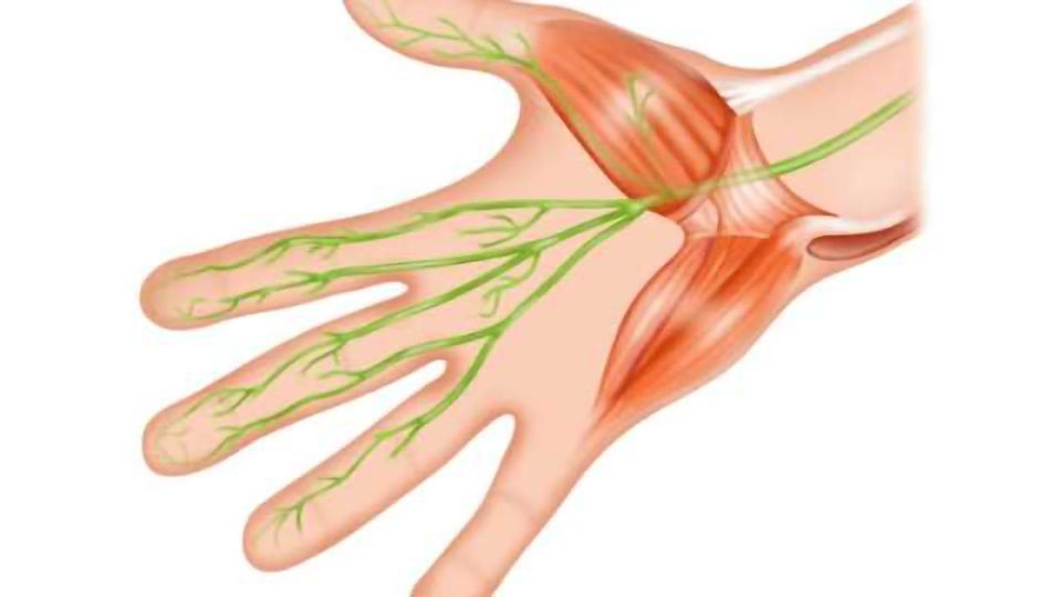
Carpal tunnel syndrome is caused by compression of the median nerve. On the hand’s palm side, the carpal tunnel is a small opening encircled by bones and ligaments. Numbness, tingling, and weakness in the hand and arm are signs of median nerve compression.

Carpal tunnel syndrome can be caused by repetitive hand motions, health issues, and wrist morphology.

The tingling and numbness are typically reduced with appropriate care, and wrist and hand function is recovered.

Symptoms

The following list of signs and symptoms of carpal tunnel syndrome includes:

Feeling tingly or numb. Numbness and tingling in the fingers or hand may be apparent. Normal afflicted fingers include the thumb, index, middle, and ring fingers, but not the little finger. In certain fingers, you might experience something like to an electric jolt.

The wrist may feel the sensation before it moves up the arm. These symptoms may awaken you from sleep and frequently happen while you are holding the phone, newspaper, or steering wheel.

To try to alleviate their symptoms, many people “shake out” their hands. Over time, the numb sensation could persist continuously.

Weakness. You can feel weak in your hands and drop things. This can be because the thumb’s pinching muscles, which are similarly regulated by the median nerve, are weak or because the hand is numb.

Migraine and carpal tunnel syndrome

When the nerve that travels from the forearm to the palm of the hand is squeezed at the wrist, carpal tunnel syndrome develops.

In comparison to 16% of those without carpal tunnel syndrome, 34% of those with the condition get migraines, according to a cross-sectional study with 25,880 participants. It might be more effective to screen patients for the disorders if it is known whether nerve compression around the head is related to nerve compression in the hands and arms.

Researchers looked at how frequently people who had nerve decompression surgery for the hands and arms were diagnosed with migraines.

According to their findings, people who have particular types of nerve compression are more prone to suffer from migraine headaches. Not a part of the study, Dr. Chantel Strachan is an internist at ColumbiaDoctors and an assistant professor of medicine at Columbia University Irving Medical Centre in New York. She said.

“I wouldn’t jump to advise carpal tunnel release in every migraine patient. The choice to proceed with surgical treatment for nerve compression is specific to the patient and should be carefully considered with the patient’s medical care team.

Journal of Plastic and Reconstructive Surgery published the findings.

Most likely to experience migraine

Data from 9,558 patients who underwent nerve decompression surgery of the hands and arms between 2009 and 2019 were analysed for the study.

Participants were also evaluated by the researchers for the presence of migraine.

Of the subjects, the median nerve was decompressed in about 71% of cases. Surgery is done on the wrist to release pressure on the nerve, which lessens carpal tunnel syndrome symptoms.

A decompression of the ulnar nerve was done on about 14% of subjects. That is an elbow nerve decompression. 6.5% of patients underwent decompression procedures at various body locations.

In the end, the researchers discovered that people with multiple nerve decompression and median nerve decompression were respectively 30% and 70% more likely to experience migraines than people with ulnar nerve decompression.

Nerve compression and migraine

Dr. Sean Ormond, a specialist in anesthesiology and interventional pain management, did a study to learn more about the potential connection between nerve decompression and migraine.

He mentioned that there are a number of possibilities, but that the causes of nerve compression in the arms and hands and migraine are not entirely known.

“Both upper extremity nerve compression syndromes and migraine may share common risk factors, such as obesity, sedentary lifestyle, poor posture, or repetitive stress injuries,” stated Dr. Ormond.

The affected area may experience inflammation as a result of nerve compression. It is also recognised that inflammation contributes to the pathophysiology of migraines. The presence of inflammation in one place of the body may cause inflammation to spread throughout the body, potentially aggravating migraines, the doctor added.

Ormond observed that although further research is need to establish this, some people may be more prone to higher nerve compression and migraine due to a hereditary tendency.

According to Dr. Strachan, migraine sufferers may become more sensitive to pain due to nerve damage.

Limitations of the migraine study

Dr. Strachan pointed out that because the study was retrospective in nature, the results suggest association rather than causality.

She stated that different providers and their subspecialties, such as primary care, neurology, and pain, may have utilised different criteria to diagnose migraine.

The association between migraine and pain from nerve compression may be the consequence of other variables, as the researchers stated in their report that there is a general overlap across chronic pain disorders.

REFERENCES:

For Migraine disease medications that have been suggested by doctors worldwide are available here https://mygenericpharmacy.com/index.php?therapy=17

Important food tips to consider for lower cholesterol.

Important food tips to consider for lower cholesterol.

What is cholesterol?

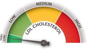
To function effectively, your body requires some cholesterol. But, if there is too much in your blood, it can adhere to the artery walls and constrict or even block them. You run the risk of developing coronary artery disease and other heart disorders as a result.

On a class of proteins known as lipoproteins, cholesterol moves through the blood. LDL, one type, is referred to as the “bad” cholesterol. The accumulation of cholesterol in your arteries is caused by a high LDL level. The “good” cholesterol is frequently referred to as another type, HDL. It transports cholesterol back to your liver from other places of your body. The cholesterol is then eliminated from your body by your liver.

You can take actions to increase your HDL (good cholesterol) and decrease your LDL (bad cholesterol). You can reduce your chance of developing heart illnesses by maintaining normal cholesterol levels.

Food tips to lower your cholesterol

These ten methods include foods that lower cholesterol, suggestions for modest exercise, and more. These can all be used to lower cholesterol without the need of medicine.

To prevent trans fats, read the nutrition labels.

Reading nutrition labels is one of the simplest things you can do to help control your diet, so you’ve definitely heard this advice repeated time and time again. You can use nutrition labels to identify the healthy elements you consume and to steer clear of trans fats, which are among the worst ingredients for your cholesterol levels.

Trans fats, commonly referred to as “hydrogenated oils” or “partially hydrogenated vegetable oil,” are cunning substances that may benefit food producers but are bad for you.

Trans fats make things more durable, which makes them simpler to ship and store. They can be found in a lot of processed foods and many baked items made with margarine or shortening. Sadly, they also increase levels of dangerous LDL cholesterol while lowering levels of good HDL cholesterol.

Thus, read labels and make an effort to avoid trans fats whenever you can if you genuinely want to lower your cholesterol. Cutting them out of your diet can have a significant impact because they are among of the worst culprits when it comes to high cholesterol.

Choose fish or chicken or other meats that contain lower saturated fats.

Lookin’ a little bit more bare in the fridge than usual? When you leave to restock, take a moment to go through your shopping list to see if there are any simple protein substitutions you can make.

Start by cutting back on the red meat. Saturated fats, which are prevalent in a lot of red meats, can cause unhealthy LDL cholesterol levels to rise. Choose skinless chicken or skinless turkey more frequently and stay away from processed meats for healthier choices. Adding additional seafood to your diet is another option.

Fish is low in saturated fats and contains a variety of omega-3 fatty acids that are beneficial for your heart and can raise your levels of the good HDL cholesterol. You can try including the following fish varieties in your diet:

  • oily fish such as tilapia, Atlantic mackerel, or salmon from the Atlantic or Pacific
  • Shellfish include crab and shrimp
  • Lake herring and trout are examples of freshwater fish.
  • Whitefish from the sea, such as cod and grouper
  • Light tuna steaks or fillets in a can

Despite this, it can be difficult to resist eating hamburger and steak. Choose thinner meat pieces when grilling outside. Like anything else, it’s acceptable to consume certain saturated fats. Just remember to consume them in moderation.

Kidney beans, quinoa, whole grain bread, and other foods are good sources of soluble fibre.

You undoubtedly already know that fibre can improve your intestinal health. But, if you believed that fibre was only useful for digestion, reconsider; it can also improve your cardiovascular health.

Soluble fibre is abundant on a list of foods low in cholesterol (fiber that can dissolve into water). To lower levels of harmful LDL cholesterol, soluble fibre absorbs cholesterol in the gut before it enters the bloodstream.

Among the foods high in soluble fibre are:

  • Oats
  • Barley
  • Quinoa
  • granola bread
  • mung beans
  • Lentils
  • Chickpeas

Adding more of these food categories to your diet is simple. For lunch, try curried lentils, and for dinner, try turkey chilli with kidney beans. For morning, try oats and whole grain toast.

But, it’s crucial to keep in mind that not all “healthy” foods are made equal. In general, the more processed a grain or bean is, the less likely it is to be nutritious and provide health advantages. Try to stock up on fresh ingredients whenever you can.

Snacking on fruits, veggies, and nuts will increase your intake of unsaturated fats and fibre.

Having a snack between meals to increase energy or calm an upset stomach is perfectly acceptable. Yet, frequent snacks like crackers, cookies, pastries, microwave popcorn, chips, and other baked goods are high in trans and saturated fats.

On the other hand, snacking on fruits, veggies, and nuts helps you receive both good fats and fibre in addition to helping you avoid toxic fats.

Unsaturated fats, the healthiest sort of fats, are abundant in raw nuts. Due to their ability to increase levels of the beneficial HDL cholesterol and decrease levels of the harmful LDL cholesterol, nuts are a fantastic addition to a heart-healthy diet. Olives and avocados are two further examples of foods high in unsaturated fat.

In addition to numerous fruits and vegetables, nuts can be a fantastic source of soluble fibre. A double dosage of cholesterol-lowering effects may result from including as many of these foods in your diet as you can.

Uncertain about where to begin? Here are a few ideas:

  • Avocados
  • Apples
  • Strawberries
  • Blueberries
  • Oranges
  • Grapes
  • Olives
  • Peas
  • Broccoli
  • Carrots
  • Okra
  • Eggplant
  • Walnuts
  • Peanuts
  • Almonds
  • Cashews
  • Pistachios

Keep in mind that more processing equals less benefit, much like with meats and whole grains. For instance, eating an apple whole will provide more benefits than eating applesauce. Hence, try to obtain raw fruits, vegetables, and nuts if you can (unsalted if you can).

Take advantage of low-fat milk, cheese, and yoghurts.

Making better choices is all it takes to lower your cholesterol; you don’t have to give up everything you love in order to accomplish it. Choosing a healthier alternative when it comes to dairy is a significant area where success can be achieved quickly.

Choose low-fat dairy products rather than the standard varieties for foods like cheese, milk, cream, and yoghurt. Try soy milk as well if you’re up for some experimentation. Exactly what? That can develop into your upcoming craving.

Because full-fat dairy products contain saturated fat as well as cholesterol, making these changes is beneficial. By choosing a low-fat (or non-fat) option, you’re boosting your blood’s cholesterol levels.

Try preparing your food in a new way.

Not only what you eat matters, but also how you eat. The same way you may alter what you purchase at the grocery store, you can also decide on healthier cooking methods that naturally lower your cholesterol. For instance:

While preparing meat or fish, take into account reducing the fat and removing the skin (either before cooking or before eating). This enables you to consume less fat while still getting the protein.

Put your attention on boiling, broiling, baking, poaching, or grilling. These preparation techniques are superior to deep-frying and breading, which might increase fat content.

Consider eating one vegetarian meal a week.

Don’t be alarmed by the phrase “vegetarian.” By selecting a carefully prepared vegetarian dinner, you can simultaneously lower your cholesterol by increasing your intake of soluble fibre and consuming healthy fats. Also, a lot of vegetarian dishes are just as tasty and filling as their meat-based counterparts.

Here is one concept for a recipe with reduced cholesterol: Consider a freshly made salad with grilled, seasoned tofu and a sesame vinaigrette. For dessert, mix low-fat vanilla yoghurt with some fresh blueberries, strawberries, and oats.

Establishing a routine is crucial in this situation; for example, make every Tuesday night vegetarian night. As that becomes customary, consider adding more nights or include a weekly vegetarian lunch as well. You can also be a “flexitarian” by just consuming less meat. These adjustments could truly pay off in the long run.

Add additional movement to your daily activities.

By keeping your body active, you’re assisting it in doing what it was designed to do, which can have benefits for your general health. This includes increasing the heart-healthy HDL cholesterol, controlling blood pressure, and many other advantages.

Do I have to start going for runs every day? Do I need to purchase a lot of home exercise equipment or join a gym? You can if you want to! There are, however, a variety of alternative options, and it’s crucial to establish a schedule that works for you. The workout you will stay with is ultimately the one that is best for your heart.

REFERENCES:

  • https://www.healthline.com/health/ways-lower-cholesterol
  • https://medlineplus.gov/howtolowercholesterol.html
  • https://www.healthpartners.com/blog/how-to-lower-cholesterol-naturally/

For more details, kindly visit below.

Quick peek on causes of hiccups and its prevention.

Quick peek on causes of hiccups and its prevention.

The diaphragm uncontrollably contracts, which results in hiccups. Drinking carbonated beverages, indulging in a heavy meal, inhaling too much air, or being stressed are a few common triggers for this spasm.

What are hiccups?

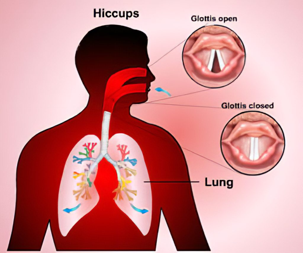
The muscle tissue just below your lungs, the diaphragm, contracts repeatedly and uncontrollably during hiccups.

In addition to defining the boundary between your chest and abdomen, the diaphragm controls respiration. The contraction of your diaphragm causes your lungs to take in oxygen. Your lungs exhale carbon dioxide as your diaphragm relaxes.

Hiccups are brought on by the diaphragm contracting irregularly. Every time the diaphragm contracts, the larynx (voice box) and vocal cords abruptly close. A quick rush of air enters the lungs as a result of this. Your body responds by gasping or chirping, which produces the hiccup-specific sound.

Hiccups can’t be predicted in advance. Prior to eliciting the unique hiccup sound, each spasm is typically preceded by a small constriction of the chest or throat.

The majority of hiccup episodes begin and terminate quickly and without apparent cause. Typically, episodes are only a few minutes long.

Causes of Hiccups

Why people experience hiccups is a mystery. Hiccups can occur for a variety of reasons, including inflamed nerves and low blood carbon dioxide levels. Important components of breathing include the vagus nerve, which connects the brain to the stomach, and the phrenic nerve, which runs from the neck to the diaphragm.

Mild hiccups (those that disappear quickly) can occur when you:

  • Drink and eat too soon.
  • ingest alcohol or fizzy beverages.
  • Eat excessively.
  • Feel anxiety, including exhilaration and terror.
  • strain your neck too far.
  • ingest drugs (particularly those for anxiety – benzodiazepines).
  • Drink anything really hot or chilly.
  • undergoing chemotherapy
  • are put to sleep before an operation.
  • inhale dangerous vapours.

Hiccups lasting for more than two days

Persistent hiccups are those that don’t go away after a few days. They are referred to as “intractable” if they persist for several months (long-lasting hiccups). Rarely do hiccups last for a long time. They could make you feel anxious and worn out. Hiccups that are difficult to control may be a symptom of a more serious underlying medical condition and may persist until that problem is resolved.

Among these more serious underlying problems are:

  • tumours and cancer.
  • Stroke.
  • stomach or esophageal conditions, such as GERD (a gastrointestinal and abdominal disorder).
  • the diaphragm’s pleurisy.
  • Uremia.
  • Pneumonia.
  • Bowel ailments.
  • irritated bladder and pancreatitis.
  • Cancer of the liver with hepatitis
  • lesions and tumours.

Additionally, hiccups might occur throughout a procedure’s recovery and after surgery. If your hiccups continue for a long time, consult a doctor.

How are hiccups diagnosed?

It’s simple to identify glitches. Simply hearing the “hic” sound will be enough for your healthcare provider.

However, your medical professional might do a physical examination to see whether an underlying condition might be the source of your hiccups. The physician may request testing including imaging tests, endoscopic tests, and blood tests if the physical examination indicates anything concerning.

How do I treat hiccups?

Some treatments could or might not be effective because the precise cause of hiccups is unknown. There is often no damage in attempting these at-home remedies because they won’t do you any harm. Home remedies consist of:

  • swift water consumption.
  • swallowing crushed ice, dry bread, or granulated sugar.
  • tugging your tongue gently.
  • Gagging (sticking a finger down your throat) (sticking a finger down your throat).
  • rubbing your eyes softly.
  • water is gargled.
  • retaining breath.
  • Taking a breath into a paper bag (do not use a plastic bag).

How are hiccups prevented?

Pre-treatment medicine can occasionally stop hiccups from occurring. For instance, taking metoclopramide before anaesthesia might stop the hiccups that can result from it. Ramosetron-containing steroids may stop chemotherapy-related hiccups.

Again, the following factors can result in minor hiccups (those that pass quickly). As a result, you might try to steer clear of the following to stop hiccups from occurring. Try to avoid:

  • Drink and eat too soon.
  • ingest alcohol or carbonated beverages
  • Eat excessively.
  • Feel anxiety, including exhilaration and terror.
  • strain your neck too far.
  • ingest drugs (particularly those for anxiety – benzodiazepines).
  • Drink anything really hot or chilly.
  • inhale dangerous vapours.

Possible complications of untreated hiccups

Hiccups that last a long time can be painful and potentially dangerous to your health. Long-lasting hiccups might interfere with your food and sleeping schedules and result in:

  • sleeplessness
  • exhaustion
  • malnutrition
  • slim down
  • dehydration

REFERENCES:

  • https://www.healthline.com/health/hiccups
  • https://www.medicalnewstoday.com/articles/181573
  • https://www.mayoclinic.org/diseases-conditions/hiccups/symptoms-causes/syc-20352613
  • https://my.clevelandclinic.org/health/diseases/17672-hiccups

For more details, kindly visit below.

Quick peek of study in the Deep Vein Thrombosis (DVT).

Quick peek of study in the Deep Vein Thrombosis (DVT).

Blood clots commonly form in a deep vein in the leg, which is where deep vein thrombosis occurs. The disorder can also affect pelvic veins in addition to leg veins. The condition known as venous thromboembolism includes the conditions of deep vein thrombosis (DVT) and pulmonary embolism (PE).

A medical emergency is DVT. According to the Centers for Disease Control and Prevention (CDC), 10–30% of persons with DVT in the leg die within a month of their diagnosis due to severe consequences.

What is Deep Vein Thrombosis (DVT)?

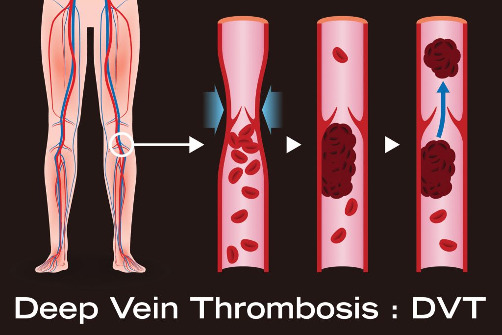
Blood clotting known as DVT occurs in deep veins, frequently in the pelvis or leg. Doctors refer to a thrombus or blood clot breaking off as an embolus. A PE can develop if emboli reach the lung.

As in the case of those who have Paget-Schotter illness, clots can also form in the arm veins. DVT is the most frequent reason for maternal death in the industrialised world, according to a 2017 analysis.

DVT in children is incredibly uncommon. The most recent statistics, according to a 2016 study, indicate that 0.30 of every 100,000 children under the age of nine and 0.64 of every 100,000 children between the ages of 10 and 19 get DVT.

DVT symptoms

Only about half of DVT patients, according to the Centers for Disease Control and Prevention (CDC), experience symptoms. Typical DVT signs include:

  • swelling on one side of your leg, ankle, or foot
  • cramping pain, which typically starts in the calf, in the affected leg.
  • acute, irrational foot and ankle pain
  • a patch of skin that feels warmer than the rest of the body
  • depending on skin tone, the skin over the affected area becomes pale, reddish, or bluish in hue.

People who have an arm blood clot or an upper extremity DVT may not exhibit any symptoms. If they do, typical signs include:

  • a stiff neck
  • shoulders hurt
  • the hand or arm swelling
  • deeper or bluer tinge to the skin
  • from the arm to the forearm moving discomfort
  • fragility in the hand

It’s possible for someone to discover they have DVT only after receiving emergency care for a pulmonary embolism (blood clot in the lung).

A DVT clot that has entered the lung from the arm or leg may cause a pulmonary embolism. It is life threatening and necessitates immediate medical attention when an artery in the lung becomes blocked.

Causes and risk factors

A blood clot is the root cause of DVT. The clot prevents blood from flowing through a vein and into your body normally. Various things can cause a DVT or raise the chance of getting one.

They consist of:

  • Injury: Blood flow can be restricted or blocked when a blood vessel’s wall is damaged. As a result, a blood clot may develop.
  • Surgery: Blood vessels may be harmed during surgery, which may cause a blood clot to form. Following surgery, bed rest with minimal to no activity may potentially raise your chance of developing a blood clot.
  • Reduced mobility or inactivity: Blood can accumulate in your legs, especially the lower ones, when you sit regularly, such on a lengthy flight. The blood circulation in your legs may dwindle if you are immobile for a lengthy amount of time. This may lead to the formation of a clot.
  • A few drugs: Some medications make it more likely for your blood to clot. These include antidepressants, glucocorticoids, hormone treatment medications, and birth control pills.
  • Age: DVT can occur at any age, however it is more common as people get older. DVT only occurs in 1 in 10,000 people under the age of 20, whereas it occurs in 1 in 100 people beyond the age of 80.
  • Trauma: A blood clot may form as a result of a wound that weakens your veins, such as a bone fracture.
  • Obesity. Being overweight might increase strain on the veins in your pelvis and legs.
  • Pregnancy: DVT risk is higher during pregnancy. Actually, compared to non-pregnant people, pregnant people have a 5–10 times higher risk of developing DVT.
  • Family history: If you have DVT, you may be more prone to get it.
  • Catheter: Inserting a catheter into a vein can make it more likely for a blood clot to form.
  • Smoking: This is linked to an increased risk of DVT.

Additionally, a number of other medical disorders can raise the risk of DVT.

These consist of

Complications of DVT

Pulmonary embolism is one of the main side effects of DVT. If a blood clot travels to your lungs and clogs a blood vessel, you could have a pulmonary embolism.

Your lungs and other organs could suffer severe harm as a result. Immediately seek medical attention if you experience any pulmonary embolism symptoms. These indicators include:

  • dizziness
  • sweating
  • coughing or taking big breaths causes chest pain that intensifies
  • quickly breathing
  • spitting blood
  • quick heartbeat

Treatments of DVT

Some DVT sufferers may require inpatient treatment. Others might be eligible for outpatient care.

Compression stockings, elevating the affected leg(s) during the day, and taking anticoagulant drugs (blood thinners) are all forms of treatment. In rare instances, intrusive therapies (catheter-based procedures) may be necessary when the DVT is severe.

The main objectives of treatment include:

  • Ensure that the clot doesn’t spread or include other veins.
  • Avoid having a venous clot fragment escape and travel to your lungs.
  • reduce the possibility of a new blood clot.
  • Prevent long-term complications from the blood clot (like chronic venous insufficiency).

Prevention of DVT

Many required modifications are included into a healthy lifestyle to stop blood clots from forming. This entails increasing physical activity, giving up smoking, and maintaining a healthy weight. Your risk of developing DVT can also be decreased by:

  • control of blood pressure
  • quitting smoking
  • keeping a healthy weight

In order to reduce your risk of getting clots following surgery, take whatever blood thinners your doctor recommends. When you sit for longer than four hours, your chance of getting DVT increases.

When you’ve been sitting for a while, moving your legs around will also keep your blood circulating. After being confined to bed, getting up and moving around can stop clots from developing. During long travels, get out of the vehicle and take regular breaks to stretch.

REFERENCES:

For more details, kindly visit below.

Quick survey on Paralysis and its different types.

Quick survey on Paralysis and its different types.

What is Paralysis?

Loss of muscle function in the body is referred to as paralysis. Sometimes paralysis is transitory, and occasionally it is permanent. While paralysis can affect any portion of the body, the majority of cases are seen in the limbs. Paralysis, both partial and total, can happen at any time. There is no immediate pain experienced by a patient who is paralysed.

The treatment plan is designed to either cure or treat the condition, depending on the nature of the underlying cause, in order to prevent the patient’s everyday lifestyle from being significantly impacted.

The most frequent underlying cause of partial or total paralysis in a patient is a stroke. Incomplete paralysis means the patient has no control over the afflicted muscle tissue while partial paralysis means the patient still has some control over the affected muscle.

Types and causes of Paralysis

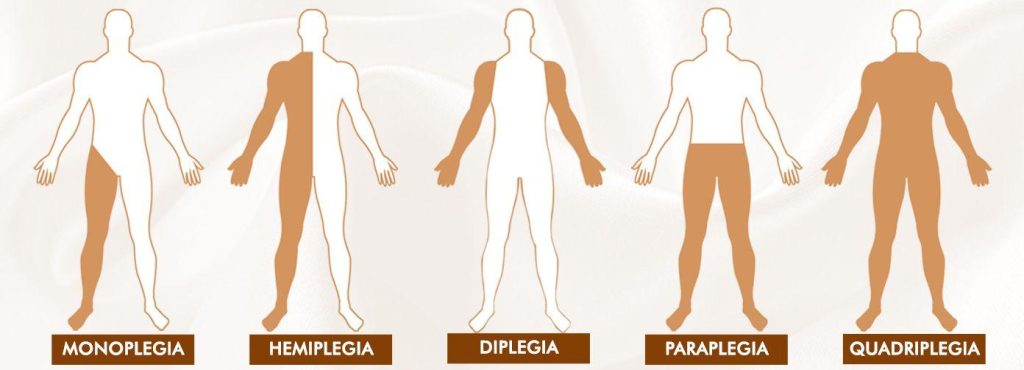
The paralysis types that are best known to the public are:

  • Monoplegia
  • Hemiplegia
  • Paraplegia
  • Quadriplegia
  • lock-in Syndrome

Monoplegia:

Monoplegia is a form of paralysis in which a person is unable to control one limb. The paralysis frequently affects just one arm, or occasionally only particular arm muscles. A common side effect of cerebral palsy is monoplegia. The brain’s capacity to control some body muscles is lost in cerebral palsy. The majority of cerebral palsy patients are young children and teenagers.

Since the patient can still do his everyday activities with only one limb paralysed, monoplegia is regarded as a positive indicator in the prognosis of this illness.

Hemiplegia:

Hemiplegia is a form of paralysis in which one side of the body loses control. This usually only has an impact on one arm or limb, though it might occasionally also have an impact on the torso. Hemiparesis is a condition in which a person’s functional use of their limbs is greatly diminished in terms of strength and endurance, but they retain some degree of functional use. In a few situations, hemiparesis progresses to hemiplegia.

When the corpus callosum between the left and right sides of the brain is damaged, hemiplegia can result. It can also result from spinal cord injury. A stroke, which impairs one side of the brain’s functionality, can also result in hemiplegia.

Left and right hemiplegia are additional divisions of hemiplegia. The diagnosis of right or left hemiplegia depends on where the injury is located.

Paraplegia:

In the case of paraplegia, the patient is unable to control their muscles below the waist. Each person experiences paraplegia differently. The fundamental cause of paraplegia typically arises in the brain or spinal cord, and people who experience it have completely normal legs. Sometimes a lower torso limb only has partial paraplegia, while other times it occurs completely. Regular physical therapy and medication are frequently responsible for partial paraplegia.

Paraplegia frequently results from a patient suffering from a brain, spinal cord, or both types of injuries. The impulses that the brain sends to the lower body are not returned to the brain through the spinal cord in paraplegic patients. Patients who suffer from this absence of brain communication not only lose their ability to move, but also their ability to feel.

A patient with partial or incomplete paraplegia may still be able to use one leg, whereas a patient with complete paraplegia loses sensation and use of both legs.

Quadriplegia

All four limbs of the body are paralysed in a quadriplegia. In this scenario, the patient’s hands and legs become impaled as a result of the brain’s signals to the areas below the neck not being returned. Sending and receiving signals from the brain is the responsibility of the spinal cord. This stops working in quadriplegia, resulting in the condition.

Quadriplegia may begin as a result of spinal cord or brain injuries. The likelihood of recovery from this condition increases as the wounds to the afflicted area heal or as the brain inflammation subsides. It is impossible to provide an accurate forecast, though.

In addition to exhaustion, sudden spasms, lack of sensation below the neck, trouble passing urine from the body, respiratory distress, bedsores, and depression, patients with quadriplegia also experience fatigue.

Locked-in Syndrome:

In a condition known as “Locked-in Syndrome,” the patient is unable to control any area of their body with their under-eye muscles. Locked-in syndrome is primarily a consequence of a serious brain damage, a stroke, or brain cancer. A person suffering from locked-in syndrome won’t be able to move any of his body’s limbs or his lips, jaw, or up and down or side to side movements in the neck. However, those who experience locked-in syndrome are still able to blink and move their eyes up and down.

Doctors rely on the moment of the eyes to make a precise diagnosis because locked-in syndrome frequently mimics the symptoms of a coma.

To ensure that the patient gets all the nutrients needed, adequate nutrition is essential. However, the patient must be fed through a stomach tube, which must either be inserted directly into the stomach through an incision made in the small intestines or through the nose.

Patients who spend a lot of time in bed can develop pressure sores, blood clots, damaged muscles, and damaged nerves. By moving the patient, rubbing the muscles, rotating the joints along their axes, and using physiotherapy, care should be given to prevent pressure sores.

Complications of paralysis

Other physiological processes including respiration and heart rate might be impacted by paralysis. Other body systems in the affected area may also be affected by the illness. Depending on the kind of paralysis you have, you could be vulnerable to:

  • breathing issues, coughing, and pneumonia risk.
  • Deep vein thrombosis (DVT) and clots in the blood.
  • issues with speech or swallowing (dysphagia).
  • both anxiety and depression.
  • Sexual difficulties and erectile dysfunction.
  • Excessively high blood pressure (autonomic dysreflexia) or low blood pressure (orthostatic hypotension) and heart problems.
  • bowel incontinence and urinary incontinence.
  • Sepsis and pressure wounds (bedsores).

How is paralysis diagnosed?

Paralysis is frequently simple to diagnose, particularly when your lack of muscle function is visible. Your doctor may employ X-rays, CT scans, MRI scans, or other imaging techniques to examine inside body parts where paralysis is more challenging to detect.

If you suffer a spinal cord injury, your doctor might perform a myelogram to determine how you are doing. In this surgery, a particular dye will be injected into the spinal cord’s nerves. This will make it easier for them to see your nerves on X-rays. In addition, they might conduct an electromyography. They will utilise sensors to assess the electrical activity of your muscles during this operation.

How is paralysis treated?

The underlying cause of the paralysis as well as the symptoms present will determine a therapy strategy. For illustration, a physician would advise:

  • operation or potential amputation
  • physical exercise
  • Workplace therapy
  • wheelchairs, braces, portable scooters, and other mobility aids
  • If you have spastic paralysis, you may take drugs like Botox or muscle relaxants.

Paralysis is frequently incurable. However, a medical team might suggest a range of medications, equipment, and tactics to assist manage symptoms.

REFERENCES:

For more details, kindly visit below.

Contrasting types of Brain aneurysm and their symptoms.

Contrasting types of Brain aneurysm and their symptoms.

An aneurysm is an artery that has enlarged due to weakened arterial wall. Even though an aneurysm rupture can result in catastrophic complications, it frequently has no symptoms. An aneurysm is a bulge or distention of the artery caused by a weakening of the artery wall.

The majority of aneurysms are not harmful and exhibit no symptoms. Some, however, have the potential to burst at their most serious state, resulting in potentially fatal internal haemorrhage.

Over 25,000 deaths in the United States (U.S.) are attributed to aortic aneurysms each year, according to the Centers for Disease Control and Prevention (CDC). Every year, some 30,000 brain aneurysms burst in the United States. About 40% of these cases result in death within 24 hours.

What is Brain aneurysm?

A brain aneurysm is a condition of the artery in which a region of the arterial wall bulges and swells with blood. It can also be referred to as a cerebral or intracranial aneurysm.

Any age can be affected by a brain aneurysm, which has the potential to be fatal. If a brain aneurysm ruptures, it is a medical emergency that, if left untreated, might result in a stroke, brain damage, and even death.

Types of Brain aneurysm

The location of an aneurysm within the body determines its classification. The two most typical locations for a severe aneurysm are the heart’s arteries and the brain’s arteries.

There are two primary shapes for the bulge:

  • Blood vessels with fusiform aneurysms bulge on all sides.
  • Saccular aneurysms only have a side that bulges.

The size of the bulge affects the chance of rupture.

Aortic aneurysm

The left ventricle of the heart is where the aorta originates. It then travels through the chest and abdominal cavities. The aorta’s diameter is from 2 to 3 centimetres (cm), but an aneurysm can cause it to swell to more than 5 cm.

Abdominal aortic aneurysms are the most prevalent aortic aneurysms (AAA). The area of the aorta that passes through the abdomen is where this happens. The annual survival rate for a AAA larger than 6 cm is 20% without surgery.

AAA can quickly turn fatal, but those who make it to the hospital have a 50% chance of surviving the rest of their lives.

Less frequently, the portion of the aorta that runs across the chest may be affected by a thoracic aortic aneurysm (TAA). Without treatment, TAA has a survival probability of 56% and an overall recovery rate of 85% after surgery. As only 25% of aortic aneurysms form in the chest, it is a rare disorder.

Cerebral aneurysm

Intracranial aneurysms are aneurysms in the blood vessels that supply the brain. They are frequently referred to as “cherry” aneurysms due to their appearance.

Within 24 hours, a brain aneurysm rupture can be fatal. Around 66 percent of people who survive brain aneurysms will have a neurological impairment or disability as a result. Brain aneurysms account for 40% of fatalities.

The most frequent cause of a form of stroke known as subarachnoid haemorrhage is ruptured brain aneurysms (SAH).

Peripheral aneurysm

An aneurysm in the popliteal region develops behind the knee. The most frequent peripheral aneurysm is this one.

  • Aneurysm of the splenic artery: This kind develops close to the spleen.
  • Splenic artery aneurysm: This type of aneurysm occurs near the spleen.
  • Mesenteric artery aneurysm: The artery that carries blood to the intestines is impacted by this.
  • Femoral artery aneurysm: The femoral artery is located in the groyne and can rupture.
  • Carotid artery aneurysm: This happens in the neck.
  • Visceral aneurysm: Aneurysm of the arteries supplying blood to the gut or kidneys is known as a visceral aneurysm.

Aortic aneurysms are more prone to burst than peripheral aneurysms.

Symptoms of Brain aneurysm

Unless it ruptures, a brain aneurysm rarely exhibits any symptoms (ruptures). If a brain aneurysm is unusually large or presses against brain tissues or nerves, it may occasionally induce symptoms even if it is unruptured.

Unruptured brain aneurysm symptoms can include:

If you have signs of a brain aneurysm that has not ruptured, you should visit a doctor as soon as possible. Although aneurysms typically do not burst, it is always crucial to have one evaluated in case medical attention is required.

An intense headache that strikes out of nowhere is typically the first sign of a ruptured brain aneurysm. It has been compared to getting hit on the head and causes blinding anguish unlike anything else ever felt.

Other signs of a ruptured brain aneurysm may also manifest suddenly and include the following:

  • an ill feeling or being
  • a sore neck or ache in the neck
  • responsiveness to light
  • double or blurry vision
  • abrupt confusion
  • consciousness is lost
  • fits (seizures)
  • weakness in any limbs or on one side of the body

Causes of Brain aneurysm

An artery in the brain experiences structural alterations that lead to brain aneurysms. The artery’s walls weaken and thin as a result of these alterations. The distortion may result from wall weakening, but it is also possible for inflammation or trauma to be the only cause of the deformity.

What specifically causes aneurysms to occur is still a mystery. But it’s thought that one or a few of the following elements might favour their development:

  • an elastic tissue breakdown inside the artery
  • stress brought on by the artery’s blood flow
  • because of an increase in inflammation, the artery’s tissue has changed.

Additionally, where an artery splits off into several directions, brain aneurysms are more likely to occur. This is due to the weaker arteries that are present there by nature.

Aneurysms can develop at any time after birth. But they typically come about throughout the course of your lifetime.

Complications of Brain aneurysm

Complications from a ruptured aneurysm could be one of its early warning indications. Instead of only the aneurysm, a rupture is more likely to cause symptoms.

The majority of persons who have an aneurysm do not have any complications. However, problems can also include the following in addition to thromboembolism and aortic rupture:

  • Severe chest or back pain: After an aortic aneurysm in the chest ruptures, severe chest or back pain may develop.
  • Angina: Another form of chest pain that can result from some aneurysms is angina. Myocardial ischemia and heart attacks can result from angina.
  • A sudden extreme headache: An unexpectedly strong headache is the primary sign of SAH caused by a brain aneurysm.

Any aneurysm rupture may result in pain, low blood pressure, a fast heartbeat, and dizziness. The majority of persons with an aneurysm won’t have any problems.

Prevention of Brain aneurysm

Since certain aneurysms are congenital—that is, existing from birth—it is not always possible to prevent them. But some lifestyle decisions can influence the risk:

  • Smoking increases the risk of developing aortic aneurysms and having an aneurysm rupture in any part of the body. Smoking cessation can lower the risk of developing a serious aneurysm.
  • Reduced aneurysm risk can also be achieved by controlling blood pressure. Dietary changes, regular exercise, and medication can all help lower blood pressure to a healthy level.
  • These actions are crucial for lowering pressure on the arterial walls since obesity can put the heart under additional strain.
  • Additionally, a healthy diet helps lower cholesterol and lower the risk of atherosclerosis. Atherosclerosis and fusiform aneurysms frequently go hand in hand.

Anyone who has been given a conservative treatment plan after being diagnosed with an aneurysm can work with a medical professional to address any risk factors.

REFERENCES:

For more details, kindly visit below.

Consequences of low cholesterol level and its treatment.

Consequences of low cholesterol level and its treatment.

High cholesterol is typically linked to cholesterol issues. This is because having high cholesterol increases your risk of developing cardiovascular disease. A fatty substance called cholesterol can block your arteries and possibly result in a heart attack or stroke by impeding blood flow through the afflicted artery.

Too little cholesterol is a possibility. This is less often than high cholesterol, though. While low cholesterol may contribute to other illnesses including cancer, depression, and anxiety, high cholesterol is firmly linked to heart disease.

What exactly is cholesterol?

Despite being linked to health issues, the body need cholesterol. Some hormones can only be produced with cholesterol. It contributes to the production of vitamin D, which aids in calcium absorption. Additionally, several of the chemicals necessary for food digestion are made in part by cholesterol.

Lipoproteins, which are small fat molecules encased in protein, are the carriers of cholesterol in the blood. High-density lipoprotein (HDL) and low-density lipoprotein (LDL) are the two main categories of cholesterol (HDL).

Some people refer to LDL as “bad” cholesterol. This is so that your arteries won’t become blocked by the particular type of cholesterol. LDL cholesterol is transported to the liver by HDL, or the “good” cholesterol. The extra LDL cholesterol is eliminated from the body via liver.

Another important factor in cholesterol is the liver. Your liver produces the majority of your cholesterol. What you eat provides the remaining energy. Only foods derived from animals, such as eggs, meat, and poultry, contain dietary cholesterol. Plants don’t contain it.

Dangers of low cholesterol

Medication, like as statins, frequent exercise, and a nutritious diet can all help lower high LDL levels. There typically isn’t a concern when your cholesterol decreases for these causes. In fact, most of the time, having lower cholesterol is preferable to having high cholesterol. When your cholesterol drops for no apparent cause, you should pay attention and talk to your doctor about it.

Researchers are concerned about how low cholesterol looks to have a harmful impact on mental health, even if the precise consequences of low cholesterol on health are still being explored.

In a 1999 Duke University research of young, healthy women, it was discovered that those with low cholesterol were more prone to have anxiety and depressive symptoms. According to research, low levels of cholesterol may have an impact on your brain’s health because they are necessary for the production of hormones and vitamin D. Cell development requires vitamin D. Anxiety or despair could be a sign that your brain cells aren’t functioning properly. Research is being done to better understand the relationship between low cholesterol and mental wellness.

A 2012 study revealed a potential link between low cholesterol and cancer risk during the American College of Cardiology Scientific Sessions. Cancer may be impacted by the process that modifies cholesterol levels, but further research is required.

Women who might get pregnant are another group who should be concerned about low cholesterol. You have an increased chance of having a premature delivery or a kid with a low birth weight if you’re pregnant and have low cholesterol. Discuss what to do in this situation with your doctor if you typically have low cholesterol.

What Causes Low Cholesterol?

You could have extremely low cholesterol as a result of:

  • an uncommon condition that runs in your family
  • Malnutrition (not eating enough, or not eating enough good foods) (not eating enough, or not eating enough healthy foods)
  • Malabsorption (insufficient fat absorption by your body)
  • Anemia (low red blood cell count) 
  • thyroid problems
  • liver illness
  • a few different infections (like hepatitis C)
  • severe damage or illness
  • Cancer

Low cholesterol symptoms

There are frequently no signs for persons with high LDL cholesterol until they have a heart attack or stroke. You could suffer chest pain if a coronary artery has a significant blockage because less blood is getting to the heart muscle.

When cholesterol levels are low, chest pain that indicates an arterial accumulation in fatty substances does not occur. Low cholesterol may be one of the many reasons of depression and anxiety. Depression and anxiety signs include:

Consult your doctor if you experience any of the symptoms listed above. If your doctor doesn’t suggest a blood test, ask whether you should have one.

Treating low cholesterol

The sort of low cholesterol a person has and what caused it will determine the course of treatment.

Changing one’s lifestyle to include things like:

  • giving up smoking
  • achieving a healthy weight
  • being more active physically

When low LDL cholesterol is present together with symptoms or a genetic disease, medication may be necessary. Taking vitamin E supplements and other fat-soluble vitamins may be used as treatment for the hereditary diseases that lower LDL cholesterol. A doctor could occasionally advise adding additional fat to the diet as a supplement.

Preventing low cholesterol

Individuals very rarely take action to prevent having a level of cholesterol that is too low because most people don’t worry about having it.

Get regular checks to keep your cholesterol levels in range. To avoid taking statins or blood pressure drugs, maintain a heart-healthy diet and an active lifestyle. Know whether there is a family history of high cholesterol. Finally, be aware of any signs of stress and anxiety, especially if they make you feel aggressive.

REFERENCES:

For more details, kindly visit below.

Important treatment option to consider for Cystic Fibrosis.

Important treatment option to consider for Cystic Fibrosis.

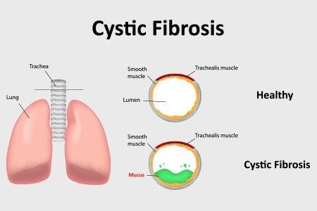
A hereditary disorder that affects the lungs, digestive system, and other organs is called cystic fibrosis (CF). When the protein responsible for producing mucus does not function normally, it develops. As a result, the body produces thick, sticky mucus that can harm or clog organs.

Although there is no known cure for the condition, medicines can help control it, ease the symptoms, and lower the chance of consequences. As a result, a person’s life expectancy and quality of life are improved.

What is Cystic fibrosis?

The lungs and digestive system are the two main organs affected by the hereditary illness CF. Additionally, it may result in consequences including diabetes and liver disease. A gene termed the cystic fibrosis transmembrane conductance regulator has a hereditary mutation in CF patients (CFTR). The CFTR protein is regulated by this gene.

Every organ that produces mucus contains the protein. Additionally, various tissues and organs, such as those in the:

  • lungs
  • pancreas
  • intestines
  • liver
  • heart
  • system of defence
  • sweat ducts

The CFTR protein does not operate as it would in a healthy organism due to the genetic mutation. The body produces mucus that is thicker and stickier than usual as a result of this dysfunction. It may close off the airways, resulting in serious lung infections and breathing problems.

The pancreatic enzymes may not be able to adequately break down food as a result of the genetic mutation, which could further affect pancreatic function. Digestion issues may result from this and result in stunted growth and malnutrition.

A chronic illness, CF can have complications that could be fatal. Treatments, however, can lengthen and improve the quality of life.

Symptoms of Cystic fibrosis.

Most frequently, CF affects the lungs, leading to symptoms of the respiratory system like:

  • wheezing
  • breathing difficulty
  • a prolonged cough that occasionally produces blood or mucous
  • further breathing problems

Additionally, the mucus that prevents lung function in CF patients provides ideal living conditions for infections. A person is therefore at a higher risk of developing lung infections including bronchitis and pneumonia.

CF symptoms might differ from person to person based on the organs that are impacted. Other potential signs and issues include:

  • sinus infections frequently
  • gastrointestinal conditions like:
    • abdomen ache
    • constipation
    • diarrhoea
    • oily, unpleasant stool
  • Small, fleshy growths inside the nose called nasal polyps
  • perspiration and salted skin
  • morning sweats
  • fever
  • jaundice
  • muscle and joint ache
  • low body mass
  • little development or weight increase in children
  • postponed puberty
  • infertility in men
  • Due to a shortage of oxygen reaching the extremities, people have clubbed fingers and toes.

In addition to increasing the risk of diabetes and osteoporosis, pancreatic blockage can result in malnutrition and stunted growth.

Causes of cystic fibrosis

The “cystic fibrosis transmembrane conductance regulator” gene, often known as the CFTR gene, is the cause of CF. This gene regulates the flow of salt and water into and out of the cells in your body.

Your mucus becomes thicker and stickier than it should be as a result of a sudden mutation, or alteration, in the CFTR gene. Your perspiration contains more salt as a result of this abnormal mucus, which also accumulates in a number of body organs, including the:

  • intestines
  • pancreas
  • liver
  • lungs

The CFTR gene is susceptible to a variety of abnormalities. The severity of CF is correlated with the kind of defect. The child inherits the faulty gene from their parents.

Cystic Fibrosis Diagnosis

Early detection translates into quicker treatment and greater long-term health. Every state in the United States uses one or more of these three tests to check neonates for cystic fibrosis:

  • Blood test. This examination measures the amount of immunoreactive trypsinogen (IRT). Blood levels of it are higher in those with CF.
  • DNA analysis. This checks for CFTR gene mutations.
  • Sweat test. Your sweat’s salt content is measured. Results that are higher than usual point to CF.

Some infants who weren’t screened for CF aren’t given the diagnosis until they are adults. If you exhibit symptoms of the disease, your doctor could do DNA or sweat testing on you.

Cystic fibrosis (CF) treatment

Your medical team will likely include a cystic fibrosis expert in addition to many other kinds of providers. Although there is no treatment for cystic fibrosis, your team will assist you in managing the condition. Keeping your airways open is the main goal of management. When necessary, your doctor will also write prescriptions for medication.

Clearing the airways

If you have cystic fibrosis, you can maintain your airways open in a number of ways:

  • You can pick up different breathing and coughing techniques.
  • You can employ treatment vests that use vibrations to loosen mucus or mouthpiece devices.

Chest physical therapy, also known as postural drainage and percussion to remove mucus, is a skill you may master. This technique involves moving into specific positions to allow your lungs to empty. To help loosen the mucus, another person may place their touch on your back or chest. You might cough while doing this.

Cystic fibrosis medications

These drugs, which won’t treat CF but will be helpful to you in some circumstances, may be prescribed by your doctor. They consist of:

  • Antibiotics to treat or prevent lung infections.
  • Breathing is made easier by inhaling bronchodilators, which widen and relax your airways.
  • medication was inhaled to thin the mucus and make it easier to expel.
  • steroids and non-steroidal anti-inflammatories, which are anti-inflammatory medications.
  • medications for those with specific gene variations to treat the underlying causes of cystic fibrosis.
  • Digestive enzymes from the pancreas.
  • Stool softeners for constipation relief.

Cystic fibrosis operations

Surgery can be required if you have cystic fibrosis or one of its side effects.

These could consist of:

  • surgery on the sinuses or nose.
  • bowel surgery to clear obstructions.
  • procedure involving organ transplantation, such as a liver or double lung transplant.

When to see a doctor

Consult your doctor about getting tested for the disease if you or your kid exhibit symptoms of cystic fibrosis or if someone in your family has the condition. Consult a medical professional who is familiar with CF.

Your doctor must be followed up with consistently and regularly, at least every three months, if you have cystic fibrosis. If you notice any new or worsening symptoms, such as more mucus or a change in the colour of your mucus, fatigue, loss of weight, or severe constipation, speak to your doctor right once.

If you experience severe stomach discomfort and distention, chest pain, difficulty breathing, or are coughing up blood, get immediate medical attention.

REFERENCES:

  • https://www.healthline.com/health/cystic-fibrosis
  • https://www.medicalnewstoday.com/articles/147960
  • https://www.mayoclinic.org/diseases-conditions/cystic-fibrosis/symptoms-causes/syc-20353700
  • https://my.clevelandclinic.org/health/diseases/9358-cystic-fibrosis
  • https://www.webmd.com/children/what-is-cystic-fibrosis

For more details, kinlty visit below.

Potential causes and symptoms of Epilepsy you must know.

Potential causes and symptoms of Epilepsy you must know.

What is Epilepsy?

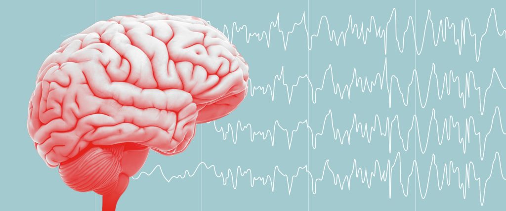
Unprovoked, repeated seizures caused by the neurological disorder are called Epilepsy. A seizure is a sudden spike in aberrant brain electrical activity. When you experience two or more seizures without another obvious explanation, a doctor will diagnose epilepsy.

According to the World Health Organization (WHO), 50 million people throughout the world have epilepsy, while the Centers for Disease Control and Prevention estimates that 3.5 million individuals in the United States have epilepsy (CDC).

Epilepsy can affect anyone, however it typically starts in young children and elderly people. Men are more likely than women to acquire epilepsy, probably as a result of higher exposure to risk factors such alcohol consumption and head trauma.

There are two primary categories of seizures:

  • universal seizures
  • focused epilepses

Your entire brain is affected by generalised seizures. Only one area of your brain is impacted by focal or partial seizures.

It could be challenging to identify a minor seizure. You might be awake during it, and it might only last a few seconds. Spasms and uncontrollable muscular twitches might result from more severe seizures. They may cause confusion or unconsciousness and last anywhere from a few seconds to many minutes. You might not remember having a seizure afterward.

Epilepsy cannot currently be cured, but it can be controlled with medicine and other methods.

What happens in your brain when you have epilepsy?

All parts of your body can communicate with and receive information from the cells in your brain. A constant electrical impulse that moves from cell to cell is used to transmit these messages.

This periodic electrical impulse pattern is disturbed by epilepsy. Instead, there are electrical activity spikes between cells in one or more regions of your brain, much like an erratic lightning storm. Your awareness (including loss of consciousness), sensations, emotions, and muscle actions are all affected by this electrical disruption.

Types and symptoms of epilepsy

The primary epilepsy symptom is seizures. The symptoms of a seizure vary from person to person and depending on the type.

Focused (partial) seizures

Previously known as a simple partial seizure, a focal aware seizure doesn’t cause unconsciousness. These signs include:

  • Sense of taste, smell, sight, hearing, or touch changes
  • dizziness
  • limb twitching and tingling

Unaware focal seizures (previously called complex partial seizures) involve a loss of consciousness or awareness. Additional signs include:

  • looking vacant
  • unresponsiveness
  • performing frequent motions

Universal seizures

The entire brain is involved in generalised seizures. Subtypes consist of:

  • Absence seizures. “Petit mal seizures” were the previous name for absence seizures. They frequently result in a brief loss of awareness, a blank stare, and occasionally repeated actions like blinking or lip-smacking.
  • Tonic seizures. Sudden stiffness in your legs, arms, or trunk muscles is a symptom of tonic seizures.
  • Atonic seizures. Muscle control is lost as a result of atonic seizures. Because a sudden loss of muscle power can cause you to fall quickly, they are also known as “drop seizures.”
  • Clonic seizures. Repeated, jerky muscular movements of the face, neck, and arms are hallmarks of clonic seizures.
  • Myoclonic seizures. Arms and legs twitch quickly and spontaneously as a result of myoclonic seizures. These seizures can occasionally group together.
  • Seizures with tonic-clonic. Grand mal seizures are another name for tonic-clonic seizures. These signs include:
    • rigidification of the body
    • shaking
    • a lack of bowel or bladder control
    • gnawing at one’s tongue
    • consciousness is lost

You might not remember having a seizure afterward or you might have mild nausea for sometime.

What causes epilepsy?

According to the WHO, the cause of epilepsy cannot be identified in roughly 50% of cases. Seizures can be brought on by a number of circumstances, including:

  • Traumatic head injury or another type of head injury
  • following a brain injury, brain scarring (post-traumatic epilepsy)
  • severe ailment or extremely high fever

According to the CDC, stroke accounts for roughly half of older people’ instances of epilepsy with no known aetiology.

  • oxygen deprivation in the brain
  • brain cyst or tumour
  • incorporating Alzheimer’s illness, dementia
  • a mother’s use of certain medicines, an accident during pregnancy, a brain abnormality, or a newborn’s lack of oxygen
  • Infectious diseases such as HIV, AIDS, and meningitis
  • genetic, developmental, or neurological conditions

Although epilepsy can manifest at any age, it is typically diagnosed in the first few years of life or after the age of 60.

What are seizure triggers?

Some people can pinpoint the events or circumstances that cause their seizures. The following are a handful of the known triggers that are most frequently used:

  • absence of sleep
  • a fever or sickness
  • stress
  • flashing or patterned lights, or both
  • Caffeine, alcohol, or alcohol withdrawal, narcotics, or prescription medications
  • skipping meals, overindulging, or certain dietary components
  • really low blood sugar
  • a brain injury

Finding triggers is not always simple. It’s not necessarily true that a single occurrence qualifies as a trigger. Frequently, a seizure is brought on by a number of circumstances. Keeping a seizure notebook can help you identify your triggers. Keep the following in mind following each seizure:

  • time and day
  • which activity you engaged in
  • what was going on in the area
  • strange sounds, scents, or sights
  • unexpected stressors
  • how much you ate or how long it
  • Depending on what you were eating or how recently you had last eaten
  • the degree of your exhaustion and the quality of your sleep the night before

Your seizure notebook can also be used to check on the effectiveness of your medication. Take note of any adverse effects and how you felt right before and right after your seizure.

When you visit the doctor, bring the journal with you. If changing your prescription dosage or looking into alternative treatments is or becomes required, it might be helpful for your doctor to know.

REFERENCE:

For more details, kindly visit below.