Browsed by
Category: Cancer

Boost skin cancer immunotherapy by targeting proteins.

Boost skin cancer immunotherapy by targeting proteins.

A protein that aids tumors in evading immune response and supports the growth of melanoma has been discovered by new research.

According to researchers, immunotherapy should be more effective with tailored medicines directed particularly at this protein.

One of the most prevalent malignancies, melanoma is typically brought on by exposure to UV light, while hereditary factors also play a part in its development.

Experts advise staying away from tanning beds and direct sunshine, as well as keeping an eye out for any moles that seem out of the ordinary.

The growth of melanoma has been the subject of recent research, which has also opened up new potential treatment options.

In a study that was published in the journal Science Advances, researchers showed how a protein called NR2F6 aids tumor growth by assisting tumours to elude the immune system.

The scientists discovered that in mice, eliminating the protein made the immune treatment work more effectively.

“This tells us that NR2F6 helps melanoma evade the immune system, and without it, the immune system can more readily suppress tumour growth,” said Dr. Hyungsoo Kim, a research assistant professor at Sanford Burnham Prebys, a research centre in La Jolla, California, and the study’s first author.

Treatments that prevent the protein’s action are thought to be twice as effective since it behaves the same way whether it is in a tumor or the tissues around it.

The scientists are currently searching for fresh medications that can particularly target NR2F6.

learning about melanoma

Melanoma develops when the DNA in skin cells is harmed, according to dermatologist Dr. Ahmad Chaudhry of the United Kingdom, who spoke to us.

According to Chaudhry, exposure to ultraviolet (UV) light from the sun or tanning booths is frequently to blame for this. “Due to this damage, the melanocytes (cells that produce melanin) proliferate out of control and aggregate into a mass of malignant cells. The development of melanoma in the eyes or internal organs does occur occasionally, but it is less frequent.”

While there are some hereditary risk factors that can potentially play a role, sunshine and tanning beds are linked to skin cancer for a reason.

We were informed by Dr. Sudarsan Kollimuttathuillam, a medical oncologist and haematologist at the City of Hope cancer research organization’s Huntington Beach and Irvine Sand Canyon locations, that 7% to 15% of people with melanoma also have a family member who has the condition.

According to him, having characteristics like pale skin, freckles, or blonde or red hair raises one’s overall risk of developing skin cancer. Atypical mole syndrome is another genetic disorder that dramatically raises the lifetime risk of melanoma and is characterized by a high number of moles with odd forms or color.

Risk can be reduced, but genetics cannot be changed. Doctors advise limiting exposure to the sun during peak hours, staying away from tanning beds in general, and wearing sun protection when outdoors to reduce your risk of acquiring skin cancer.

In the words of Kollimuttathuillam, “regular skin examinations by both you and a dermatologist will help detect melanoma at an early stage, when it is more treatable.”

Experiencing melanoma

One of the most prevalent types of cancer are skin malignancies like melanoma.

More than 97,000 Americans are expected to receive melanoma diagnoses in the US in 2023, according to the American Cancer Society.

As previously mentioned, melanoma can be detected early by a number of telling indications, including genetics and moles. The following procedure usually entails removing and then examining the mole if a doctor suspects it may be malignant. Melanoma presence or absence can be assessed by a range of tests.

It’s crucial to get an early diagnosis of melanoma because it spreads quickly.

According to Kollimuttathuillam, melanoma is the type of skin cancer that is most likely to spread to distant organs or bones. Because of this, imaging technologies may be utilized to spot cancer cells that have done so.

After receiving a melanoma diagnosis, a patient has a variety of treatment choices at their disposal, including radiation therapy, surgery, and immunotherapy.

In the earliest stages of melanoma, patients typically do not require imaging tests because, as Kollimuttathuillam noted, “we know that the best way to stop cancer is to prevent it.” “I cannot emphasize enough how crucial it is for patients to be advocates for their skin health to avoid advanced stages of this disease,” the doctor said.

Types of Immunotherapy

Medication is used in immunotherapy to boost your immune system. This might aid in its attack on cancer cells.

Severe melanoma is treated with a variety of immunotherapies, including:

Checkpoint blockers. The PD-1 blockers nivolumab (Opdivo) and pembrolizumab (Keytruda) as well as the CTL4-blocker ipilimumab (Yervoy) are among these drugs. These medications could aid T cells in your immune system in identifying and eliminating melanoma cancer cells.

Oncolytic virus therapy. In this procedure, melanoma tumors are injected with talimogene laherparepvec (T-VEC, Imylgic), a modified virus. In addition to killing cancer cells, this virus may also cause your immune system to fight cancer cells.

Cytokine therapy. Immune cells can interact with one another with the aid of a class of proteins called cytokines. Interleukin-2 (aldesleukin, proleukin) therapy may enhance your immune system’s defense against cancer.

Your doctor may recommend a single immunotherapy treatment or a cocktail of immunotherapy medications. They might prescribe Yervoy and Opdivo combined, for instance.

Individuals with stage 4 melanoma now have better survival rates thanks to immunotherapy. However, there is a chance that this treatment will have negative side effects.

Contact your doctor straight away if you suspect any potential side effects.

REFERENCES:

For Skin cancer medications that have been suggested by doctors worldwide are available here https://mygenericpharmacy.com/index.php?therapy=10

Experimental drugs could improve several cancer therapies.

Experimental drugs could improve several cancer therapies.

In order to combine medication with diagnostics, researchers have found a novel radioactive compound named CB-2PA-NT as a possible possibility.

The chemical exhibits strong uptake and retention in tumors while maintaining a distinct difference from surrounding tissues. It works by targeting neurotensin receptors, which are prevalent in numerous malignancies.

Shortly, researchers intend to carry out human imaging investigations utilizing CB-2PA-NT, which may have an impact on personalized cancer treatment for patients. Regulatory approval is still waiting.

The University of Wisconsin and the University of North Carolina worked together to create the anti-cancer medication candidate CB-2PA-NT, which has the potential to be used widely.

This study lays the framework for future investigations that will use CB-2PA-NT in human imaging, albeit these studies still require regulatory approval in order to start.

New research positions CB-2PA-NT as a promising candidate for an original theranostics method, according to data presented at the Society of Nuclear Medicine and Molecular Imaging Annual Meeting (SNMMI 2023).

Therapy and diagnostics are combined in theranostics.

Correct diagnosis is a prerequisite to choosing the best course of action. A precise diagnosis is even more crucial in the era of personalized medicine. This is where treatments can be tailored to a person’s unique biomarkers.

This is especially true in the field of cancer, where it is crucial to get a precise diagnosis.

Theranostics offers a potent method for battling cancer by fusing two crucial components. It entails locating cancer cells throughout the body and removing them with specialized radiation.

Positron emission tomography (PET) is used to localise the malignancy, and then medicine is given to kill it. Theranostics’ astounding accuracy greatly reduces the possibility of damaging nearby healthy tissues.

CB-2PA-NT has the potential to significantly advance the field of precision medicine by particularly targeting neurotensin receptors (NTSRs) present in diverse cancer types.

Researchers pursue the receptors on cancer cells

When it comes to many malignancies, including lung, colorectal, breast, pancreatic, and prostate cancers, NTSRs are more prevalent receptors.

A radioactive chemical that can precisely bind to NTSR1, one of these receptors, has been developed by scientists.

In terms of how well they are absorbed and retained by tumors, earlier attempts to synthesize these compounds have had mixed results.

One of the study’s authors and doctorate candidate at the University of North Carolina in Chapel Hill, Xinrui Ma, gave us an explanation of the main conclusions.

“This [study] abstract reports] a novel theranostic agent targeting neurotensin receptor 1 (NTSR1) and its application in cancer imaging and therapy,” said Ma.

The optimization of NTSR1-related compounds has already been attempted by numerous organizations, including our own. In fact, we have been working on this topic since 2012. Challenges include limited serum stability, significant liver absorption, some ligands’ agonistic character, and/or quick washout, she added.

The cross-linked propylamine moiety, Ma continued, “can significantly improve tumor uptake and retention, building on previous research and experience.”

“The tumor absorption increased 10-fold as compared to the peptide-based ligand while still maintaining the high contrast. The high uptake was also sustained at 24 and 48 hours following injection, which is more significant, the researcher said.

We have a rare chance to create theranostic drugs for patient treatment because of the much better tumor absorption and retention. Indeed, to investigate the theranostic potential of these novel compounds in a variety of cancer types, such as lung cancer, colorectal cancer, and PSMA-negative prostate cancer, we have forged a close partnership with Prof. Jonathan Engle’s team at the University of Wisconsin, said Xinrui Ma.

Which NTSR1 inhibitor is most effective?

The best NTSR1 antagonist for imaging and therapy was determined by the researchers’ investigation, which looked at a variety of NTSR1 antagonists.

They then carried out studies to radioactively designate these compounds. They verified that the NTSR1 receptor was in fact present in the lung cancer cells (H1299 cells) using a procedure known as western blot.

The compounds were also examined for stability in test tubes and on living lung cancer cells. Also, for their capacity to bind to lung cancer cells in laboratory settings and on animals.

Finally, to examine how the chemicals were dispersed throughout the body, they used PET and CT imaging on tiny animals.

Western blot analysis of the results revealed that the NTSR1 receptor was substantially expressed in the H1299 lung cancer cells. The chemical known as CB-2PA-NT has shown a potent ability to bind to the H1299 lung cancer cells among the NTSR1 antagonists.

Small animal imaging provided proof that CB-2PA-NT was taken up by the tumor in large numbers. Clearly contrasted with the surrounding tissues, and stayed in the tumor for a considerable amount of time.

CB-2PA-NT was chosen for additional research because it stood out as the most promising compound when compared to the other NTSR1 antagonists.

There is a “need to confirm application in humans”

If this theranostic strategy is successful, it may provide a reliable method for imaging to detect the presence of NTSR1 in many cancer types.

This would be helpful for making diagnoses, selecting patients, and keeping track of how well treatments were working. It might also function as a radioactive material used in therapy.

We were informed by Yale resident doctor Dr. Tejasav Sehrawat. He was unrelated to the experiment, that “theranostics is a young discipline for diagnosing and treating malignancies. The growth of the field overall is quite intriguing and has a lot of potential. The competent execution of this investigation and the good preclinical findings are encouraging.

“While the authors have already demonstrated their findings in animal models, application in people still has to be verified. We should all be eagerly awaiting the findings of the authors’ next human investigations. Because there is significant inter-species heterogeneity in these studies, according to Dr. Tejasav Sehrawat.

Possibly negative future effects for cancer patients

This finding, according to Ma, “is important because it could offer personalised medicine for cancer patients,”

She informed us that, in terms of the disease, NTSR was discovered to be overexpressed in prostate cancer tissues. Specifically in PSMA-negative prostate cancer tissues.

This shows that NTSR1-targeted theranostics may be a prostate cancer treatment option in populations that are ineligible for PSMA-based methods. Lung, colorectal, breast, and pancreatic cancers are only a few of the tumors that could potentially benefit from NTSR-targeted theranostics, says Ma, Xinrui

“A broad spectrum of patients may benefit from the newly developed agents,” Ma noted. The researchers are still awaiting regulatory authorization to carry out the first-ever human imaging tests with CB-2PA-NT, and more study is required.

REFERENCES:

For Cancer disease medications that have been suggested by doctors worldwide are available here https://mygenericpharmacy.com/index.php?therapy=10

Important signs to consider to avoid Colorectal cancer.

Important signs to consider to avoid Colorectal cancer.

The incidence of colorectal cancer, which ranks third among all cancers in the globe, is on the decline in older people and is largely avoidable. However, the prevalence of colorectal cancer among younger people is rising.

Seven risk factors that raise the likelihood of colorectal cancer in younger males have been found in a recent study. Men under 45 who are at increased risk should get screened for colorectal cancer, according to the authors.

Colorectal cancer is the third most prevalent cancer in the world, behind breast and lung cancer. Only lung cancer accounts for more fatalities. It is estimated that each year, more than 150,000 people in the United States alone receive a colorectal cancer diagnosis.

The median age of diagnosis for colorectal cancer is 66, and persons over the age of 45 are most likely to develop it. According to the American Cancer Society, widespread screening of adults over 50 and the removal of pre-cancerous growths known as polyps are contributing to a decline in colorectal cancer occurrences and deaths in older people.

Those under 45 have a low chance of developing colorectal cancer, the number of cases and fatalities in this age group has been rising since the 1990s.

Currently, a research of American veterans has identified certain elements that are linked to a higher risk of early-onset colorectal cancer in males. The authors contend that in some situations, targeted screening may aid in case prevention.

What exactly is colorectal cancer?

Cancer that starts in the colon or rectum is called colorectal cancer. These cancers may also be known as colon cancer or rectal cancer depending on where they first appear.

The majority of colorectal malignancies begin as polyps, which are growths on the colon’s inner lining. Even though not all polyps develop into cancer, some of them can over time.

In the United States, colorectal cancer ranks third among all cancers, excluding skin cancer, according to the American Cancer Society.

Causes of colorectal cancer risk

The researchers located 956 men between the ages of 35 and 49 who had received a non-hereditary colorectal cancer diagnosis between 2008 and 2015 from the National Veterans Affairs database in the United States. 600 of them satisfied the requirements for study inclusion.

These were then matched with 1,200 controls who underwent colonoscopies but did not develop colorectal cancer and 1,200 controls who had not.

For both cases and controls, the researchers examined sociodemographic and lifestyle characteristics, family and personal medical histories, physical measurements, vital signs, medications, and laboratory results. For people with colorectal cancer, data from the six to eighteen months before diagnosis were utilized to identify characteristics that might vary with the development of the disease.

Age, cohabitation, employment, BMI, malignancies in first- or second-degree relatives, comorbidities, alcohol use, hyperlipidemia, and usage of statins, non-steroidal anti-inflammatory medications (NSAIDs), or multivitamins were all examined to find risk variables.

From this, scientists discovered 15 variables that were each independently linked to a higher chance of developing colorectal cancer at an early stage. They singled out seven of these that offered equivalent precision and whose data are easily obtainable:

  • elder age (between the ages of 35 and 49)
  • NSAIDs (such as aspirin or ibuprofen) should not be regularly used.
  • no consistent statin use
  • usage of alcohol today
  • relatives in the first or second degree who have colorectal cancer
  • a greater burden of sickness

According to corresponding author Dr. Thomas F. Imperiale, a research scientist at the Regenstrief Institute, several characteristics raised risk more than others:

It was more significant to have a first- or second-degree family who had colorectal cancer. Non-steroidal anti-inflammatory drug use and statin use were two additional factors with a stronger impact.

Risk of colorectal cancer and lifestyle

The risk of colorectal cancer may be increased by a number of lifestyle variables, according to the Centres for Disease Control and Prevention (CDC). These consist of:

The study’s author, Dr. Bilchik, who was not involved in it, emphasized the importance of lifestyle factors:

According to this study, lifestyle choices have a significant impact on the development of colorectal cancer. For instance, taking statins is connected to elevated cholesterol, and food and alcohol have also been linked to a number of different cancers.

Screening for high-risk individuals

The researchers pointed out that although their study only included men, men are still twice as likely to develop colorectal cancer as women of any age. However, they are currently conducting research along these lines on female risk factors.

They emphasised that not all younger men should have colorectal cancer screenings, but those who are at higher risk may benefit.

“Only high-risk men between the ages of 35 and 44 should be screened. Guidelines now recommend colorectal cancer screening for men between the ages of 45 and 49 but do not specify how (i.e., which test),” Thomas F. Imperiale, M.D.

“The risk factors may be helpful in deciding whether noninvasive testing (with the faecal immunochemical test or with the multi-target stool DNA test) or a colonoscopy is more appropriate,” Dr. Imperiale said to us.

Additionally, he hoped that highlighting these risk factors will prompt males between the ages of 45 and 49 who are already eligible for screening to request testing.

The findings indicating that colorectal cancer screening is advised starting at age 45 are particularly pertinent to male veterans under the age of 35. Dr. Imperiale continued, “However, the results may also be valuable for persuading 45-49 year old male veterans that they should be examined.

How is colon cancer identified?

Your doctor would advise exams and testing to determine the cause if you have colorectal cancer symptoms or if a screening test result was abnormal.

The American College of Physicians advises colorectal cancer screening using one of the following methods for those with a typical risk of developing the disease:

  • a colonoscopy every ten years
  • every two years, a high sensitivity guaiac-based faecal occult blood test (gFOBT) or faecal immunochemical test (FIT)
  • FIT every two years in addition to flexible sigmoidoscopy every ten years.

People who have a higher risk of developing colorectal cancer should discuss the type and frequency of screening that is most appropriate for them with their doctor.

To determine your personal risk level, use this risk calculator. Additional testing is necessary to stage colorectal cancer and determine the best course of therapy if it is discovered.

Physical examination and medical history

To ascertain whether you have any risk factors, such as a family history of colorectal cancer, your doctor will inquire about your medical history. Additionally, the duration of your symptoms will be questioned of you.

During a physical examination, your abdomen will be felt for lumps or enlarged organs, and a digital rectal exam (DRE) may be performed.

A gloved finger is inserted into your rectum by the doctor during a DRE to look for any anomalies.

Stool tests

Tests to look for blood in your stool may be advised by your doctor. To the naked eye, blood in the faeces is not always obvious. These examinations aid in finding blood that cannot be seen.

These tests, such as a FOBT or FIT, are carried out at home with the aid of a kit that is provided. You can use the kit to gather one to three stool samples for testing.

A blood test

Blood tests may be required to look for colorectal cancer symptoms include anaemia, which is a condition when there are not enough red blood cells in the body.

Additionally, your doctor can request tests for liver function and tumour markers such carcinoembryonic antigen (CEA) and CA 19-9. Colorectal cancer cannot be identified by blood testing alone.

Signoidoscopy

Your doctor can view the interior of your sigmoid colon via a sigmoidoscopy, also known as a flexible sigmoidoscopy. If a colonoscopy is not possible for any reason, this less intrusive technique could be suggested instead.

Colonoscopy

A diagnostic colonoscopy is carried out as a result of symptoms you are experiencing or an anomaly discovered during a screening test. The test is performed to observe your colon and rectum in their entirety.

A colonoscope, which is put into the body through the anus, is used to perform it because it is a small, flexible tube with a camera on the end.

The colonoscope can be used to insert specialised tools that can be used to remove polyps and collect tissue samples for biopsies.

Proctoscopy

During a proctoscopy, the anus is used to introduce a proctoscope. To observe the interior of the rectum, a proctoscope is a short, rigid tube with a camera at one end. It is used to examine the rectum for cancer.

Biopsy

A biopsy is a lab test that looks at a tissue sample. Polyps or questionable spots are typically removed during a colonoscopy, but if necessary, they can also be removed surgically.

The tissue is delivered to a lab where a microscope is used to examine it. The samples might also be examined for gene alterations if cancer is discovered. To help categorise the malignancy, more lab tests could be carried out.

Imaging exams

Imaging examinations can:

  • view potential cancerous regions that are suspicious
  • determine the extent of the cancer’s spread
  • test the efficacy of the treatment

REFERENCES:

For Cancer disease medications that have been suggested by doctors worldwide are available here https://mygenericpharmacy.com/index.php?therapy=10

Ultrasonic sound helps chemo drugs might treat brain cancer

Ultrasonic sound helps chemo drugs might treat brain cancer

Because chemotherapy medications cannot cross the blood-brain barrier, brain cancer can be challenging to treat. According to researchers, an ultrasound device has the potential to momentarily remove that barrier and allow chemotherapy medications to get through.

According to experts, this technique could revolutionize the way brain cancer is treated. The most lethal type of brain cancer, glioblastoma, may now be treatable according to recent studies.

Treatment for brain tumors is very challenging. The blood-brain barrier, which regulates what can travel from the bloodstream to the brain, is a contributing factor in that the majority of chemotherapy medications are inhibited by this barrier.

The issue was overcome by Northwestern Medicine doctors who implanted an ultrasonic device in the brain to momentarily break down the blood-brain barrier, enabling chemotherapy medications to be injected intravenously into the brain.

Finding that a new technology can safely and effectively open the blood-brain barrier to deliver chemotherapy is potentially a game-changing step forward in brain cancer research and treatment,” Dr. Jason Salsamendi, the lead interventional radiologist at the City of Hope Orange County Lennar Foundation Cancer Centre in California, told us.

Throughout a 4-month period, the 4-minute procedure which is performed on awake patients was repeated once every few weeks for a total of six sessions.

According to the study, which was written up in the journal Lancet Oncology, the surgery caused the concentration of chemotherapy medications in the brain to virtually multiply by four to six.

New brain cancer treatments importance

Dr. Adam Sonabend, a neurosurgeon at the Feinberg School of Medicine at Northwestern University in Illinois, is the study’s primary investigator and neurosurgeon. He also serves as an associate professor of neurological surgery. “While we have concentrated on brain cancer, this provides an opportunity to explore novel drug-based therapies for the millions of patients who are affected by a variety of brain diseases,” the authors write.

Dr Albert Kim, director of the brain tumour centre at Washington University in St. Louis’ Siteman Cancer Centre, who was not involved in this trial, told us that systemic distribution via IV is typical and simple to carry out.

Although the blood-brain barrier had previously been opened with ultrasound, Kim stated that “the implantable device allows for repeated openings, which could enable the delivery of multiple cycles of systemic drugs.”

The trial included paclitaxel and carboplatin, two powerful chemotherapy medications that are typically ineffective in the treatment of glioblastoma.

Temozolomide, the major chemotherapeutic agent now being used to treat glioblastoma, can cross the blood-brain barrier, however it is somewhat ineffective.

Injecting paclitaxel directly into the brain has been shown in prior studies to be beneficial, but it also carries the risk of meningitis and irritated brain tissue.

The five-year survival rate for glioblastoma is now at around 10%, and patients have not benefited from recent developments in cancer treatment, such as targeted medicines and immunotherapy, according to Salsamendi. An option to administeradminister medication directly into the brain each time a dose is needed is the ability to distribute chemotherapy across the blood-brain barrier.

Blood-brain barrier being opened

The blood-brain barrier quickly closed after being forced open, often within 30 to 60 minutes, according to the study’s researchers.

Salsamendi noticed that there is a greater possibility of dangerous substances entering the brain if the blood-brain barrier is breached for a longer period. In terms of treatment planning and risk minimization, it would be important to know how long the barrier may be open with as much accuracy as feasible.

The French biotech company Carthera created the ultrasound device, which breaks down the blood-brain barrier via a stream of tiny bubbles.

One hour later, the blood-brain barrier recloses.

The researchers found that the blood-brain barrier may be temporarily opened in people using ultrasound and microbubble technology and that most of its integrity returns an hour later.

The brain is permeable to medications circulating in the bloodstream for a critical period of time following sonification, according to Sonabend, who is also a member of Northwestern University’s Robert H. Lurie Comprehensive Cancer Centre.

The blood-brain barrier is fully restored 24 hours after brain sonication, according to prior human investigations. However, the field previously made the assumption that the blood-brain barrier is open during the first six to eight hours. According to the Northwestern study, this window may be smaller.

Another first is that the blood-brain barrier is opened in a brain volume that is nine times greater than the initial device (a modest single-ultrasound emitter implant) when a revolutionary skull-implantable grid of nine ultrasound emitters created by the French biotech company Carthera is used. This is crucial because for this method to be effective, a sizable area of the brain close to the cavity that remains after the excision of glioblastoma tumours must be covered.

REFERENCES:

For Cancer disease medications that have been suggested by doctors worldwide are available here https://mygenericpharmacy.com/index.php?therapy=10

New drug combination reduces Lung cancer tumors.

New drug combination reduces Lung cancer tumors.

The second most frequent type of cancer in the world is lung cancer. Around 2 million people are given a lung cancer diagnosis each year, and 1.8 million people pass away from the condition.

Small cell lung cancer (SCLC) and non-small cell lung cancer (NSCLC) are the two forms of lung cancer, with NSCLC accounting for the majority of instances. Surgery, radiation, and chemotherapy are all effective treatments for NSCLC, although it is rarely curable.

Today, studies have discovered that a combination of two medications reduces the size and quantity of tumours in mice with NSCLC, a finding that could result in human clinical trials.

Lung cancer is the second most prevalent cancer diagnosed in the United States, according to the American Cancer Society. Moreover, it is the main cause of cancer mortality, accounting for one in five cancer-related deaths, more than colon, breast, and prostate cancers put together.

The typical age of diagnosis for lung cancer is 70 years old, with most cases being found in older patients. The good news is that cases are declining as fewer individuals smoke tobacco, which is responsible for more than 80% of lung cancer cases. A diagnosis of lung cancer is still given to over 2 million people annually worldwide.

Lung tumours other than small cell comprise 85% of cases (NSCLCs). There is an urgent need for novel therapies because immunotherapy and chemotherapy are not very successful against this kind of lung cancer.

Now, researchers from the Salk Institute and Northwestern University have discovered that treating NSCLC-affected mice with a cocktail of two drugs—one of which is already approved by the Food and Drug Administration (FDA) and the other is undergoing clinical trials—reduced the size and frequency of the tumours.

Need for new treatments

According to Dr. Lillian Eichner, a principal author of the study and an assistant professor of biochemistry and molecular genetics at Northwestern University, “This medication might be helpful for patients with KRAS/LKB1 mutant lung adenocarcinoma.”

About 20,000 new cases of this disease’s molecular subtype are reported each year in the United States, she said. “Patients with this terrible disease currently have an average survival duration of 10 months after diagnosis, and improved therapeutic techniques are desperately needed.”

Histone deacetylase (HDAC) inhibitors have been suggested as a possible therapy for this particular form of lung cancer by the researchers. Animal tumour growth has been demonstrated to be slowed by HDAC inhibitors, which are epigenetic regulators.

After proving that HDAC3 was essential for the development of difficult-to-treat LKB1-mutant cancers, the study’s authors investigated if pharmacologically inhibiting HDAC3 may have an impact on tumour growth.

In this study, they treated KRAS/LKB1 mutant NSCLC in mice with two different medications: the FDA-approved MEK inhibitor trametinib and the HDAC1/HDAC3 inhibitor entinostat, which is currently in clinical development.

Lung tumor study

The LKB1 genetic mutation is present in NSCLCs, and Salk researchers were interested in investigating a novel targeted therapeutic option.

According to Dr. Andrew McKenzie, vice president of personalised medicine at Tennessee’s Sarah Cannon Research Institute and scientific director at Genospace, targeted therapies are medicines created for certain molecular subtypes of NSCLCs.

Since that these treatments are “tailored,” he explained, “it is preferable to administer a targeted therapy rather than immunotherapy or immunotherapy and chemotherapy if you test a patient and discover a mutation.

Initially, the Salk team demonstrated that the body’s histone deacetylase 3 (HDAC3) protein is essential for the development of NSCLCs with the LKB1 mutation.

This was unexpected, according to co-lead of the study Lillian Eichner, PhD, a professor at Northwestern University in Illinois who was a postdoctoral researcher at Salk during the research.

She said, “We believed the entire HDAC enzyme class was intimately related to the origin of LKB1 mutant lung cancer.

“We didn’t know the exact involvement of HDAC3 in lung tumour formation,” Eichner stated. She then moved to two drugs with the assistance of the team.

The potent drug combination

Entinostat was the first medication. Although the Food and Drug Administration (FDA) has not yet approved this medication, clinical tests have demonstrated that it targets HDACs.

Trametinib was the second medication and it works by preventing the growth of cancer cells. Trametinib is FDA-approved for the treatment of NSCLCs, but it must be used in conjunction with the medication dabrafenib, McKenzie added.

These two medications are only permitted for use in NSCLCs with the BRAF V600E mutation, the author continued.

According to McKenzie, “Trametinib on its own has not been very effective and requires to be paired with dabrafenib to see the clinical outcomes associated with FDA approval.” Because trametinib might cause tumours to become resistant, dabrafenib is often used in conjunction with it.

The goal of the study was to determine whether trametinib and the HDAC3-targeting drug entinostat would reduce resistance in the same way. Mice with LKB1-mutated NSCLC were treated with the medication cocktail for 42 days, and then the tumours were examined again.

Tumors in recipient mice have shrunk by 79% in size compared to mice not receiving the medication treatment. The researchers also noted a 63% decrease in lung cancers in the treated animals.

Human trials needed

Cancers are already being treated with these medications. For the first time, the FDA approved trametinib in 2013 to treat metastatic malignant melanoma. In 2017, it received approval for the treatment of NSCLC.

Entinostat has undergone phase 1 and phase 2 clinical trials but has not yet received FDA approval for clinical usage. Phase 3 trials in people with breast cancer are also still being conducted. People have typically tolerated the medication well during the studies.

The medications have not yet been combined in human subjects. The following stages in evaluating the combined therapy were described by Dr. Eichner.

She said that in order to determine the safety of this combined therapeutic method, a phase 1 clinical research would be conducted first.

“Based on the known safety profiles of both medications, we are hoping that this would also be the case in people,” said Dr. Eichner. “Our preclinical investigations were extremely encouraging with regard to the safety of this pharmacological combination.”

A phase 2 research would then determine whether this combination inhibits tumour growth and lengthens the patients’ lives, she continued.

New hope for cancer patients

On average $1.3 billion is spent to bring a new treatment to market, according to a recent study, making drug development a time-consuming and expensive process.

Also, it often takes 6 to 12 years for new cancer medications to be approved. So, it is quicker and more cost-effective to identify new ways to use existing medications.

According to our research, cancer treatments that were previously unsuccessful might be successful if they are modified. In some cases, understanding basic tumour biology can result in novel cancer therapy strategies without the need to first create new medications, which can be a lengthy process, according to Dr. Lillian Eichner.

Although it is still early, their discoveries might result in new lung cancer medicines for this difficult-to-treat disease. Dr. Eichner is upbeat, but more study is required to validate the results.

According to “our findings,” treating patients concurrently with both of these already-approved medications “may significantly limit the growth of lung tumours for this set of patients.”

REFREENCES:

For more details, kindly visit below.

New combination could reduce the risk of Prostate cancer.

New combination could reduce the risk of Prostate cancer.

The effectiveness of talazoparib plus enzalutamide in treating metastatic castration-resistant prostate cancer in adult males was investigated in the TALAPRO-2 international phase 3 clinical trial.

Comparing talazoparib and enzalutamide therapy to placebo and enzalutamide therapy, a 37% lower risk of cancer progression or death was observed.

In 2023, the Food and Drug Administration (FDA) is anticipated to make a decision on the use of this combination therapy to treat men with metastatic castration-resistant prostate cancer.

Prostate cancer affects one in eight men in the United States and is the second most frequent cancer in males after skin cancer, according to the American Cancer Society.

Male hormones called androgens, such testosterone, promote the growth of prostate cancer cells. Even when blood testosterone levels are controlled, prostate cancer occasionally still progresses. Castration-resistant prostate cancer is the term for this.

Metastatic castration-resistant prostate cancer is the term used to describe a type of cancer that has migrated from the prostate gland to other bodily tissues like the lymph nodes and bones.

Treatment for metastatic castration-resistant prostate cancer has greatly advanced in recent years. Despite these advancements, cancer might recur after therapy because existing medicines only have a temporary impact.

Pfizer researchers have combined the drugs talazoparib (Talzenna) and enzalutamide to create a breakthrough treatment for metastatic castration-resistant prostate cancer (Xtandi). In the phase 3 trial of TALAPRO-2, they evaluated the effectiveness and safety of this combination medication.

Dr. Neeraj Agarwal, professor of oncology and Presidential Endowed Chair of Cancer Research at Huntsman Cancer Institute, University of Utah, and principal investigator for TALAPRO-2, delivered the trial’s findings at the 2023 ASCO Genitourinary Cancers Conference.

Why this combination therapy?

Enzalutamide is a type of hormone therapy that has been approved for the treatment of prostate cancer in males. It functions by preventing testosterone from growing prostate cancer cells. Even after they have migrated to other parts of the body, without which they cannot proliferate.

The group of cancer medications known as poly-ADP ribose polymerase (PARP) inhibitors includes talazoparib. An enzyme (protein) called PARP is present in all cells and aids in the self-healing of injured cells. The repair activity of PARP in cancer cells is blocked by PARP inhibitors, which leads to the death of the cancer cells.

The FDA has authorised the PARP inhibitor talazoparib to treat germline (inherited) HER2-negative advanced breast cancer. However, has not yet licenced it to treat prostate cancer.

When combined with medications that restrict testosterone, PARP inhibitors may be beneficial for the treatment of advanced prostate cancer, according to earlier research.

This inspired Pfizer researchers to create a combination therapy that combines the testosterone-blocking drug enzalutamide with the PARP inhibitor talazoparib.

Study

Adult men from 26 different countries who had metastatic castration-resistant prostate cancer were included in the trial in December 2017.

At random, the participants were given one of the following:

  • Enzalutamide 160 mg once daily and talazoparib 0.5 mg were given to 402 individuals.
  • Or, for 403 individuals, a placebo and enzalutamide 160 mg once daily.

The TALAPRO-2 trial’s main goal was to determine whether adding talazoparib to enzalutamide extends radiographic progression-free survival (rPFS)—the period of time patients remain cancer-free—in comparison to placebo plus enzalutamide.

To see if any study participants had defective DNA repair genes, the researchers also analysed the DNA from the cancer cells of all study participants.

Drug combo lowers cancer progression risk

The median follow-up period for the combination therapy group was 24.9 months. However, the group receiving placebo + enzalutamide experienced a median follow-up period of 24.6 months.

According to the findings, talazoparib plus enzalutamide significantly decreased the risk of disease progression or mortality compared to placebo and enzalutamide by 37%. This was true whether “homologous recombination repair,” or DNA repair gene mutations, were present or not (HRR).

Dr. Andrew J. noted that TALAPRO-2, which joins the PROPEL research, is the second randomised phase 3 trial to show a benefit with combination [androgen receptor] plus PARP inhibition in delaying rPFS in the first line [metastatic castration-resistant prostate cancer] context.

According to Dr. Armstronf, “the delays in rPFS range from > 50% relative improvements in HRR+ patients to 30-40% improvements in HRR-undetected individuals.

The results of TALAPRO-2 “differ from what was seen in the MAGNITUDE study with niraparib and abiraterone. Those without HRR deficiency (biomarker negative) group were stopped early due to lack of efficacy,” added Dr. Cora N. Sternberg, a genitourinary cancer specialist at Weill Cornell Medicine who was not involved in the study.

Data on overall survival were “immature” when the trial findings were announced. This indicates that more research is required to evaluate whether combination therapy with talazoparib and enzalutamide extends patient survival when compared to placebo and enzalutamide.

Is the combination therapy safe?

The study assessed any negative effects that men may have had from combination therapy.

The most frequent negative consequences were:

  • (65.8%) Anemia
  • reduction in neutrophil count (35.7%)
  • exhaustion (33.7%)
  • reduction in platelet count (24.6%)
  • Leukocyte count dropped (22.1%).
  • a backache (22.1%)
  • loss of appetite (21.6%
  • sickness (20.6%).

According to Dr. Zorko, the severe anaemia and neutropenia in the combination therapy group are not surprising given what is known about the side effects of PARP inhibitors.

Also, he advised that “before beginning combination therapy, consideration should be given to the necessity for transfusions and dose cessation. Particularly since 49% of patients had anaemia previous to therapy.”

The time toxicity required to obtain transfusions and supportive care in the clinic may further lessen patients’ enthusiasm for this oral combo therapy, the doctor added.

According to Dr. Armstrong, “there is higher toxicity and cost to patients getting combination [treatment], but these are tolerable for most patients and do not seem to impede quality of life in the long run in most patients with [dose] changes and side effect control.”

Study limitations and next steps

The primary limitations of this trial, according to Dr. Scott T. Tagawa, professor of medicine and urology at Weill Cornell Medicine who was not involved in it, include “early data for overall survival as well as [unknown] long-term adverse events.”

Dr. Zorko added: “In the trial, only 5.2% of patients had received abiraterone treatment in the past. We will see more patients in this area as they become castration-resistant as [triple therapy with] androgen-deprivation therapy, docetaxel, abiraterone/prednisone is used more frequently in the metastatic hormone-sensitive prostate cancer setting, but whether this specific subgroup benefits will be interesting to see.

The final stage of medication development was the phase 3 clinical trial. The FDA must now analyse the results of the clinical trials and make a determination regarding the applicability of this therapy to patients with metastatic castration-resistant prostate cancer. In 2023, the FDA is anticipated to make a decision regarding the clinical application.

REFERENCES:

For more details, kindly visit below.

Can consuming red meat really cause you cancer?

Can consuming red meat really cause you cancer?

Years have been spent by nutritionists and health professionals arguing the advantages and disadvantages of consuming red meat in an effort to discover whether it is beneficial or harmful to health. Results have been inconsistent so far.

Red meat, according to researchers, supplies vital elements like protein, vitamin B-12, and iron. Yet, research indicates that consuming large amounts of red meat may increase one’s risk of developing some malignancies, heart disease, and other illnesses.

The amount of red meat that may be healthy is examined in this article along with what the science and official dietary recommendations have to say.

The effects of red meat on health are the main topic of this essay. The ethical and environmental concerns surrounding the consumption of red meat are not addressed.

Is red meat nutritious?

Iron, vitamin B-12, and zinc are among the healthy components found in red meat.

The primary nutritional sources of vitamin B-12 are animal-based foods like meat and dairy. Because of this, individuals who consume a vegetarian or vegan diet may need to supplement their B-12 to avoid developing B-12 deficiency anaemia.

The United States Department of Agriculture reports that one 3.5-ounce (oz) or 100-gram (g) serving of raw ground beef provides the following nutrients:

  • calories in 247
  • Fat weight 19.07 g
  • Protein content: 17.44 g
  • Iron 1.97 milligrammes (mg)
  • Potassium 274 mg
  • zinc 4.23 mg
  • B-12 vitamin dosage of 2.15 micrograms

A piece of meat’s nutritional value might vary depending on a variety of circumstances. The amount of calories and fat, for instance, differs between cuts from various animal parts. The food of the animal, how the farmer grew the animal, and even the animal’s age and sex can have an impact on the nutritional value of the meat.

Several varieties of red meat are recommended as good sources of heme iron by the National Institutes of Health (NIH). The only foods that contain heme iron are meat, poultry, and shellfish. Plants and meals enriched with iron, such as cereals and plant milk, contain nonheme iron.

Heme iron is more bioavailable, which implies that the body can utilise it more readily, according to the NIH. Despite the fact that many people consume adequate amounts of iron, the NIH notes that some people are at risk of iron deficiency, including:

  • infants
  • little children
  • those with frequent menstruation
  • expecting mothers

Difference between unprocessed and processed red meat

It’s crucial to comprehend the many varieties of red meat before delving into the studies on the connection between red meat and cancer.

Unprocessed

Red meats that haven’t been altered or manipulated are known as unprocessed red meats. Examples comprise:

  • steak
  • Lamb chops
  • lamb ribs

Unprocessed red meat can be healthy on its own. It frequently contains high levels of protein, vitamins, minerals, and other vital components. When processed, red meat loses some of its original value.

Processed

Meat that has undergone some sort of modification—often for taste, texture, or shelf life—is referred to as processed meat. Meat that has been salted, cured, or smoked can accomplish this.

Red meats that have been processed include:

  • hotdogs
  • the salami and pepperoni
  • ham with bacon
  • dinner meats
  • sausage
  • bologna
  • jerky
  • cans of meat

Processed red meat typically has fewer healthy elements and is higher in fat and salt than unprocessed red meat. When taken in large quantities, red meat is regarded by experts as a potential cause of cancer. Processed meat and the chance of developing cancer are more closely related.

Processed meat has been identified as a carcinogen by experts. As a result, its link to cancer is now established.

What the research says

Many studies have examined the link between eating unprocessed and processed red meat and health outcomes over time.

There is some evidence that eating a lot of red meat may increase your chance of developing some malignancies, while the data are still conflicting.

IARC methodology

A division of the World Health Organization is the International Agency for Research on Cancer (IARC). It is made up of professionals from throughout the world who work to categorise potential carcinogens (cancer-causing agents).

IARC members analyse scientific papers regarding a potential carcinogen for several days when there is a lot of evidence to suggest that it may cause cancer.

They take into account many aspects of the data, such as how people and animals react to a potential carcinogen and how cancer might grow following exposure to it.

The classification of the probable carcinogen according to its propensity to cause cancer in humans is one step in this procedure.

Group 1 agents are those that have been shown to cause cancer in people. On the other hand, group 4 agents consist of substances that most likely do not cause cancer.

Remember that this classification does not indicate the risk a carcinogen poses. It simply shows the amount of data that supports the association between particular carcinogens and cancer.

IARC conclusions

22 specialists from ten different countries gathered in 2015 to assess the body of knowledge regarding the connection between red meat and cancer.

Almost 800 studies over the previous 20 years were reviewed. Just processed or unprocessed red meat was the focus of certain investigations. Others observed them both.

To reduce cancer risk, avoid processed meat

Avoid eating processed meats if you want to lower your risk of colorectal cancer and perhaps other cancers as well.

Processed meat is a Category 1 carcinogen, according to the IARC. In other words, there is sufficient study to demonstrate its link to human cancer. Here are some additional Group 1 carcinogens for context:

  • tobacco
  • ultraviolet rays
  • alcohol

Once more, the basis for this classification is the evidence that links a certain agent to cancer. Although there is compelling evidence that all Group 1 agents cause cancer in humans, not all of them necessarily carry the same degree of risk.

For instance, when it comes to the danger of developing cancer, eating a hot dog isn’t necessarily equivalent to smoking a cigarette.

Be mindful about red meat consumption

For many people, unprocessed red meat is a crucial component of a healthy diet. It provides ample amounts of:

  • protein
  • vitamin B-6 and vitamin B-12
  • minerals, such as selenium, iron, and zinc

Yet, the IARC analysis came to the conclusion that frequently consuming red meat probably raises the risk of developing several malignancies.

Therefore, there’s no need to exclude all red meet from your diet. Simply be mindful of how you prepare it and how much you consume.

Cooking techniques

In their analysis, IARC specialists also mentioned that the manner red meat is prepared can affect your chance of developing cancer.

High-temperature meat grilling, burning, smoking, or cooking appears to enhance risk. But, the IARC specialists stated that there wasn’t enough data to issue any official advice.

Serving suggestion

There is no requirement to completely give up unprocessed red meat, according to the authors of the IARC analysis. So it’s recommended to stick to three servings per week.

The bottom line

Red meat has come under fire for its alleged associations with a number of diseases, including cancer. According to experts, routinely consuming red meat may up your risk of developing colorectal cancer.

A consensus among experts also exists that consuming a lot of processed meat does raise your chance of developing cancer. Therefore, you don’t have to completely eliminate red meat from your diet. Just make an effort to stick to eating only a few servings of high-quality, unprocessed red meat per week.

REFERENCES:

For more details, kindly visit below.

How gut bacteria can boost cancer immunotherapy efficacy?

How gut bacteria can boost cancer immunotherapy efficacy?

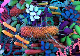
Researchers looked into how gut bacteria affected mice’s response to immune checkpoint inhibitor (ICI) therapy. They discovered that ICIs enable specific gut bacteria to get through tumor locations. It then stimulates the immune system, which then destroys cancer cells.

To confirm whether these results may apply to humans, more research is required.

Immunotherapy includes the use of immune checkpoint inhibitors (ICIs). They function by “taking the brakes off” of the immune system so it can eliminate cancer cells by blocking certain proteins that restrict immune function, such as CTLA-4 or PD-1.

Unfortunately, ICI therapies are ineffective in up to 50% of cancer patients. The effectiveness of ICI treatment may be influenced by the gut flora, according to a growing body of research.

According to research, animals with impaired gut flora or those given antibiotic treatment react to ICI less favourably. Studies have also shown that faecal transplants of new microbiota may improve ICI responsiveness.

The best gut bacteria for boosting ICI response and the mechanism by which gut bacteria enhance immune response are still unknown.

Immune Checkpoint Inhibitors(ICI) and gut bacteria

Recent studies examined the relationship between gut bacterial diversity and ICI effectiveness in a mouse model of melanoma.

They discovered that ICI treatment results in gastrointestinal inflammation. This allows bacteria to get through the intestines. Thereby moves to lymph nodes close to tumors where they activate immune cells.

The research is published in Science Immunology. Even though checkpoint inhibitor treatment has demonstrated unheard-of clinical success, a sizable portion of responders will later develop acquired resistance. As previously mentioned, the gut microbiota has a significant impact on host anti-tumor immunity in several ways. This affects the clinical reactions and outcomes of cancer immunotherapy patients.

Dr. Anton Bilchik, chief of medicine and director of the Gastrointestinal and Hepatobiliary Program at Saint John’s Cancer Institute in Santa Monica, California, as well as a surgical oncologist and division chair of general surgery at Providence Saint John’s Health Center, did not take part in the study.

Investigating ICI efficacy

Mice with and without melanoma tumours received ICI therapy as part of the study.

They discovered that ICI treatment exacerbated gastrointestinal inflammation, allowing certain bacteria to pass from the gut to lymph nodes close to the tumour as well as the tumour site. In that location, the bacteria triggered a group of immune cells that destroyed tumour cells.

The effectiveness of ICI may be impacted by antibiotic exposure, according to the study. To do this, mice were first given antibiotic treatment. Further followed by melanoma tumor implantation and ICI treatment a week later.

They discovered that exposure to antibiotics lowered the number of immune cells and the migration of the gut microbiota to the lymph nodes.

Finally, they looked at whether giving out certain bacteria may counteract the effect of the antibiotics on the effectiveness of the ICI. They discovered that using Escherichia coli and Enterococcus faecalis in treatments increased ICI effectiveness.

Fecal microbiome transplantation

FMT is the most direct way to change the microbiota. Feces from one donor is given to another by lyophilized or frozen pills that are taken orally. Also, they can be delivered directly via colonoscopy or gastroscopy.

With almost 300 registered clinical trials as of now, FMTs are being investigated as a treatment alternative for an increasing range of illnesses (clinicaltrials.gov, accessed Aug 2021). Over the past ten years, it has been clear that FMTs are extraordinarily effective at treating resistant and recurring Clostridium difficile infections. This helps patients feel better and get rid of their clinical symptoms.

Dietary intervention and lifestyle

The relationship between diet and the microbiota has been studied for numerous years at various resolution levels because gut microbes have a role in food digestion. In fact, distinct microbial communities are closely involved in the sequential host digestion and nutrient extraction, with the gut microbiota playing the major role.

On the one hand, the host’s inability to digest a large number of chemicals released by the gut microbiota affects the food’s ability to provide nutrients. Contrarily, both short- and long-term dietary modifications can affect the microbial transcriptome and metabolomic profiles, especially for newborn nutrition. This may have long-term effects through microbial modulation of the immune system. For instance, high-fat diets are linked to significant changes in the makeup of the colonic microbiota. This includes decreases in both Gram-positive.

Study restrictions

Dr. Andrew Koh, senior author of the present work and associate professor at the Harold C. Simmons Comprehensive Cancer Center at UT Southwestern, was contacted by MNT to discuss its limitations.

They only employed one preclinical cancer model, which, according to Dr. Koh, is a significant restriction, necessitating additional research to see whether the results also apply to other cancers.

Although we have not yet produced evidence to support that notion, we think that our findings may also be applicable to other tumours, he said.

According to published research, various human cancers include specific or unique tumour microbiomes, and many of the prominent taxa are bacteria that normally live in the gut. Dr. Bilchik stated that it is still unclear whether the results apply to people when asked about the study’s other limitations.

Interventional gastroenterologist Dr. Lance Uradomo, who is not affiliated with the study and practice in Irvine, California at the City of Hope Orange County Lennar Foundation Cancer Center, stated that “the type of therapy applied for testing melanoma can be linked to adverse side effects, such as colitis.”

Before it is known if microbiome therapy — and the proper administration — is genuinely successful, more research is required, he continued.

Conclusion

The gut microbiome appears to have a significant impact on host immunity and therapeutic response in cancer, either locally within the tumour microenvironment or via systemic antiviral immune responses, according to strong evidence from preclinical and clinical research. The latter is most likely the reason why the gut microbiota is able to control how the body reacts to immunotherapy and traditional chemotherapeutic drugs, eventually having a variety of effects on patient outcomes.

REFERENCES:

For more details, kindly visit below.

Is Toilet Paper a Source of Cancer-PFAS in Wastewater?

Is Toilet Paper a Source of Cancer-PFAS in Wastewater?

According to researchers, PFAS—also known as potentially dangerous substances—may be released into wastewater systems by using toilet paper. In addition to cosmetics and cleansers, PFAS are also present in paper products.

According to them, a variety of health problems, including cancer, infertility, and liver disease, are thought to be exacerbated by the chemicals. The presence of potentially dangerous compounds known as PFAS in groundwater has been linked to the use of toilet paper.

Cosmetics, cleansers, and firefighting foams are only a few examples of the numerous consumer goods that include per- and polyfluoroalkyl substances (PFAS).

PFAS are suspected of contributing to a number of illnesses, including cancer, lowered immunity, and issues with reproduction and development. However the study is not conclusive in this regard.

What researchers found?

The most frequently found PFAS in sewage sludge samples, while at low levels, was one specific molecule, termed 6:2 diPAP. According to University of Florida researchers who were examining the occurrence of PFAS in wastewater.

Also, samples of toilet paper sold in North America, South America, Africa, and western Europe were found to contain the highest concentration of this PFAS. In the online journal of the American Chemical Society, they today published the results of their research.

In their investigation, the researchers calculated that toilet paper supplied roughly 4% of the 6:2 diPAP in sewage in the United States and Canada. As well as 35% in Sweden and as much as 89% in France.

Jake Thompson, a senior research author and doctoral student at the University of Florida, said that while it isn’t the entire issue, it is undoubtedly a component of it. Data indicate that there are geographical disparities in contamination, he said, adding that.

What are PFAS?

The word “PFAS” describes a family of more than 9,000 different kinds of synthetic compounds. PFAS, which were first discovered in the late 1930s, can still be found in a wide range of items, such as:

  • Carpets and clothing with stain resistance
  • cleaning supplies
  • goods for personal care and cosmetics
  • burning foam
  • a nonstick pan
  • garment that repels water
  • goods that are resistant to water, oil, or grease.

American blood PFAS levels have been tested by the National Health and Nutrition Examination Survey for more than 20 years. According to the survey, the majority of Americans had PFAS in their bloodstreams.

Past studies have connected PFAS exposure to a number of potential health problems, such as:

Where PFAS come from?

According to the study, when turning wood into pulp, certain paper makers inject PFAS. Moreover, fibres from products containing PFAS may be used to make recycled toilet paper.

Timothy Townsend, PhD, a professor of the University of Florida’s Department of Environmental Engineering Sciences and a principal author of the study told, “We believe it comes from the pulping process and is put on instruments to keep paper from adhering.

“PFAS discovered in toilet paper at parts per billion levels are most likely pollutants that emerge from the packaging and/or production process,” concurred Pelch.

According to research, the majority of 6:2 diPAP contamination comes from other consumer products. Due to the comparatively low concentration of 6:2 diPAP in wastewater collected in the United States and the fact that Americans use more toilet paper per capita than individuals in other countries.

It is somewhat misinterpreted, according to Townsend, to think that the landfill or the wastewater treatment facility are the issue.

The health threats from PFAS

According to Craig Butt, PhD, manager of applied markets in the division of Strategic Global Technical Marketing at the biomedical and environmental company SCIEX, a growing body of research studies have demonstrated that PFAS represent serious health and environmental dangers.

According to Butt, PFAS have been linked to a wide range of health issues, including cancer and fertility issues. Also, high cholesterol and liver damage are associated in it. In recent years, regulatory authorities in Europe and the US have started establishing legislative limitations for the presence of PFAS in drinking water and consumer products. There are also no acceptable levels of PFAS exposure for humans, according to recent epidemiological and toxicological studies, meaning that even minute amounts of contamination can have a big impact.

There are 5,000 PFAS compounds, according to Butt, “many of which are not well described or understood.”

Toilet paper and PFAS

Prof. Townsend and his team decided to investigate the possible effects that toilet paper might have on the concentrations of PFAS in wastewater for this study.

While not all studies look for this, he said to MNT, “we recently released a study on PFAS in biosolids, which points to 6:2 diPAP as one of the primary PFAS in wastewater residuals.

“We looked into frequent uses for this chemical, and paper was one of them. That’s why we’re looking at toilet paper,” he said.

Rolls of toilet paper that are sold in North, South, and Central America, Western Europe, and Africa were gathered by researchers. Also, they obtained sewage samples from American wastewater treatment facilities. Scientists discovered that the PFAS type that was most prevalent in both the sewage and paper samples was 6:2 diPAP.

The research group then merged their findings with those from other studies that assessed the concentrations of PFAS in sewage and the usage of toilet paper in various nations.

REFERENCES:

For more details, kindly visit below.

Important parameters of Cervical cancer women need to know.

Important parameters of Cervical cancer women need to know.

What Is Cervical Cancer?

Women’s cervix, which connects the uterus and vagina, is where cervical cancer develops when cells in the cervix alter. The deeper tissues of their cervix may be affected by this cancer, and it has the potential to metastasis (spread to other parts of the body), most frequently the lungs, liver, bladder, vagina, and rectum.

Human papillomavirus (HPV) infection, which is avoidable with a vaccine, is the main cause of cervical cancer. Since cervical cancer develops slowly, it is typically detectable and treatable before it poses a major threat. Thanks to better screening through Pap tests, it claims fewer and fewer lives of women every year.

The majority of cases are women between the ages of 35 and 44. However, women over 65 make up more than 15% of new cases, particularly those who haven’t been undergoing routine exams.

Different Types of Cervical Cancer

Cervical cancer comes in several different forms.

  • Squamous cell carcinoma. This develops in your cervix’s lining. Up to 90% of cases have it.
  • Adenocarcinoma. This develops in the mucus-producing cells.
  • Mixed cancercarcinoma. This possesses traits from the other two categories.

Cervical cancer stages

Your doctor will determine the stage of your cancer after a diagnosis has been made. The stage reveals if and how far the cancer has spread if it has. Your doctor can identify the best course of treatment for you by staging your cancer.

There are four phases of cervical cancer:

  • Stage 1: A little cancer. There’s a chance the lymph nodes were affected. It hasn’t spread to other body areas.
  • Stage 2: The tumour has grown. It can have reached the lymph nodes or spread beyond the uterus and cervix. It hasn’t yet spread to other areas of your body.
  • Stage 3: The malignancy has gone to the pelvic or the lower vagina. The ureters, which are tubes that transfer urine from the kidneys to the bladder, may be blocked as a result. It hasn’t spread to other body areas.
  • Stage 4: The cancer may have spread to other organs, such as your lungs, bones, or liver, from the pelvis.

Signs and symptoms of cervical cancer

Early stages of cervical cancer are typically difficult to diagnose because they lack symptoms. It may take several years before cervical cancer symptoms appear. The greatest strategy to prevent cervical cancer is to find abnormal cells during testing for the disease.

Stage 1 cervical cancer symptoms and signs might include:

  • Vaginal discharge that is either bloody or watery, may be heavy, and may smell bad.
  • Vaginal bleeding following sex, in between cycles, or following menopause.
  • Periods of menstruation could be heavier and longer than usual.

Symptoms of cancer that has spread to adjacent tissues or organs include:

  • urination that is painful or difficult, occasionally with blood in the pee.
  • diarrhoea, abdominal pain, or bleeding when you poop.
  • fatigue, weight loss, and appetite loss
  • a state of general disease
  • a dull backache or leg swelling.
  • abdominal and pelvic pain

You should have a comprehensive gynaecological exam, which includes a Pap test, if you suffer abnormal bleeding, vaginal discharge, or any other unexplained symptoms.

Cervical cancer causes

The sexually transmitted human papillomavirus is the primary factor in most occurrences of cervical cancer (HPV). Genital warts are brought on by the same virus.

There are over 100 distinct HPV strains. Cervical cancer is only caused by specific types. HPV-16 and HPV-18 are the two strains that cause cancer the most frequently.

Cervical cancer is not a guarantee even if you have an HPV cancer-causing strain. Most HPV infections are cleared up by your immune system, frequently within two years.

In both men and women, HPV can lead to other malignancies. These consist of:

  • vulvar cancer
  • vaginal cancer
  • penile cancer
  • anal cancer
  • rectal cancer
  • throat cancer

Cervical cancer risk factors

The greatest risk factor for cervical cancer is HPV. Additional elements that may raise your risk include:

  • HIV
  • chlamydia
  • smoking
  • obesity
  • a history of cervical cancer in the family
  • consuming little fruit and veg
  • using contraceptive tablets
  • being pregnant three times at term
  • being under the age of 17 when you first became pregnant

You are not destined to develop cervical cancer even if you have one or more of these risk factors.

How is cervical cancer treated?

One member of the team treating cervical cancer is a gynecologic oncologist (a doctor who specialises in cancers of female reproductive organs). The stage of the disease, your age and general health, and whether or not you intend to have children in the future all play a role in the recommended course of therapy for cervical cancer.

Radiation, chemotherapy, surgery, targeted therapy, and immunotherapy are all options for treating cervical cancer.

Radiation Therapy

Your cervix’s cancerous cells are destroyed by energy beams used in radiation therapy. Radiation therapy is available in two different forms:

  • External beam radiation therapy (EBRT) uses a machine outside the body to direct powerful radiation towards tumours.
  • Radiation is applied directly to or near a malignancy during brachytherapy.

Chemotherapy

Chemotherapy (chemo) kills cancer cells by administering medications by injection into your veins or oral ingestion. It enters your circulation and kills cells effectively throughout your body. Chemotherapy uses a variety of medications, some of which can be combined. Cycles of chemotherapy are frequently administered.

Surgery

Cervical cancer is treated with a variety of surgical procedures. The most typical procedures used to treat cervical cancer include:

  • Laser procedure
  • conical biopsy
  • an easy hysterectomy
  • Trachelectomy
  • Pelvic enlargement
  • Targeted treatment

Specific cancer cells are eliminated by targeted medication therapy without harming healthy cells. It functions by focusing on proteins that regulate how cancer cells proliferate and spread.

Immunotherapy

In immunotherapy, drugs are used to activate your immune system’s capacity to detect and eliminate cancer cells. Cancer cells can also signal to avoid being attacked by your immune system. Targeting these signals with immunotherapy makes it so cancer cells can’t deceive your body into believing they are healthy cells.

Clinical trials are yet another form of treatment. Some people supplement their cancer therapy with complementary therapies like nutrition, herbs, acupuncture, and other practises. Speak with your healthcare practitioner about alternative practises that promise to lessen the symptoms of cancer. Some may be beneficial, while others may be dangerous.

Cervical cancer prevention

Screening with a Pap smear or a hrHPV test on a regular basis is one of the simplest strategies to avoid cervical cancer. Precancerous cells are detected during screening so they can be treated before they progress to malignancy.

Most occurrences of cervical cancer are caused by HPV infection. With the help of the vaccines Gardasil and Cervarix, the illness can be avoided. The best time for vaccination is before a person starts acting sexually. Boys and girls can both receive the HPV vaccine.

You can lessen your risk of HPV and cervical cancer by doing the following additional things:

  • Do not have too many sexual partners.
  • When engaging in vaginal, oral, or anal intercourse, you should always use a condom or another barrier device.

You may have precancerous cells in your cervix if your Pap smear results are abnormal.

REFERENCES:

For more details, kindly visit below.