Browsed by
Tag: Breast Cancer

Breast cancer: Can a wearable device help to detect it?

Breast cancer: Can a wearable device help to detect it?

To screen for breast cancer at home, researchers created a wearable gadget.

Initial testing reveal that it is capable of detecting tiny cysts that are comparable in size to early-stage breast tumours. To confirm the effectiveness of the gadget, clinical trials are required.

One day, a cutting-edge piece of wearable technology that can be fastened to a bra might enable individuals to identify breast cancer symptoms while lounging at home.

With approximately 1 in 6 cancer-related deaths among women, breast cancer is the most common cancer in the world. Women make up the great majority of breast cancer patients. Men experience 0.5% to 1% of cases, though.

If discovered when the cancer has not yet moved outside of the breast, breast cancer has a 5-year relative survival rate of up to 99%. The 5-year survival rate drops to 30% if discovered later, such as when cancer has spread to distant bodily areas like the lungs, liver, or bones.

Currently, the most popular approach for detecting breast cancer is a mammography, an X-ray imaging procedure.

Mammograms must be performed in an imaging center even though they are typically efficient at detecting 87% of malignancies. This limits access for many people, especially those with low incomes, who could postpone screening as a result of the high associated expenses and challenges in organizing transportation.

The health outcomes for those diagnosed with the condition globally could be improved by initiatives to increase accessibility and lower the cost of breast cancer screening.

Researchers recently created a wearable ultrasound breast patch that could enable patients to scan for breast cancer at home.

Bard-certified doctor Dr. Kamila Seilhan, stated: “This wearable ultrasound device may help patients at high risk of breast cancer in the interim between routine mammograms by enabling early tumor detection.”

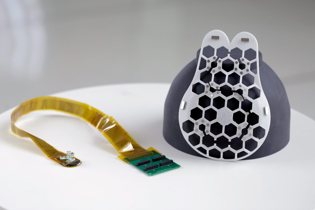
A breast-attached wearable breast cancer detector

The same ultrasound technology that imaging centres utilise is the foundation of the device. However, because of the piezoelectric materials used, it can be made smaller to function as a portable ultrasound scanner. Through a mechanism called piezoelectricity, crystals transform mechanical energy into electrical energy, which in this instance can be used to interpret ultrasound measurements.

The device sends sound waves into the breast tissue, and as it moves across the breast, it produces high-quality images identifying cysts that may need to be investigated by a breast cancer specialist,” said Dr. Jennifer Tseng, F.A.C.S., medical director of breast surgery and a double board-certified surgical oncologist specializing in breast cancer at City of Hope Orange County Lennar Foundation Cancer Centre in Irvine, California, who was not involved in the research.

To make the device wearable, the researchers created a flexible, 3D-printed patch with honeycomb-like holes. The patch fastens to a bra with holes so it may touch the skin and scan breast tissue there.

The entire breast may be imaged thanks to the scanner’s six various positioning options. In order to capture photographs from various perspectives, it can also be rotated.

The scanner has already been tested on a 71-year-old woman who has a history of breast cysts. They were able to identify cysts with the gadget that were as small as early-stage tumours, or 0.3 centimetres in diameter. According to their findings, the images produced had an 80mm depth and a resolution comparable to that of conventional ultrasounds.

Senior author of the study Canan Dagdeviren, Ph.D., Associate Professor of Media Arts and Science at Massachusetts Institute of Technology (MIT), stated that the technology makes it simple to repeatedly take pictures from the same location.

Increased screening availability for breast cancer

Dr. Dagdeviren stated that the ultimate purpose of the device is to reach underrepresented women, including those living in less economically developed nations, and to make breast cancer screening more accessible and affordable.

Dr. Seilhan remarked that if successful, the device could be especially helpful in isolated locations without simple access to medical facilities.

Healthcare facilities and organizations with limited funds can purchase the device more easily because of its low cost,” she said.

She continued by saying that while the gadget is simple to operate, it might be useful in settings where medical personnel have limited technical expertise.

Dr. Tseng pointed out, however, that in order for patients in less developed nations to benefit fully from diagnostic technologies, it is equally critical for them to have better access to those tools.

However, she added, “patients still need to have the data reviewed by an expert who can recommend what to do next.” This device may assist patients identify potential problem areas that they were unable to identify before.

When will the breast cancer wearable device be obtainable?

According to Dr. Dagdeviren, the device might be usable for 4-5 years. She is starting a business to achieve this goal and is looking for partners and investors. For mass production and FDA certification, she stated, “we will need about $40 million.”

In spite of the fact that the device now needs a “bulky computer interface” to process photographs, the author continued, her team is currently working on a more portable design and will soon release an iPhone-size image processor.

The researchers are also creating a workflow that will enable artificial intelligence to examine data and produce diagnostic evaluations that might be more precise than those made by a radiologist comparing photos that were obtained over some time.

Experts spoke with Dr. Richard Reitherman, Ph.D., a board-certified radiologist and medical director of breast imaging at MemorialCare Breast Centre at Orange Coast Medical Centre in Fountain Valley, California, who was not involved in the study to learn more about potential future uses for the device.

This kind of product will be a welcomed supplemental addition to women’s health care,” the doctor said. “If it can be demonstrated to be on par with mammography and dedicated breast ultrasound for breast cancer screening, it will be a welcome alternative.”

However, he pointed out that one of the biggest obstacles for any new gadget is completing successful clinical trials, which will probably require collaboration with the American College of Radiology.

This is a complex and difficult proposition,” he said. “The jump from translational science to clinical efficacy remains to be seen.”

What are the limitations of the study?

Its limits stem from the fact that the technology is still in its early stages of development.

The University of Kansas Cancer Centre breast radiologist Dr. Onalisa Winblad, who was not part of the study, stated that she does not currently support its use because it “does not have scientific data to prove utility.”

The linked article’s photographs are of low quality when compared to those from our normal breast ultrasound. In patients with dense breast tissue, ultrasonography is a tool that is beneficial in addition to mammography.

Dr. Tseng concurred that although ultrasonography is a useful tool for breast cancer screening, it cannot take the place of mammography and other types of preventive treatment provided by a breast cancer specialist.

Different technologies are more effective than others at detecting various breast alterations. For instance, while some calcifications cannot be seen with ultrasonography, others can be seen with mammography, she noted.

She stated that two of the most crucial elements in how effectively the tool performs as a breast cancer screening tool are “the device’s ability to find true positive cases and avoid fake positive cases.

She continued by saying that even while the tool might be simple to use, how well it works may still rely on the user. Also, she added that because the human breast varies from person to person and even among individuals, large-area and deep-tissue imaging can be challenging.

When challenged about the device’s limits, Dr. Reitherman stated that in order to maintain good quality metrics, the scanner must be operated under medical supervision, such as through “virtual supervision by a radiologist.”

Therefore, the existing medical community and physicians that would be interpreting and recommending actions based on this device’s information would need to be on board,” he said.

REFERENCES:

For Breast Cancer medications that have been suggested by doctors worldwide are avalaible here (https://mygenericpharmacy.com/index.php?therapy=10)

Does the contraceptive pill increase risk of breast cancer?

Does the contraceptive pill increase risk of breast cancer?

Researchers looked into the connection between breast cancer risk and the usage of hormonal contraceptives. They discovered that using progestin-only contraceptives raises breast cancer risk in a manner similar to using both types of contraceptives.

According to them, doctors should consider the advantages and disadvantages of patients using hormonal contraceptives.

Almost 65% of women between the ages of 15 and 49 used some form of contraception between 2017 and 2019, with 14% of them using the pill. The progestin-only pill and the combined pill, which contains both progestin and estrogen-based components. These are the two types of contraceptive tablets available.

Progesterone is a naturally occurring hormone, and progestin, or progestogen, is a synthetic version of it. Those who use combination contraceptives have a marginally higher chance of developing breast cancer within 10 years of use, according to a meta-analysis from 1996.

The use of contraceptives that exclusively contain progestin, such as pills, injectables, implants, and intrauterine devices (IUDs), has increased recently. From 1.9 million in 2010 to 3.3 million in 2020, England saw an increase in prescriptions for medications. However, the effect of progestin-only contraceptives on breast cancer risk has received little attention in the past.

Researchers recently looked into the connection between the usage of hormonal contraceptives and the risk of breast cancer. They discovered that progestin-only contraceptives marginally raise the risk of breast cancer, similar to combination tablets.

What is the contraceptive pill?

The term “the pill” also refers to the oral contraceptive. In the UK, it is a widely used method of birth control. To avoid getting pregnant, take the contraceptive pill orally. The body’s hormones regulate the menstrual cycle (periods). By altering these hormones’ concentrations, the pill stops conception.

You may learn more about the two major forms of contraceptive pills on the NHS website:

  • The combination pill is a medication that combines both oestrogen and progestogen.
  • The little tablet (also called the progestogen-only pill or POP). Just progestogen is present in this tablet.

There are additional hormonal contraceptives on the market. On them and the risk of cancer, there is limited research. You can learn more about the various forms of contraception from your doctor.

Data on hormonal contraceptives use

The study’s authors examined medical information from a primary care database in the UK. They included information from 18,171 women without breast cancer and 9,498 women under 50 who had the disease.

Together, 44% of breast cancer survivors and 39% of non-survivors had a prescription for hormonal contraception. They were for progestin-only preparations around half of the time.

In the end, the researchers discovered that taking combination contraceptive pills elevated the risk of breast cancer by 23%. They also discovered that oral contraceptives that contain solely progestin raised the incidence of breast cancer by 29%.

Injectables, implants, and intrauterine devices (IUDs) are examples of other progestin-only formulations that raised the risk of breast cancer by 18%, 28%, and 21%, respectively.

According to the study, breast cancer incidence increased after five years of oral contraceptive usage in women between the ages of 35 and 39, rising from 265 per 100,000 users to 8 per 100,000 in those between 16 and 20.

Oral birth control and breast cancer risk

City of Hope Orange County’s medical director for women’s health and medical oncology, Dr. Irene M. Kang, was not involved in the research.

“Because all medications have dangers and advantages, talking to your doctor about any worries you have is very important. A few adverse consequences of oral contraceptives are blood clots, heart attacks, and strokes. In altering the levels of oestrogen and progesterone, research has revealed that oral contraceptives can affect a person’s chance of developing specific malignancies – in some circumstances, such as ovarian and endometrial cancers, downwardly; in other situations, upwardly. Family planning and more controlled menstrual cycles are additional advantages.

Medical oncologist Dr. Lilian Harris, who was not involved in the study, concurred that hormonal contraceptives offer advantages and disadvantages:

For instance, they can aid in the treatment of acne, fibroids, endometriosis, and menstrual pain in addition to protecting against pelvic inflammatory disease. Also, it has been demonstrated that they lower the risk of uterine cancer. On the other hand, any medicine could have hazards. These side effects for oral contraceptives can include higher risk for breast cancer as well as nausea, headaches, and breast discomfort.

Study limitations 

Dr. Kang said that the study’s shortcomings include that it only explains short-term risk associations rather than long-term danger because of the way it was designed.

Not a part of the study, according to Dr. Kristina Shaffer, a breast surgery oncologist at Novant Health Cancer Institute:

“In addition, the study included premenopausal women, a population whose incidence of breast cancer is lower, suggesting that other variables could have been to blame for the study’s findings of a slightly greater risk. While the study did take into account some of the recognised risk factors for breast cancer, it did not take into account family history, genetic predisposition, or history of atypical breast cells, all of which are known to have an impact on the risk of breast cancer.

Implications for contraceptives use

As a non-participant in the study, Dr. Parvin Peddi is a board-certified medical oncologist who serves as the Margie Petersen Breast Center’s director of breast medical oncology at Providence Saint John’s Health Center and an associate professor of medical oncology at Saint John’s Cancer Institute in Santa Monica, California.

The important takeaway from this study is that despite the perception of a lower risk of perinatal cancer, women do not always need to choose a birth control pill that only contains progesterone. – Doctor Peddie

However, Dr. Peddie emphasised that the absolute risk of breast cancer from any of these drugs is relatively low, and this study should not discourage women from using birth control methods that contain hormones.

Less than 0.5% of women aged 35 to 39 who used these drugs showed a risk of breast cancer, and even fewer women who took them earlier.

For instance, a relative rise of 20% would raise the risk of breast cancer in a 30-year-old woman from 5% to 6%. And for this reason, the study came to the conclusion that there was a modest increase in the risk of breast cancer,” she said.

Dr. Kang also pointed out: “Like all cancers, the risk of developing breast cancer rises with age and, in this case, also with the duration of hormonal contraception use.

It may be more advantageous for you to switch to a hormone-free birth control if you have a higher chance of developing breast cancer. Get treatment from a doctor who specialises in your particular type of cancer if you are diagnosed with breast cancer.

Self-exams and screenings are crucial because “early detection of breast cancer is one of the most significant factors in successfully treating this disease,” Dr. Kang said.

REFERENCES:

For more details, kindly visit below.

What are the different types of Breast Cancer?

What are the different types of Breast Cancer?

About Breast

The breast is composed of a variety of tissues, from extremely fatty tissue to extremely dense tissue. There is a network of lobes within this tissue. Each lobe is composed of lobules, which are tiny, tube-like structures that house milk glands. Milk is transported from the lobes to the nipple via tiny ducts that connect the glands, lobules, and lobes. The areola, the darker region that encircles the nipple, contains the nipple in the centre.

Additionally, the breast is covered in lymphatic and blood arteries. By bringing oxygen and nutrition to the cells as well as eliminating waste and carbon dioxide, blood arteries nurture the cells. Unlike blood arteries, lymph vessels only transport fluid away from tissues. They join the lymphatic system, which removes bodily waste, and the lymph nodes. The tiny, bean-shaped organs known as lymph nodes aid in the defence against infection. The body has several locations for groups of lymph nodes, including the neck, groyne, and abdomen. Regional lymph nodes of the breast are those that are close to the breast, such as the axillary lymph nodes under the arm.

What is a breast cancer?

Healthy cells in the breast begin to alter and expand out of control to form a tumour, which is a mass or sheet of cells. A tumour may be benign or malignant. Malignant refers to the ability of a cancerous tumour to develop and metastasize to different body regions. A benign tumour is one that is still growing and has not yet spread.

Breast cancer can spread through the blood vessels and/or lymph nodes to places including the bones, lungs, liver, and brain. Though it most frequently travels to surrounding lymph nodes, in which case it is still regarded as a local or regional disease.

This is the most advanced stage of the illness and is known as metastatic or stage IV breast cancer. The presence of adjacent lymph nodes alone, however, does not typically indicate stage IV breast cancer.

After initial therapy, breast cancer may return locally, which refers to the same breast and/or nearby lymph nodes. Additionally, it may return in another location on the body; this is known as a distant or metastatic recurrence.

DIfferent types of breast cancer

Breast cancer comes in a variety of forms, each of which can be described in a variety of ways. It’s simple to become perplexed. The specific breast cells that develop into cancer dictate the type of breast cancer.

The types of breast cancer are generally classified into two categories i.e. Invasive cancer and Non-ivasive cancer.

Noninvasive (in-situ) types of breast cancer

Breast cancer cells that are in situ are non-invasive and stay in one area of the breast without spreading to neighbouring tissue, lobules, or ducts.

In situ breast cancer refers to cancer that only affects the milk ducts or lobules. Ductal carcinoma and lobular carcinoma are the two forms of in situ malignancies.

Ductal carcinoma in situ (DCIS)

The ACS estimates that 20% of newly discovered breast tumours fall into the DCIS category. A lump that develops in a milk duct, which transports milk from lobules, or glands, to the nipple, is the beginning of DCIS. There isn’t any body-wide spread of a DCIS. The likelihood that the tumour will eventually penetrate the ductal walls and enter the breast’s surrounding fat and tissue increases with time. However, thanks to improvements in diagnosis and therapy, the majority of women with DCIS, also known as stage 0 breast cancer, benefit from treatment.

Lobular carcinoma in situ (LCIS)

Technically speaking, an LCIS is not a type of cancer but rather an alteration in the breast. To create breast milk, the breast has tens of thousands of tiny clusters called lobules. These lobules may develop cells that resemble cancer cells. LCIS usually stays put and doesn’t spread. Your care team may want to watch you in order to quickly address any changes because LCIS increases your chance for developing invasive breast cancer.

Invasive types of breast cancer

The majority of breast cancers are invasive, which means the disease has progressed from the primary site to surrounding breast tissue, lymph nodes, or other parts of the body.

Breast cancer cells that are invasive (infiltrating) breach the protective boundaries of healthy breast tissue and disseminate to other parts of the body via the circulation and lymph nodes. The two most prevalent kinds of invasive breast cancer are Invasive ductal carcinoma and invasive lobular carcinoma.

Invasive ductal carcinoma

Invasive ductal carcinoma is the most prevalent kind of breast cancer, accounting for around 70–80 percent of all occurrences (IDC). IDC is a cancer that begins in a milk duct and spreads to other breast tissues. Milk ducts are the tubes in the breast that bring milk to the nipple. It may eventually expand farther, or metastatically, to other bodily regions.

Invasive lobular carcinoma

The second most frequent kind of breast cancer is invasive lobular carcinoma (ILC), which makes up between 5 and 10 percent of all cases. Breast tissue nearby is first affected by ILC, which originates in the lobules (where breast milk is produced). It might spread laterally like IDC. In contrast to IDC, this cancer is more difficult to find on mammograms and other tests. Both breasts are afflicted by ILC in one out of every five women.

Inflammatory breast cancer

Compared to other types of breast cancer, inflammatory breast cancer has a tendency to spread more quickly and can be found in the ducts or lobules. According to the NCI, this fast-moving, aggressive illness accounts for 1 to 5 percent of breast cancer cases in the country. Its name comes from the inflammatory symptoms it produces, which typically include redness and swelling on the breast’s surface.

Due to these symptoms, a breast infection is frequently misdiagnosed. According to the American Cancer Society, one in three patients with this type of cancer do not receive a diagnosis until the disease has spread to other parts of the body and is at an advanced stage. Inflammatory breast cancer has a decreased survival rate due to these factors.

Paget’s disease of the breast

Paget’s disease of the breast, sometimes called Paget’s disease of the nipple, is a far less typical form of breast cancer. According to the NCI, it primarily affects patients who have been diagnosed with another breast cancer at a ratio of 1 to 4%. Paget cells, a particular type of tumour cell, are produced as it progresses in the skin of the nipple and areola.

Angiosarcoma of the breast

Breast cancer called angiosarcoma develops in the lining of lymphatic or blood vessels. According to the NCI, it is uncommon, making up just 1 to 2 percent of all sarcomas (including those found elsewhere in the body). Angiosarcoma can affect anyone, however it most frequently affects adults over the age of 70.

It frequently results from side effects of radiation therapy to the breast, but it may take up to eight or ten years before it does. Angiosarcoma is a fast-growing kind of cancer that frequently isn’t discovered until it has spread to other parts of the body.

Phyllodes tumors

Phyllodes tumours are uncommon and develop in the breast’s connective tissues. Despite the fact that it can afflict patients of any age, this form of tumour primarily affects women in their 40s. This kind of tumour is more common in people with Li-Fraumeni syndrome, an inherited genetic disorder. The ACS estimates that 25 percent of phyllodes tumours are malignant.

Adenoid cystic carcinoma, low-grade adenosquamous carcinoma, medullary carcinoma, mucinous carcinoma, papillary carcinoma, and tubular carcinoma are further, even more uncommon varieties of invasive breast cancer.

REFERENCES:

For more details, kindly visit below.