Browsed by
Tag: Causes and risk factors

Lets Explore the types of Cardiomyopathy and its treatment.

Lets Explore the types of Cardiomyopathy and its treatment.

Cardiomyopathy is a condition that affects the heart muscle that gets worse with time and can be fatal. As the heart muscle ages, it becomes less effective in pumping blood.

Cardiomyopathy can be brought on by a genetic trait that is inherited or it can develop as a result of a number of different medical disorders, including heart disease, hyperthyroidism, or alcohol abuse.

Weakness, lightheadedness, shortness of breath, elevated blood pressure, and fluid retention, or edoema, are among the symptoms. Cardiomyopathy can be treated with lifestyle modifications, medicines, and surgery.

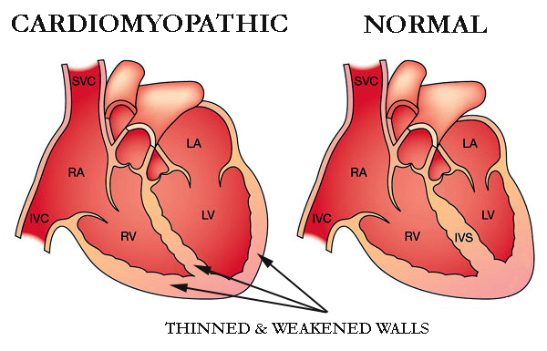
Cardiomyopathy is a condition that progressively affects the heart muscle, or myocardium. The heart muscle typically weakens and becomes less capable of properly pumping blood to the rest of the body.

Cardiomyopathy comes in a variety of forms and is brought on by a number of conditions and medications, including coronary heart disease. Heart failure, an irregular pulse, and other consequences can result from cardiomyopathy.

It’s crucial to receive medical care and continue receiving it. They can lessen the risk of heart disease or heart failure.

Types of Cardiomyopathy

Cardiomyopathy comes in a variety of forms, including the following:

Dilated

Heart palpitations and shortness of breath are two symptoms of cardiomyopathy. The most prevalent type of the condition is dilated cardiomyopathy. Adults between the ages of 20 and 60 are the typical victims.

Although the disease frequently begins in the left ventricle, it can later spread to the right ventricle as well. Additionally, the atria’s structure and operation might be impacted by dilated cardiomyopathy.

Hypertrophic

A hereditary disorder known as hypertrophic cardiomyopathy causes the heart muscle fibres to develop abnormally, thickening or “hypertrophying” as a result. The thickening restricts blood flow and stiffens the heart’s chambers. Additionally, it may raise the chance of electrical disturbances, called arrhythmias.

It is the second most prevalent kind of cardiomyopathy in children, according to the Children’s Cardiomyopathy Foundation. Before the age of one year, a diagnosis is made in around one-third of the affected children.

Restrictive

When the tissues in the ventricles stiffen and cannot properly fill with blood, restrictive cardiomyopathy develops. It might eventually result in cardiac failure. It is more prevalent in elderly persons and may be brought on by infiltrative disorders, such as amyloidosis, which involve the buildup of aberrant substances in physiological tissues.

Arrhythmogenic

In arrhythmogenic cardiomyopathy, the healthy tissues of the right ventricle are replaced by fibrotic and fatty tissue, which may lead to irregular heart beats. This procedure may occasionally take place in the left ventricle as well.

Arrhythmogenic cardiomyopathy increases the risk of sudden cardiac mortality, particularly in young individuals and sports, according to study published in the journal Circulation Research. It is a genetic inherited disorder.

Other types of cardiomyopathy

The majority of the following forms of cardiomyopathy fall under one of the previous four categories, however each has different causes or side effects:

Peripartum cardiomyopathy. Cardiomyopathy peripartum can happen before, during, or after pregnancy. This uncommon kind happens in the final month of pregnancy or within five months after delivery when the heart weakens. Postpartum cardiomyopathy is the term sometimes used to describe it after delivery. This dilated cardiomyopathy variant poses a serious risk to life. One instance raises your chances for subsequent pregnancies.

Alcoholic Cardiomyopathy. Alcoholic cardiomyopathy is caused by consuming excessive amounts of alcohol over time, which can weaken your heart and make it less effective at pumping blood. Additionally, your heart may grow. It’s a type of dilated .cardiomyopathy

Ischemic cardiomyopathy. Ischemic cardiomyopathy happens when coronary artery disease prevents your heart from pumping blood to the rest of your body. Heart muscle blood vessels constrict and become clogged. The cardiac muscle cannot receive oxygen as a result. The most common cause of heart failure is ischemic cardiomyopathy. Alternatively, any form of cardiomyopathy that is unrelated to coronary artery disease is referred to as nonischemic cardiomyopathy.

Noncompaction cardiomyopathy. The medical term for this is spongiform cardiomyopathy. It is a rare condition that manifests at birth and is brought on by faulty heart muscle development during pregnancy. Any stage of life may experience a diagnosis.

Pediatric cardiomyopathy. Pediatric cardiomyopathy is the term used to describe cardiomyopathy that affects children.

Idiopathic cardiomyopathy. Idiopathic cardiomyopathy indicates the reason is unknown.

Symptoms of cardiomyopathy

Early on in the development of cardiomyopathy, there may be neither indications nor symptoms. However, when the illness worsens, several indications and symptoms, such as:

  • a lack of breath during exercise or even while at rest
  • Legs, ankles, and feet swelling
  • abdominal bloating brought on by a fluid buildup
  • Cough when seated
  • difficulty falling asleep flat
  • Fatigue
  • fast, hammering, or fluttering heartbeats
  • chest pressure or discomfort
  • Unsteadiness, fainting, and dizziness

If not treated, symptoms and signs frequently worsen. It depends on the individual whether the illness becomes worse swiftly or gradually.

Causes and risk factors

There are certain well-established risk factors for cardiomyopathy, albeit its exact aetiology is not always known. For instance, disorders that cause the heart to inflame or sustain damage can raise a person’s risk of cardiomyopathy.

Cardiomyopathy can also be brought on by heart failure, which can happen as a result of a heart attack or other medical issues.

Additional risk elements consist of:

  • a history of cardiomyopathy, sudden cardiac death, or heart illness in the family
  • blood pressure is high.
  • cardiovascular disease
  • The diseases sarcoidosis and amyloidosis, which can harm the heart
  • viruses that affect the heart
  • diabetes
  • drinking disorder

After giving birth, certain women may be at an increased risk for cardiomyopathy.

Research from 2016 indicates, cardiomyopathy risk is also increased by HIV and HIV therapies. Heart failure and dilated cardiomyopathy in particular are risks that are increased by HIV.

Talk to your doctor if you have HIV about obtaining frequent testing to monitor the condition of your heart. Additionally, you ought to make an effort to maintain an active and heart-healthy diet.

Diagnosis of cardiomyopathy

To confirm cardiomyopathy, doctors will do a physical examination and diagnostic procedures. They may employ one or more of the diagnostic tests listed below:

  • Chest X-ray: A chest X-ray can assist diagnose some medical disorders by revealing whether the heart has enlarged.
  • Electrocardiogram (EKG): A heart’s electrical activity, including how quickly it beats, is measured by an electrocardiogram (EKG). Additionally, it demonstrates if the heart rhythm is normal or irregular.
  • Echocardiogram: An echocardiogram produces a moving image of the heart using sound waves. It displays the heart’s size and shape.
  • Cardiac catheterization: This procedure examines the blood flow through the heart’s chambers.

How is cardiomyopathy treated?

Treatment for cardiomyopathy focuses on controlling your symptoms. The course of the disease is also slowed by treatment. You will get routine examinations to monitor the condition of your heart.

Your healthcare provider might advise:

Medication: Heart drugs can increase blood flow, address underlying issues, or manage symptoms. You may use beta blockers like propranolol (Inderal), blood thinners like warfarin (Coumadin), or drugs to decrease cholesterol.

Arrhythmia-treating equipment: Pacemakers and implantable cardioverter defibrillators (ICDs) are used to treat erratic heartbeats. Your heartbeat is monitored by these gadgets. When an arrhythmia begins, they send electrical impulses to your heart.

Devices that improve blood flow: Some gadgets make your heart’s blood-pumping process more effective. Devices used in cardiac resynchronization therapy (CRT) regulate the heart’s left and right side contractions. Your heart is assisted in pumping blood with a left ventricular assist device (LVAD).

Surgery: Your doctor might advise heart surgery if you have serious symptoms or underlying heart issues. Open heart surgery or a heart transplant are typically only suggested by healthcare professionals after all other options have failed to provide relief.

REFERENCES:

For more details, kindy visit below.

Lets explore the appendicitis types and its treatment.

Lets explore the appendicitis types and its treatment.

What is appendicitis?

Your appendix can develop appendicitis when it gets inflamed, most frequently as a result of a blockage. It could be acute or ongoing. The most typical cause of stomach discomfort requiring surgery in the US is appendicitis. It affects up to 9% of Americans at some time in their lives.

A little pouch connected to the intestine, the appendix. Your lower right abdomen is where it’s positioned. Bacteria can grow inside your appendix when it becomes obstructed. Pus and edoema may grow as a result, putting pressure on your abdomen that hurts. Blood flow can also be blocked by appendicitis.

Your appendix may rupture if appendicitis is not treated. Bacteria could enter your abdominal cavity as a result, which could be dangerous and occasionally fatal.

Types of appendicitis

Acute appendicitis

A severe and sudden bout of appendicitis is referred to as acute. Between the ages of 10 and 30, children and young adults are the most susceptible and affects men more often than women. Over the course of a day, pain frequently starts out mild and quickly gets worse.

It needs emergency medical attention. It can result in the rupture of your appendix if untreated. This issue has the potential to be lethal.

About 7 to 9 percent of all Americans will experience acute appendicitis in their lifetime, which is more prevalent than chronic appendicitis.

Chronic appendicitis

Less frequently than acute appendicitis, chronic appendicitis. Only 1.5% of persons who have already experienced chronic appendicitis experience it.

The symptoms of chronic appendicitis might be somewhat modest and are generally assumed to follow an episode of acute appendicitis. Over the course of weeks, months, or even years, symptoms may stop and then come back.

The diagnosis of this kind of appendicitis might be difficult. Occasionally, it doesn’t get diagnosed until it turns into acute appendicitis. Appendicitis that persists can be dangerous.

Symptoms of appendicitis

In the early stages of appendicitis, you may have light abdominal cramping that gradually spreads to your lower right abdominal quadrant. This ache frequently:

  • begins abruptly
  • worsens when you cough or move
  • is so strong it wakes you up from sleep
  • is intense and distinct from any prior stomach discomfort that you may have had
  • within a few hours becomes worse

These are some other signs of appendicitis:

  • reduced appetite
  • indigestion
  • nausea
  • vomiting
  • abdominal enlargement
  • minimal fever

You might suffer gastrointestinal issues less frequently, such as:

  • diarrhoea
  • constipation
  • the want to go to the bathroom
  • inability to exhale

Avoid using laxatives or an enema if you have constipation and think you could have appendicitis. Your appendix may rupture as a result of these procedures.

If you experience any other appendicitis symptoms in addition to soreness on the right side of your abdomen, call your doctor straight once. A medical emergency can arise from appendicitis very quickly. Find out all you need to know to identify this dangerous disease.

Call your child’s doctor as soon as you see any appendicitis symptoms in your child.

Causes and risk factors

Appendicitis’ precise cause is frequently unknown. According to experts, it arises when a portion of the appendix becomes clogged or obstructed.

Your appendix may get blocked for a variety of reasons, including:

  • a mass of hardened faeces
  • increased lymphoid follicle size
  • digestive worms
  • a serious injury
  • tumours

Abdominal pain can be brought on by numerous medical conditions. For information on more possible causes of pain in your lower right abdomen, go here.

Anyone can get appendicitis. However, some people can be more prone to this illness than others. Appendicitis risk factors include:

  • Age. Although it can happen at any age, appendicitis most frequently affects adolescents and persons in their 20sTrusted Source.
  • Sex. Males are more likely than females to develop appendicitis.
  • Family background. An increased chance of having appendicitis exists in people with a family history of the condition.

How is Appendicitis Diagnosed?

Appendicitis can be difficult to diagnose. Many times, the symptoms of an illness, such as gallbladder issues, bladder or urinary tract infections, Crohn’s disease, gastritis, kidney stones, intestinal infection, and ovary issues, are vague or resemble those of other conditions.

Appendicitis can be identified using these tests:

  • Checking your abdomen for any irritation
  • To rule out a urinary tract infection, use a urine (pee) test.
  • Rectal examination
  • To determine whether your body is battling an infection, have a blood test
  • A CT scan
  • Ultrasound

Treatment of appendicitis

According to the NIDDK, a doctor will recommend antibiotics to anyone who has appendicitis. This can successfully treat appendicitis in some instances without the need for surgery. Nevertheless, the appendix must typically be removed by a surgeon.

Appendicitis surgery possibilities include:

  • Laparoscopy: This is a precise procedure that only requires a little incision and little blood loss. As a result, there is less scarring and the recovery period is shorter than with open surgery. The following steps are involved in laparoscopic, keyhole, or minimally invasive surgery (MIS):
    • Through a hollow instrument called as a cannula, the surgeon inserts a laparoscope—a very thin tube with a tiny video camera and light—into the abdomen.
    • On a monitor, the surgeon can see a magnified image of the abdomen.
    • Small abdominal incisions are used to remove the appendix, which is done with the aid of tiny instruments that are controlled by the surgeon’s hands.
  • Open surgery: In extremely rare circumstances, a wider incision will be made to allow for thorough cleaning of the abdominal cavity. If the following occurs:
    • An infection has spread after the appendix ruptured.
    • The abscess was brought on by the appendix.
    • patient has digestive system tumours.
    • The patient is a woman who is nine months pregnant.
    • The patient has undergone numerous abdominal procedures.

Therefore, the patient will get intravenous antibiotics following open surgery.

Recovery time for appendicitis

With keyhole surgery, the patient can typically return home after 24 hours. The patient may feel some constipation, soreness, and bruising over the first few days.

Additionally, there could be soreness near the shoulder’s point. The surgeon inflates the abdomen with gas while performing the procedure. This may result in phrenic nerve stimulation at the diaphragm, which brings on referred pain. The real pain cause is not present where the referred pain arises.

Painkillers sold over-the-counter (OTC) may ease postsurgical discomfort. The patient might need to stay in the hospital for up to a week if open surgery is required, or if peritonitis or another complication is present.

For three to five days following a laparoscopic procedure, one should avoid strenuous activities. After a laparotomy, they should refrain from physical exertion for 10–14 days. At each stage of rehabilitation, the doctor will provide guidance on the appropriate level of exercise. It is crucial to call the doctor if there are any infection-related symptoms.

Infection warning signs include:

  • swelling and discomfort getting worse
  • recurring vomiting
  • a high degree
  • It is too hot to touch the operation site.
  • the surgical site is discharged with pus or another material

REFERENCES:

For more details, kindly visit below.

Important note on causes and treatment for brain cancer.

Important note on causes and treatment for brain cancer.

The development of malignant cells in the brain leads to brain cancer. Depending on the type of tumour, the cancer cells produce slow- or fast-growing tumours.

The goal of brain cancer treatment is to remove the tumour and then eradicate any cancer cells that may still be present. In particular for slow-growing tumours, new advancements in brain cancer therapies are increasing survival rates.

What is brain cancer?

Primary brain cancer, commonly referred to as brain cancer, is characterised by an excess of brain cells that develops into masses known as brain tumours. Cancer, on the other hand, begins in another place of the body and progresses to the brain. It is referred to as secondary or metastasized brain cancer when that occurs.

Some malignant brain tumours have a rapid rate of growth. These cancerous tumours may interfere with how your body functions. Brain tumours should be treated as soon as they are discovered since they can be fatal.

Brain tumours are rather rare. People have a less than 1% lifetime chance of getting a malignant brain tumour, according to estimates from the American Cancer Society.

Types of brain tumors

Names for brain tumours are determined by their location within the brain or upper spine. A grade is also assigned to tumours. You can determine the projected rate of growth of a tumour by looking at its grade. Grades range from one to four, with four being the fastest-growing grades and one being the slowest.

The following are some of the most prevalent kinds of brain tumours:

  • Glioma. About 3 out of 10 occurrences of brain cancer are gliomas, which are brain tumours that start in the glial cells.
  • Astrocytoma. Glioblastomas, the kind of quickly-expanding brain tumour, are a subtype of astrocytomas.
  • Meningioma. Meningioma tumours, the most prevalent kind of brain tumour in adults, develop in the tissue that surrounds your brain and spinal cord and are frequently benign and slow-growing.
  • Ganglioglioma. Surgery is typically used to treat the slow-growing tumours known as gangliogliomas that are present in glial and neuronal cells.
  • Craniopharyngiomas. Craniopharyngiomas are slow-growing tumours that develop between the pituitary gland and the brain. Because they frequently encroach on the optic nerves, they can impair eyesight.
  • Schwannomas. Almost typically benign, schwannomas are slow-growing tumours that develop around the cranial nerves.
  • Medulloblastoma. Children are more likely to develop medulloblastomas, which are rapidly developing tumours that develop on the nerve cells in the brain.

Symptoms of brain cancer

The size and location of the brain tumour affect the symptoms of brain cancer. Particularly in its early stages, brain cancer exhibits many of the same symptoms as a number of less dangerous diseases.

Numerous of these symptoms are extremely typical and are not likely to be signs of brain cancer. However, it’s a good idea to see a doctor if you’ve had any of these symptoms for longer than a week, if they came on suddenly, if they don’t go away with over-the-counter painkillers, or if any of them worry you.

Typical signs of brain cancer include:

  • morning headaches that are typically worse
  • nausea
  • vomiting
  • a miscommunication
  • a loss of equilibrium
  • having trouble walking
  • Memory problems
  • having trouble thinking
  • speech issues
  • vision issues
  • personality alters
  • inconsistent eye motions
  • body jerking
  • muscle jerking
  • unexpected fainting or syncope
  • drowsiness
  • tingling or numbness in the arms or legs
  • seizures

Early diagnosis significantly improves the prognosis for brain cancer. If you frequently suffer any of the aforementioned symptoms or suspect that your symptoms may be more serious, schedule an appointment with a doctor right away for a diagnosis.

Causes and risk factors

Primary brain cancer has an unidentified specific cause. However, studies have connected excessive ionising radiation exposure to a higher chance of developing brain cancer. The most frequent sources of ionising radiation include radiation therapy treatments, frequent medical imaging tests (CT scans and X-rays), and potential employment exposure.

Additional factors that could increase the risk of acquiring brain cancer include:

  • greater age
  • a history of brain cancer in the family
  • chronic smoking
  • being exposed to fertilisers, pesticides, and herbicides
  • working with substances like lead, plastic, rubber, petroleum, and some textiles that might cause cancer
  • having mononucleosis or Epstein-Barr virus infection

Secondary brain cancer Some types of cancer are more likely than others to cause brain cancer, which develops when cancer that started in another part of your body travels to your brain.

The following cancers frequently metastasis, or spread, to the brain:

  • lung disease
  • mammary cancer
  • renal cancer
  • urethral cancer
  • melanoma, a form of skin cancer

How is brain cancer treated?

Brain cancer can be treated in a number of ways. A primary brain cancer will be treated differently than a cancer that has spread to other organs.

The kind, size, and location of your brain tumour will determine whether you receive one therapy or more. There will also be considerations for your age and general health.

Treatments for brain tumours include:

Surgery.

The most frequent form of treatment for brain tumours is brain surgery. Depending on the tumor’s position, it might be possible to remove it whole, partially, or not at all.

Chemotherapy.

These medications can reduce your tumour and kill brain cancer cells. Chemotherapy can be administered orally or intravenously.

Radiation treatment. 

Using high-energy waves like X-rays, this procedure eliminates cancer cells and tumour tissue that can’t be removed surgically.

Combination treatment.

Combination therapy refers to the simultaneous administration of chemotherapy and radiation therapy.

Biologic medicines

These medications support, guide, or restore your body’s natural tumour defences. For instance, immunotherapy is a class of biologic medication that is frequently prescribed and increases your immune system’s capacity to recognise and combat cancer.

Other medicines.

To treat symptoms and adverse effects brought on by your brain tumour and brain cancer therapies, your doctor may prescribe drugs.

Clinical studies.

Clinical trial medicines and drugs may be employed in advanced brain cancer instances that don’t respond to treatment. These are medications that are still being tested. An immunotherapy trial and a CAR T cell therapy trial may be part of clinical trials for brain cancer.

Rehabilitation.

If your disease or treatment have made it difficult for you to speak, walk, or perform other daily tasks, you might need to go to rehabilitation sessions. Physical therapy, occupational therapy, and other types of therapies are all included in rehabilitation. These treatments can assist you in relearnng activities.

Various forms of treatment.

There isn’t much evidence to back up the use of complementary medicines to treat brain cancer. To make up for the nutrients lost during cancer treatment, some medical specialists do advise taking measures like following a bland diet and taking vitamin and mineral supplements. 

Before making any dietary changes, using any herbs or supplements, or pursuing any alternative treatments, see your doctor.

How to reduce your risk of brain cancer?

Although there is no known way to prevent brain cancer, you can lower your risk by staying away from:

  • pesticide and insecticide exposure
  • exposure to cancer-causing substances
  • smoking
  • radiation exposure that is not essential

REFERENCES:

  • https://www.healthline.com/health/brain-cancer
  • https://www.cancercenter.com/cancer-types/brain-cancer
  • https://www.mayoclinic.org/diseases-conditions/brain-tumor/symptoms-causes/syc-20350084

For more details, kindly visit below.